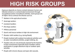
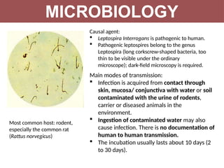
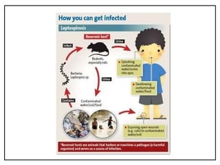
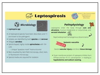
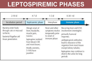
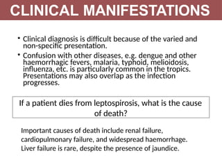
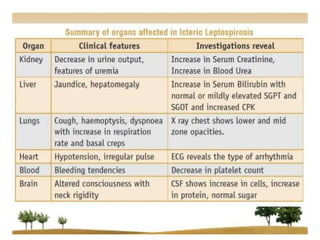
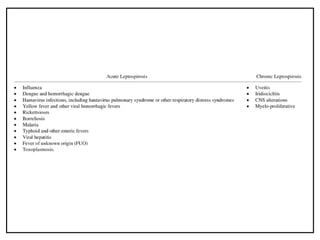
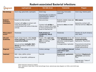
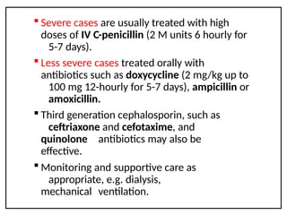
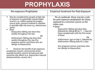
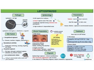
LEPTOSPIROSIS
INTRODUCTION
• Leptospirosis is an infectious
disease caused by pathogenic
bacteria called leptospires, that
are transmitted directly or
indirectly from animals to
humans. It is therefore a
zoonosis. Human-to- human
transmission occurs only very
rarely.
• It often peaks seasonally,
sometimes in outbreaks, and is
often linked to climate changes,
to poor urban slum
communities, to occupation
or to recreational activities.
• The clinical course in humans ranges
from mild to lethal with a broad
spectrum of symptoms and
clinical signs.
Leptospirosis occurs worldwide but is most common in
tropical and subtropical areas with high rainfall. The disease is
found mainly wherever humans come into contact with the
urine of infected animals or a urine-polluted environment.
DISTRIBUTION
INCIDENCE
 The number of human cases worldwide is not known precisely.
 Estimated annual incidence (WHO)
—0.1 to 1 per 100 000 per year in temperate climates
—10 or more per 100 000 per year in the humid tropics
 Estimated case-fatality rates in different parts of the world have been
reported to range from <5% to 30%
 Seasonal – peak in summer, during rainy/monsoon season
Why is there a lack of
recognition of leptospirosis?
 Clinical manifestation wide and varied
 May mimic many other diseases, e.g.
dengue fever and other viral
haemorrhagic diseases
 Diagnostic capabilities are not readily
available (especially in countries
where the disease is highly endemic)
 poor surveillance and reporting of
cases
Exposure depends on chance contacts between human and
infected animals or a contaminated environment through
occupational and/or recreational activities. Some groups are
at higher risk to contract the disease such as:
 Workers in the agricultural sectors
 Sewerage workers
 Livestock handlers
 Pet shops workers
 Military personnel
 Search and rescue workers in high risk environment
 Disaster relief workers (e.g. during floods)
 People involved with outdoor/recreational activities such
as water recreational activities, jungle trekking, etc.
 Travelers who are not previously exposed to the bacteria
in their environment especially those travellers and/or
participants in jungle adventure trips or outdoor sport
activities
 People with chronic disease and open skin wounds.
HIGH RISK GROUPS
MICROBIOLOGY
Causal agent:
 Leptospira Interrogans is pathogenic to human.
 Pathogenic leptospires belong to the genus
Leptospira (long corkscrew-shaped bacteria, too
thin to be visible under the ordinary
microscope); dark-field microscopy is required.
Main modes of transmission:
 Infection is acquired from contact through
skin, mucosa/ conjunctiva with water or soil
contaminated with the urine of rodents,
carrier or diseased animals in the
environment.
 Ingestion of contaminated water may also
cause infection. There is no documentation of
human to human transmission.
 The incubation usually lasts about 10 days (2
to 30 days).
Most common host: rodent,
especially the common rat
(Rattus norvegicus)
• Infection  leptospires appear in the blood 
invade all tissues and organs particularly
affecting the liver and kidney  cleared from
the body by the host's immune response
• May also settle in the convoluted tubules of the
kidneys  shed in the urine for a few weeks
to several months or longer
• Subsequently cleared from the kidneys and other
organs (may persist in the eyes for much
longer)
• Produces endotoxin  attach onto the endothelial
cells  capillary vasculitis (endothelial necrosis
and lymphocytic infiltration)
PATHOPHYSIOLOGY
• Vasculitis and leakage  petechiae,
intraparenchymal bleeding and bleeding along
serosa and mucosa
• Lost of fluids into the third space  hypovolaemic
shock and vascular collapse
• Humans react to an infection by producing specific
anti-Leptospira antibodies
• Seroconversion – as early as 5–7 days after the
onset of disease – sometimes only after 10 days or
longer – IgM appear somewhat earlier than IgG
and generally remain detectable for months or
even years but at low titre.
PATHOPHYSIOLOGY
CLASSIFICATION
LEPTOSPIREMIC PHASES
Figure 1: Leptospiremic phases in conjunction with the laboratory methods of
diagnosis.
Note: Biphasic nature of leptospirosis and relevant investigations at different stages
of disease. Specimens 1 and 2 for serology are acute-phase specimens, 3 is a
convalescent-phase sample which may facilitate detection of a delayed immune
response, and 4 and 5 are follow-up samples which can provide epidemiological
information, such as the presumptive infecting serogroup
• The incubation period is usually 10 days, with a
range of 2 to 30 days.
• The clinical manifestations are highly variable.
Typically, the disease presents in four broad clinical
categories:
(i) a mild, influenza-like illness (ILI);
(ii)Weil's syndrome characterized by jaundice, renal
failure, haemorrhage and myocarditis with arrhythmias;
(iii) meningitis / meningoencephalitis;
(iv) pulmonary haemorrhage with respiratory failure.
CLINICAL MANIFESTATIONS
• Clinical diagnosis is difficult because of the varied and
non-specific presentation.
• Confusion with other diseases, e.g. dengue and other
haemorrhagic fevers, malaria, typhoid, melioidosis,
influenza, etc. is particularly common in the tropics.
Presentations may also overlap as the infection
progresses.
CLINICAL MANIFESTATIONS
If a patient dies from leptospirosis, what is the cause
of death?
Important causes of death include renal failure,
cardiopulmonary failure, and widespread haemorrhage.
Liver failure is rare, despite the presence of jaundice.
Multiorgan involvement
 Ocular
—Suffusion – dilation of the conjunctival vasculature,
subconjuctival haemorrhage, uveitis
—Icterus scleral with conjunctival suffusion-pathognomic of
Weil’s disease
 GI
– Jaundice not associated with hepatocellular necrosis.
Bilirubin, ALT, AST will normalise
 Renal
– Acute tubular necrosis (direct leptospire injury)
– Interstitial nephritis (relate to Ag-Ab complexes)
CLINICAL MANIFESTATIONS
 Cardiac – Myocarditis, 1st degree heart block, coronary
arteritis
 Pulmonary
–Spectrum ranging from cough, dyspnoea,
haemoptysis to ARDS
– Pulmonary haemorrhage may cause death
–Radiology reveals diffuse small opacities which
may disseminate or coalesce
– a sign of intra-alveolar and interstitial haemorrhage
CLINICAL MANIFESTATIONS
If patients survive, do they
fully recover from
leptospirosis?
 Most patients recover completely from leptospirosis. In some patients, however,
recovery may take months or even years. Late sequelae may occur.
What are the late sequelae
in leptospirosis?
 Late sequelae include chronic fatigue and other neuropsychiatric symptoms such
as headache, paresis, paralysis, mood swings and depression. In some cases,
uveitis and iridocyclitis may be a late presentation of leptospirosis.
 Ocular symptoms are probably attributable to the persistence of leptospires in the
eyes, where they are sheltered from the patient's immune response.
 Apart from eye involvement, the pathogenesis of alleged late or persistent
symptoms is unknown. The existence of persistent or chronic infections has not
been confirmed and "scars" caused during the acute disease have not been
demonstrated.
HOW TO
DIAGNOSE ?
CASE CLASSIFICATION
Leptospirosis is difficult to distinguish from a number of other diseases on clinical
grounds alone. History of possible exposure is paramount to aid clinical diagnosis.
CLINICAL CASE
A case that is compatible with the following clinical description:
 Acute febrile illness with history of exposure to water and/or environment
possibly contaminated with infected animal urine with ANY of the following
symptoms: Headache
 Myalgia particularly associated with the calf muscles and lumbar region
 Arthralgia
 Conjunctival suffusion
 Meningeal irritation
 Anuria or oliguria and/or proteinuria
 Jaundice
 Hemorrhages (from the intestines and lungs)
 Cardiac arrhythmia or failure
 Skin rash
 Gastrointestinal symptoms such as nausea, vomiting, abdominal pain,
diarrhoea
CASE CLASSIFICATION
PROBABLE CASE
A clinical case AND positive ELISA/other Rapid tests.
CONFIRMED CASE
A confirmed case of leptospirosis is a suspected OR probable case with any one of
the following laboratory tests:
 Microscopic Agglutination Test (MAT),
For single serum specimen - titre ≥1:400
For paired sera - four fold or greater rise
in titre
 Positive PCR (samples should be taken
within 10 days of disease onset)
 Positive culture for pathogenic leptospires (blood samples should be taken
within 7 days of onset and urine sample after the 10 th day)
 Demonstration of leptospires in tissues using immunohistochemical
staining
(e.g. in post mortem cases)
 In places where the laboratory capacity is not well established, a case can be
considered as confirmed if the result is positive by 2 different rapid diagnostic
tests.
 Cases that require confirmation are:
i. Hospitalized cases
ii. All suspected leptospirosis death cases
DIFFERENTIAL DIAGNOSIS?
TREATMENT
 Severe cases are usually treated with high
doses of IV C-penicillin (2 M units 6 hourly for
5-7 days).
 Less severe cases treated orally with
antibiotics such as doxycycline (2 mg/kg up to
100 mg 12-hourly for 5-7 days), ampicillin or
amoxicillin.
 Third generation cephalosporin, such as
ceftriaxone and cefotaxime, and
quinolone antibiotics may also be
effective.
 Monitoring and supportive care as
appropriate, e.g. dialysis,
mechanical ventilation.
Pre-exposure Prophylaxis Empirical treatment for Post-Exposure
 May be considered for people at high risk
of exposure to potentially contaminated
sources e.g. soldiers going into jungles,
rescue team, persons involved in activities
in possible high risk areas e.g. adventurous
sports.
 Dose:
Doxycycline 200mg stat dose then
weekly throughout the stay
OR
Azithromycin 500mg stat dose then
weekly throughout the stay (For
pregnant women and those who are
allergic to Doxycycline)
 However the benefit of pre-exposure
prophylaxis remains controversial where
possible benefits need to be balanced with
potential side effects (e.g. doxycycline induced
photosensitivity, nausea, etc.)
In an outbreak, there may be a role
for post exposure prophylaxis for those
exposed to a common source as the
index case.
 Dose:
Doxycycline 200mg stat dose then
followed by 100mg BD for 5 – 7 days for
those symptomatic with the first onset
of fever.
OR
Azithromycin 1gm on Day-1, followed by
Azithromycin 500mg daily for 2 days
(For pregnant women and those who
are allergic to Doxycycline)
PROPHYLAXIS
Leptospirosis notes IM full notes with clinical

Leptospirosis notes IM full notes with clinical

  • 1.
  • 2.
    INTRODUCTION • Leptospirosis isan infectious disease caused by pathogenic bacteria called leptospires, that are transmitted directly or indirectly from animals to humans. It is therefore a zoonosis. Human-to- human transmission occurs only very rarely. • It often peaks seasonally, sometimes in outbreaks, and is often linked to climate changes, to poor urban slum communities, to occupation or to recreational activities. • The clinical course in humans ranges from mild to lethal with a broad spectrum of symptoms and clinical signs.
  • 3.
    Leptospirosis occurs worldwidebut is most common in tropical and subtropical areas with high rainfall. The disease is found mainly wherever humans come into contact with the urine of infected animals or a urine-polluted environment. DISTRIBUTION INCIDENCE  The number of human cases worldwide is not known precisely.  Estimated annual incidence (WHO) —0.1 to 1 per 100 000 per year in temperate climates —10 or more per 100 000 per year in the humid tropics  Estimated case-fatality rates in different parts of the world have been reported to range from <5% to 30%  Seasonal – peak in summer, during rainy/monsoon season
  • 4.
    Why is therea lack of recognition of leptospirosis?  Clinical manifestation wide and varied  May mimic many other diseases, e.g. dengue fever and other viral haemorrhagic diseases  Diagnostic capabilities are not readily available (especially in countries where the disease is highly endemic)  poor surveillance and reporting of cases
  • 5.
    Exposure depends onchance contacts between human and infected animals or a contaminated environment through occupational and/or recreational activities. Some groups are at higher risk to contract the disease such as:  Workers in the agricultural sectors  Sewerage workers  Livestock handlers  Pet shops workers  Military personnel  Search and rescue workers in high risk environment  Disaster relief workers (e.g. during floods)  People involved with outdoor/recreational activities such as water recreational activities, jungle trekking, etc.  Travelers who are not previously exposed to the bacteria in their environment especially those travellers and/or participants in jungle adventure trips or outdoor sport activities  People with chronic disease and open skin wounds. HIGH RISK GROUPS
  • 6.
    MICROBIOLOGY Causal agent:  LeptospiraInterrogans is pathogenic to human.  Pathogenic leptospires belong to the genus Leptospira (long corkscrew-shaped bacteria, too thin to be visible under the ordinary microscope); dark-field microscopy is required. Main modes of transmission:  Infection is acquired from contact through skin, mucosa/ conjunctiva with water or soil contaminated with the urine of rodents, carrier or diseased animals in the environment.  Ingestion of contaminated water may also cause infection. There is no documentation of human to human transmission.  The incubation usually lasts about 10 days (2 to 30 days). Most common host: rodent, especially the common rat (Rattus norvegicus)
  • 9.
    • Infection leptospires appear in the blood  invade all tissues and organs particularly affecting the liver and kidney  cleared from the body by the host's immune response • May also settle in the convoluted tubules of the kidneys  shed in the urine for a few weeks to several months or longer • Subsequently cleared from the kidneys and other organs (may persist in the eyes for much longer) • Produces endotoxin  attach onto the endothelial cells  capillary vasculitis (endothelial necrosis and lymphocytic infiltration) PATHOPHYSIOLOGY
  • 10.
    • Vasculitis andleakage  petechiae, intraparenchymal bleeding and bleeding along serosa and mucosa • Lost of fluids into the third space  hypovolaemic shock and vascular collapse • Humans react to an infection by producing specific anti-Leptospira antibodies • Seroconversion – as early as 5–7 days after the onset of disease – sometimes only after 10 days or longer – IgM appear somewhat earlier than IgG and generally remain detectable for months or even years but at low titre. PATHOPHYSIOLOGY
  • 12.
  • 13.
  • 14.
    Figure 1: Leptospiremicphases in conjunction with the laboratory methods of diagnosis. Note: Biphasic nature of leptospirosis and relevant investigations at different stages of disease. Specimens 1 and 2 for serology are acute-phase specimens, 3 is a convalescent-phase sample which may facilitate detection of a delayed immune response, and 4 and 5 are follow-up samples which can provide epidemiological information, such as the presumptive infecting serogroup
  • 15.
    • The incubationperiod is usually 10 days, with a range of 2 to 30 days. • The clinical manifestations are highly variable. Typically, the disease presents in four broad clinical categories: (i) a mild, influenza-like illness (ILI); (ii)Weil's syndrome characterized by jaundice, renal failure, haemorrhage and myocarditis with arrhythmias; (iii) meningitis / meningoencephalitis; (iv) pulmonary haemorrhage with respiratory failure. CLINICAL MANIFESTATIONS
  • 17.
    • Clinical diagnosisis difficult because of the varied and non-specific presentation. • Confusion with other diseases, e.g. dengue and other haemorrhagic fevers, malaria, typhoid, melioidosis, influenza, etc. is particularly common in the tropics. Presentations may also overlap as the infection progresses. CLINICAL MANIFESTATIONS If a patient dies from leptospirosis, what is the cause of death? Important causes of death include renal failure, cardiopulmonary failure, and widespread haemorrhage. Liver failure is rare, despite the presence of jaundice.
  • 18.
    Multiorgan involvement  Ocular —Suffusion– dilation of the conjunctival vasculature, subconjuctival haemorrhage, uveitis —Icterus scleral with conjunctival suffusion-pathognomic of Weil’s disease  GI – Jaundice not associated with hepatocellular necrosis. Bilirubin, ALT, AST will normalise  Renal – Acute tubular necrosis (direct leptospire injury) – Interstitial nephritis (relate to Ag-Ab complexes) CLINICAL MANIFESTATIONS
  • 19.
     Cardiac –Myocarditis, 1st degree heart block, coronary arteritis  Pulmonary –Spectrum ranging from cough, dyspnoea, haemoptysis to ARDS – Pulmonary haemorrhage may cause death –Radiology reveals diffuse small opacities which may disseminate or coalesce – a sign of intra-alveolar and interstitial haemorrhage CLINICAL MANIFESTATIONS
  • 21.
    If patients survive,do they fully recover from leptospirosis?  Most patients recover completely from leptospirosis. In some patients, however, recovery may take months or even years. Late sequelae may occur. What are the late sequelae in leptospirosis?  Late sequelae include chronic fatigue and other neuropsychiatric symptoms such as headache, paresis, paralysis, mood swings and depression. In some cases, uveitis and iridocyclitis may be a late presentation of leptospirosis.  Ocular symptoms are probably attributable to the persistence of leptospires in the eyes, where they are sheltered from the patient's immune response.  Apart from eye involvement, the pathogenesis of alleged late or persistent symptoms is unknown. The existence of persistent or chronic infections has not been confirmed and "scars" caused during the acute disease have not been demonstrated.
  • 22.
  • 23.
    CASE CLASSIFICATION Leptospirosis isdifficult to distinguish from a number of other diseases on clinical grounds alone. History of possible exposure is paramount to aid clinical diagnosis. CLINICAL CASE A case that is compatible with the following clinical description:  Acute febrile illness with history of exposure to water and/or environment possibly contaminated with infected animal urine with ANY of the following symptoms: Headache  Myalgia particularly associated with the calf muscles and lumbar region  Arthralgia  Conjunctival suffusion  Meningeal irritation  Anuria or oliguria and/or proteinuria  Jaundice  Hemorrhages (from the intestines and lungs)  Cardiac arrhythmia or failure  Skin rash  Gastrointestinal symptoms such as nausea, vomiting, abdominal pain, diarrhoea
  • 24.
    CASE CLASSIFICATION PROBABLE CASE Aclinical case AND positive ELISA/other Rapid tests. CONFIRMED CASE A confirmed case of leptospirosis is a suspected OR probable case with any one of the following laboratory tests:  Microscopic Agglutination Test (MAT), For single serum specimen - titre ≥1:400 For paired sera - four fold or greater rise in titre  Positive PCR (samples should be taken within 10 days of disease onset)  Positive culture for pathogenic leptospires (blood samples should be taken within 7 days of onset and urine sample after the 10 th day)  Demonstration of leptospires in tissues using immunohistochemical staining (e.g. in post mortem cases)  In places where the laboratory capacity is not well established, a case can be considered as confirmed if the result is positive by 2 different rapid diagnostic tests.  Cases that require confirmation are: i. Hospitalized cases ii. All suspected leptospirosis death cases
  • 28.
  • 31.
  • 32.
     Severe casesare usually treated with high doses of IV C-penicillin (2 M units 6 hourly for 5-7 days).  Less severe cases treated orally with antibiotics such as doxycycline (2 mg/kg up to 100 mg 12-hourly for 5-7 days), ampicillin or amoxicillin.  Third generation cephalosporin, such as ceftriaxone and cefotaxime, and quinolone antibiotics may also be effective.  Monitoring and supportive care as appropriate, e.g. dialysis, mechanical ventilation.
  • 33.
    Pre-exposure Prophylaxis Empiricaltreatment for Post-Exposure  May be considered for people at high risk of exposure to potentially contaminated sources e.g. soldiers going into jungles, rescue team, persons involved in activities in possible high risk areas e.g. adventurous sports.  Dose: Doxycycline 200mg stat dose then weekly throughout the stay OR Azithromycin 500mg stat dose then weekly throughout the stay (For pregnant women and those who are allergic to Doxycycline)  However the benefit of pre-exposure prophylaxis remains controversial where possible benefits need to be balanced with potential side effects (e.g. doxycycline induced photosensitivity, nausea, etc.) In an outbreak, there may be a role for post exposure prophylaxis for those exposed to a common source as the index case.  Dose: Doxycycline 200mg stat dose then followed by 100mg BD for 5 – 7 days for those symptomatic with the first onset of fever. OR Azithromycin 1gm on Day-1, followed by Azithromycin 500mg daily for 2 days (For pregnant women and those who are allergic to Doxycycline) PROPHYLAXIS