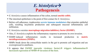
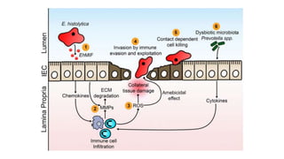
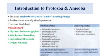
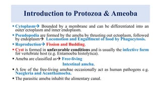
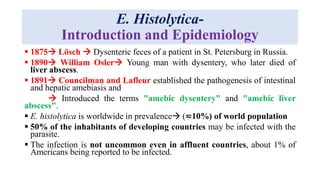
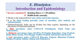
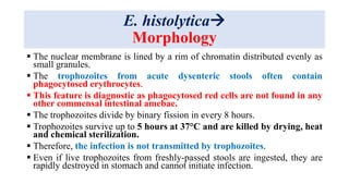
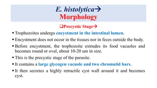
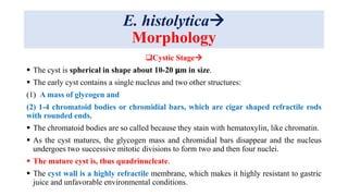
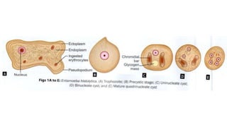
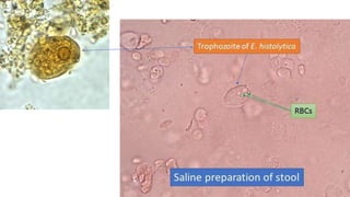
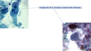
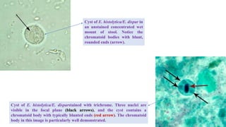
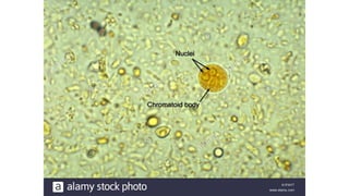
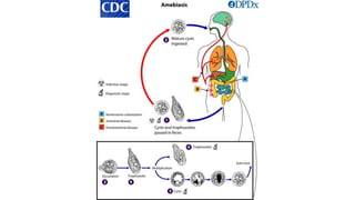
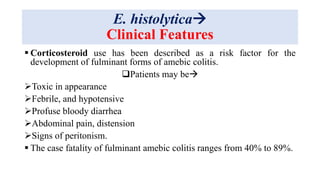
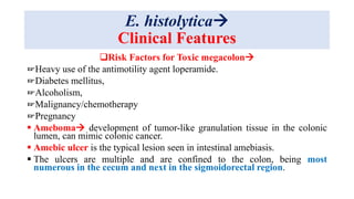
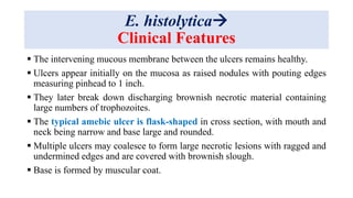
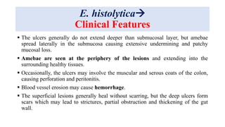
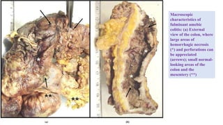
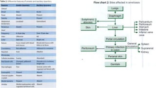
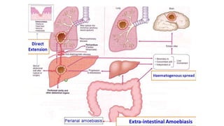
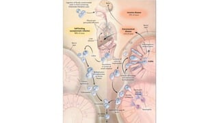
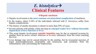
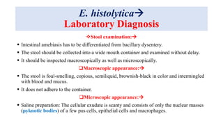
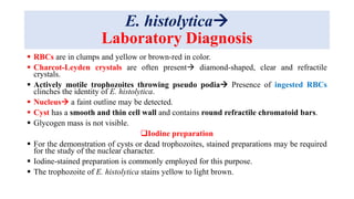
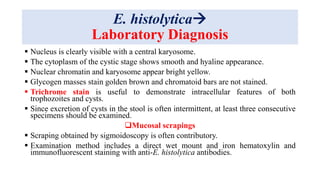
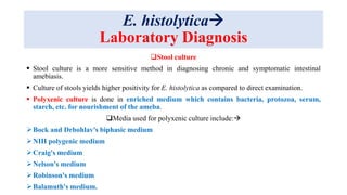
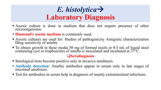
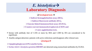
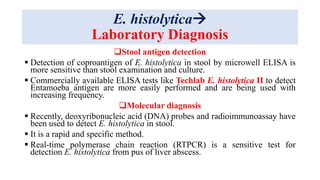
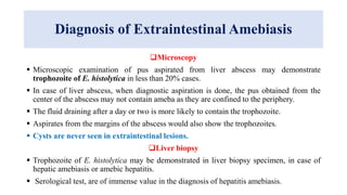
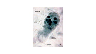
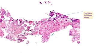
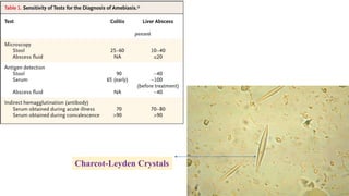
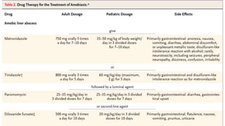
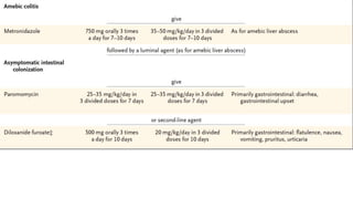
E. histolytica
Dr. Suprakash Das
Assist. Prof.
Introduction to Protozoa & Ameoba
 Single-celled eukaryotic microorganisms belonging to kingdom Protista are classified
as Protozoa (Greek protos: first; zoon: animal).
 Parasitic protozoa are adapted to different host species. Out of 10,000 species of parasitic
protozoa, man harbours only about 70 species.
GENERAL FEATURES
 The single protozoal cell performs all functions.
 Most of the protozoa are completely nonpathogenic but few may cause major diseases
such as malaria, leishmaniasis and sleeping sickness.
 Protozoa like Cryptosporidium parvum and Toxoplasma gondii are being recognized as
opportunistic pathogens in patients affected with human immunodeficiency virus (HIV)
and in those undergoing immunosuppressive therapy.
 Protozoa exhibit wide range of size (1-150 um), shape and structure; yet all possess
essential common features.
Introduction to Protozoa & Ameoba
 The word amebaGreek word "amibe” meaning change.
 Amebae are structurally simple protozoan.
 Have no fixed shape.
Taxonomy
Phylum: Sarcomastigophora,
Subphylum: Sarcodina,
Superclass: Rhizopoda
Order: Amebida
Introduction to Protozoa & Ameoba
 Cytoplasm Bounded by a membrane and can be differentiated into an
outer ectoplasm and inner endoplasm.
 Pseudopodia are formed by the ameba by thrusting out ectoplasm, followed
by endoplasm Locomotion and Engulfment of food by Phagocytosis.
 Reproduction Fission and Budding.
 Cyst is formed in unfavorable conditions and is usually the infective form
for vertebrate host (e.g. Entamoeba histolytica).
 Ameba are classified as Free-living
Intestinal ameba.
 A few of the free-living amebae occasionally act as human pathogens e.g.
Naegleria and Acanthamoeba.
 The parasitic ameba inhabit the alimentary canal.
E. Histolytica-
Introduction and Epidemiology
 1875 Lösch  Dysenteric feces of a patient in St. Petersburg in Russia.
 1890 William Osler Young man with dysentery, who later died of
liver abscess.
 1891 Councilman and Lafleur established the pathogenesis of intestinal
and hepatic amebiasis and
 Introduced the terms "amebic dysentery" and "amebic liver
abscess".
 E. histolytica is worldwide in prevalence (≂10%) of world population
 50% of the inhabitants of developing countries may be infected with the
parasite.
 The infection is not uncommon even in affluent countries, about 1% of
Americans being reported to be infected.
E. Histolytica-
Introduction and Epidemiology
 Invasive amebiasis disabling illness in (≂50 million)
 50,000 deaths annually.
 Mostly in the tropical belt of Asia, Africa and Latin America.
 It is the third leading parasitic cause of mortality, after malaria and
schistosomiasis.
 Epidemiologically, India can be divided into three regions, depending on the
prevalence of intestinal amebiasis:
1. High prevalence states (>30%): Chandigarh, Tamil Nadu and
Maharashtra.
2. Moderate prevalence states (10-30%): Punjab, Rajasthan, Uttar Pradesh,
Delhi, Bihar, Assam, West Bengal, Andhra Pradesh, Karnataka and Kerala.
3. Low prevalence states (<10%): Haryana, Gujarat, Himachal Pradesh,
Madhya Pradesh, Odisha, Sikkim and Puducherry.
W.T Councilman
E. histolytica
Morphology
E. histolytica occurs in three forms:
1. Trophozoite
2. Precyst
3. Cyst.
Trophozoite
 Trophozoite is the vegetative or growing stage of the parasite.
 It is the only form present in tissues.
 It is irregular in shape and varies in size from 12-60 𝜇m; average being 20 𝜇m.
 It is large and actively motile in freshly-passed dysenteric stool, while smaller in
convalescents and carriers.
 The parasite, as it occurs free in the lumen as a commensal is generally smaller in
size, about 15-20 𝜇m and has been called the minuta form.
E. histolytica
Morphology
Cytoplasm
 Outer ectoplasm is clear, transparent and refractile.
 Inner endoplasm is finely granular, having a ground glass appearance.
 The endoplasm contains nucleus, food vacuoles, erythrocytes, occasionally
leukocytes and tissue debris.
 Pseudopodia are finger-like projections formed by sudden jerky
movements of ectoplasm in one direction, followed by the streaming in
of the whole endoplasm.
 Typical ameboid motility is a crawling or gliding movement.
 The direction of movement may be changed suddenly, with another
pseudopodium being formed at a different site, when the whole cytoplasm
flows in the direction of the new pseudopodium.
E. histolytica
Morphology
 The cell has to be attached to some surface or particle for it to move.
 In culture tubes, the trophozoites may be seen crawling up the side of the
glass tube.
 Pseudopodia formation and motility are inhibited at low temperatures.
Nucleus
 Spherical 4-6 um in size and contains central karyosome, surrounded by
clear halo.
 Anchored to the nuclear membrane by fine radiating fibrils called the
limin network, giving a cartwheel appearance.
 The nucleus is not clearly seen in the living trophozoites, but can be clearly
demonstrated in preparations stained with iron hematoxylin.
E. histolytica
Morphology
 The nuclear membrane is lined by a rim of chromatin distributed evenly as
small granules.
 The trophozoites from acute dysenteric stools often contain
phagocytosed erythrocytes.
 This feature is diagnostic as phagocytosed red cells are not found in any
other commensal intestinal amebae.
 The trophozoites divide by binary fission in every 8 hours.
 Trophozoites survive up to 5 hours at 37°C and are killed by drying, heat
and chemical sterilization.
 Therefore, the infection is not transmitted by trophozoites.
 Even if live trophozoites from freshly-passed stools are ingested, they are
rapidly destroyed in stomach and cannot initiate infection.
E. histolytica
Morphology
Precystic Stage
 Trophozoites undergo encystment in the intestinal lumen.
 Encystment does not occur in the tissues nor in feces outside the body.
 Before encystment, the trophozoite extrudes its food vacuoles and
becomes round or oval, about 10-20 um in size.
 This is the precystic stage of the parasite.
 It contains a large glycogen vacuole and two chromatid bars.
 It then secretes a highly retractile cyst wall around it and becomes
cyst.
E. histolytica
Morphology
Cystic Stage
 The cyst is spherical in shape about 10-20 𝛍m in size.
 The early cyst contains a single nucleus and two other structures:
(1) A mass of glycogen and
(2) 1-4 chromatoid bodies or chromidial bars, which are cigar shaped refractile rods
with rounded ends.
 The chromatoid bodies are so called because they stain with hematoxylin, like chromatin.
 As the cyst matures, the glycogen mass and chromidial bars disappear and the nucleus
undergoes two successive mitotic divisions to form two and then four nuclei.
 The mature cyst is, thus quadrinucleate.
 The cyst wall is a highly refractile membrane, which makes it highly resistant to gastric
juice and unfavorable environmental conditions.
Trophozoite of E. histolytica stained with trichrome.
Cyst of E. histolytica/E. dispar in
an unstained concentrated wet
mount of stool. Notice the
chromatoid bodies with blunt,
rounded ends (arrow).
Cyst of E. histolytica/E. disparstained with trichrome. Three nuclei are
visible in the focal plane (black arrows), and the cyst contains a
chromatoid body with typically blunted ends (red arrow). The chromatoid
body in this image is particularly well demonstrated.
E. histolytica
Life Cycle
 E. histolytica passes its life cycle only in one host Man.
Infective Form
 Mature quadrinucleate cyst passed in feces of convalescents and carriers, remain viable under
moist conditions for about 10 days.
Mode of Transmission
 Man acquires infection by swallowing food and water contaminated with cysts.
 Cyst wall is resistant to action of gastric juice, pass through the stomach undamaged and enter the
small intestine.
Excystation:
 When the cyst reaches cecum or lower part of the ileum, due to the alkaline medium, the cyst
wall is damaged by trypsin, leading to excystation.
 The cytoplasm gets detached from the cyst wall through which quadrinucleate ameba is
liberated.
 This stage is called the metacyst.
E. histolytica
Life Cycle
 The nuclei in the metacyst immediately undergo division to form eight nuclei, each of
which gets surrounded by its own cytoplasm to become eight small amebulae or
metacystic trophozoites.
 The optimal habitat for the metacystic trophozoite is the submucosal tissue of cecum
and colon, where they lodge in the glandular crypts and grow by binary fission.
 Some develop into precystic forms and cysts, which are passed in feces to repeat the
cycle.
 In most of the cases, E. histolytica remains as a commensal in the large intestine without
causing any ill effects.
 Such persons become carriers or symptomatic cyst passers and are responsible for
maintenance and spread of infection in the community.
 Sometimes, the infection may be activated and clinical disease ensues.
 Latency and reactivation are the characteristics of amebiasis.
E. histolytica
Pathogenesis
 E. histolytica pathogenesis can be characterized by 3 events:
Host cell death,
Inflammation, and
Parasite invasion.
 Contact with host cells is required for parasite-induced death.
 Adherence is mediated through the parasite Gal/GalNAc lectin adhesion molecule.
 Trophozoites are able to kill host cells by several different mechanisms
Inducing programmed cell death,
Phagocytosis
Trogocytosis(“trogo-” means “nibble”).
 During trogocytosis, amoebae kill human cells by extracting and ingesting “bites” of human cell
membrane and intracellular contents.
E. histolytica
Pathogenesis
 E. histolytica causes inflammation of the colon, termed amebic colitis.
 The intestinal epithelium is the point of first contact for E. histolytica.
 Before cell adherence, trophozoites secrete immune modulators that stimulate epithelial
cells, resulting incytokine production and subsequent mucosal inflammatory cell
infiltration.
 E. histolytica secretes macrophage migration inhibitory factor (EhMIF).
 Also, E. histolytica exploits the inflammatory response to promote its own invasion.
 EhMIF-induced inflammation results in increased production in matrix
metalloproteinases (MMPs).
 MMPs break down the extracellular matrix in the gut to promote cell migration and are
overexpressed in amebiasis.
 It appears that EhMIF (parasite virulence factor) triggers inflammation
increased expression of MMPs invasion of the host.
E. histolytica
Clinical Features
Intraluminal Amebiasis
 Asymptomatic infection is referred to as luminal amebiasis.
 Screening for asymptomatic infection
History of travel to/immigration from an endemic area or a
History of this in either a household or
Sexual contact.
 Gender difference in amebiasis
Not seen in children
Invasive disease is more common in adult males than females.
Amebic Colitis
 Symptomatic intestinal infection can have a wide spectrum of manifestations.
 Hallmark of amebic colitis is diarrhea, which may be watery or bloody and present with
abdominal cramps, pain/tenderness, and weight loss.
E. histolytica
Clinical Features
Presentation may be
 Acute or more gradual
 May also present similarly to inflammatory bowel disease (IBD).
 It may not be possible to distinguish amebic colitis from IBD even by imaging,
inflammatory markers, or endoscopy, and the colon may appear friable, with
diffuse ulceration by gross examination.
 Disease may be limited to the ascending colon or cecum.
 Life-threatening manifestations of amebic colitis include 
Massive areas of colonic involvement
Perforation and peritonitis,
Bowel necrosis
Toxic megacolon.
E. histolytica
Clinical Features
 Corticosteroid use has been described as a risk factor for the
development of fulminant forms of amebic colitis.
Patients may be
Toxic in appearance
Febrile, and hypotensive
Profuse bloody diarrhea
Abdominal pain, distension
Signs of peritonism.
 The case fatality of fulminant amebic colitis ranges from 40% to 89%.
E. histolytica
Clinical Features
Risk Factors for Toxic megacolon
☞Heavy use of the antimotility agent loperamide.
☞Diabetes mellitus,
☞Alcoholism,
☞Malignancy/chemotherapy
☞Pregnancy
 Ameboma development of tumor-like granulation tissue in the colonic
lumen, can mimic colonic cancer.
 Amebic ulcer is the typical lesion seen in intestinal amebiasis.
 The ulcers are multiple and are confined to the colon, being most
numerous in the cecum and next in the sigmoidorectal region.
E. histolytica
Clinical Features
 The intervening mucous membrane between the ulcers remains healthy.
 Ulcers appear initially on the mucosa as raised nodules with pouting edges
measuring pinhead to 1 inch.
 They later break down discharging brownish necrotic material containing
large numbers of trophozoites.
 The typical amebic ulcer is flask-shaped in cross section, with mouth and
neck being narrow and base large and rounded.
 Multiple ulcers may coalesce to form large necrotic lesions with ragged and
undermined edges and are covered with brownish slough.
 Base is formed by muscular coat.
E. histolytica
Clinical Features
 The ulcers generally do not extend deeper than submucosal layer, but amebae
spread laterally in the submucosa causing extensive undermining and patchy
mucosal loss.
 Amebae are seen at the periphery of the lesions and extending into the
surrounding healthy tissues.
 Occasionally, the ulcers may involve the muscular and serous coats of the colon,
causing perforation and peritonitis.
 Blood vessel erosion may cause hemorrhage.
 The superficial lesions generally heal without scarring, but the deep ulcers form
scars which may lead to strictures, partial obstruction and thickening of the gut
wall.
E. histolytica
Clinical Features
The differential diagnosis of amebic colitis
 Bacterial enteric infection, such as
Salmonella spp.,
Shigella spp.,
Campylobacter spp.,
Enterohemorrhagic E. coli,
Enteroinvasive E. coli,
Clostridium difficile infection,
IBD
Noninfectious causes of colitis.
Amebic colitis may also present as acute appendicitis.
Macroscopic
characteristics of
fulminant amebic
colitis: (a) External
view of the colon, where
large areas of
hemorrhagic necrosis
(*) and perforations can
be appreciated
(arrows); small normal-
looking areas of the
colon and the
mesentery (**)
E. histolytica
Clinical Features
Hepatic amebiasis
 Hepatic involvement is the most common extraintestinal complication of amebiasis.
 In the tropics, about 2-10% of the individuals infected with E. histolytica suffer from
hepatic complications.
 The history of amebic dysentery is absent in more than 50% of cases.
 Several patients with amebic colitis develop an enlarged tender liver without detectable
impairment of liver function or fever.
 This acute hepatic involvement (amebic hepatitis) may be due to repeated invasion by
amebae from an active colonic infection or to toxic substances from the colon reaching
the liver.
 Liver damage may not be caused directly by the amebae, but by lysosomal enzymes of
lysed polymorphonuclear neutrophils and monocytes and cytokines from the
inflammatory cells surrounding the trophozoites.
E. histolytica
Clinical Features
Amebic liver abscess
 In about 5-10% of persons with intestinal amebiasis.
 The center of the abscess contains thick chocolate brown pus (anchovy
sauce pus), which is liquefied necrotic liver tissue.
 Its bacteriologically sterile and free of ameba.
 At the periphery almost normal liver tissue contains invading ameba.
 Liver abscess may be multiple or more often solitary,
 Located in the upper right lobe of the liver.
 Cardinal sign painful hepatomegaly.
E. histolytica
Clinical Features
Other Signs & Symptoms
Fever,
Anorexia,
Nausea,
Weight loss and fatigue,
leukocytosis (>10,000/ AL) and increased serum transaminases.
E. histolytica
Clinical Features
 Jaundice develops only when lesions are multiple or when they press on the biliary tract.
 Untreated abscesses tend to rupture into the adjacent tissues through the diaphragm into
the
Lung or pleural cavity
Pericardium
Peritoneal cavity
Stomach
Intestine
Inferior vena cava
Externally through abdominal wall and skin.
 Amebic liver abscess is 10 times more frequent in adults than in children and three times
more frequent in males than in females.
E. histolytica
Clinical Features
Pulmonary amebiasis:
 Very rarely, primary amebiasis of the lung may occur by direct hematogenous spread from the
colon bypassing the liver, but
 it most often follows extension of hepatic abscess through the diaphragm and therefore, the lower
part of the right lung is the usual area affected.
 Hepatobronchial fistula usually results with expectoration of chocolate brown sputum.
 Amebic empyema develops less often.
 The patient presents with
Severe pleuritic chest pain,
Dyspnea
Nonproductive cough.
E. histolytica
Clinical Features
Cutaneous amebiasis:
 It occurs by direct extension around
Anus,
Colostomy site, or
discharging sinuses from amebic abscesses.
 Extensive gangrenous destruction of the skin occurs.
 The lesion may be mistaken for condyloma or epithelioma.
Genitourinary amebiasis:
 The prepuce and glans are affected in penile amebiasis Anal intercourse.
 In females may occur on vulva, vagina, or cervix by spread from perineum.
 The destructive ulcerative lesions resemble carcinoma.
E. histolytica
Clinical Features
Metastatic amebiasis:
 Involvement of distant organs is by hematogenous spread and through
lymphatics.
Abscesses in
Kidney,
Brain,
Spleen and
Adrenals have been noticed.
 Spread to brain leads to severe destruction of brain tissue and is fatal.
Macroscopic and microscopic study.
(A) Macroscopic lesions in patient 1
1. A view of the dorsal region of the penis showing a large ulcerative lesion with irregular borders.
2. Part of the ulcer is covered with a fibrinoid discharge.
3. Anterior view of the genital region showing the ulcerative lesion on the pubis, an ulcerative
lesion on the penis with necrotic lesions at the borders and the total absence of the prepuce.
(B) Microphotography of the biopsy specimen obtained after 10 days of metronidazole treatment
1. The tissue slides were stained with periodic acid-Schiff (PAS).
2. An infiltration of mononuclear inflammatory cells and the presence of red stained trophozoites of
Entamoeba histolytica/ Entamoeba dispar were observed.
(C) Macroscopic lesions in patient 2
1. The arrows show the exposition of testes, and upper rows indicate sites of necrotic lesions of the
skin.
(D) Slide of an imprint of ulcerative lesions showing the presence of trophozoites.
The slide was stained using the PAS technique.
E. histolytica
Laboratory Diagnosis
Stool examination:
 Intestinal amebiasis has to be differentiated from bacillary dysentery.
 The stool should be collected into a wide mouth container and examined without delay.
 It should be inspected macroscopically as well as microscopically.
Macroscopic appearance:
 The stool is foul-smelling, copious, semiliquid, brownish-black in color and intermingled
with blood and mucus.
 It does not adhere to the container.
Microscopic appearance:
 Saline preparation: The cellular exudate is scanty and consists of only the nuclear masses
(pyknotic bodies) of a few pus cells, epithelial cells and macrophages.
E. histolytica
Laboratory Diagnosis
 RBCs are in clumps and yellow or brown-red in color.
 Charcot-Leyden crystals are often present diamond-shaped, clear and refractile
crystals.
 Actively motile trophozoites throwing pseudo podia Presence of ingested RBCs
clinches the identity of E. histolytica.
 Nucleus a faint outline may be detected.
 Cyst has a smooth and thin cell wall and contains round refractile chromatoid bars.
 Glycogen mass is not visible.
Iodine preparation
 For the demonstration of cysts or dead trophozoites, stained preparations may be required
for the study of the nuclear character.
 Iodine-stained preparation is commonly employed for this purpose.
 The trophozoite of E. histolytica stains yellow to light brown.
E. histolytica
Laboratory Diagnosis
 Nucleus is clearly visible with a central karyosome.
 The cytoplasm of the cystic stage shows smooth and hyaline appearance.
 Nuclear chromatin and karyosome appear bright yellow.
 Glycogen masses stain golden brown and chromatoid bars are not stained.
 Trichrome stain is useful to demonstrate intracellular features of both
trophozoites and cysts.
 Since excretion of cysts in the stool is often intermittent, at least three consecutive
specimens should be examined.
Mucosal scrapings
 Scraping obtained by sigmoidoscopy is often contributory.
 Examination method includes a direct wet mount and iron hematoxylin and
immunofluorescent staining with anti-E. histolytica antibodies.
E. histolytica
Laboratory Diagnosis
Stool culture
 Stool culture is a more sensitive method in diagnosing chronic and symptomatic intestinal
amebiasis.
 Culture of stools yields higher positivity for E. histolytica as compared to direct examination.
 Polyxenic culture is done in enriched medium which contains bacteria, protozoa, serum,
starch, etc. for nourishment of the ameba.
Media used for polyxenic culture include:
Bock and Drbohlav's biphasic medium
NIH polygenic medium
Craig's medium
Nelson's medium
Robinson's medium
Balamuth's medium.
E. histolytica
Laboratory Diagnosis
 Axenic culture is done in medium that does not require presence of other
microorganisms.
 Diamond's axenic medium is commonly used.
 Axenic cultures are used for: Studies of pathogenicity Antigenic characterization
Drug sensitivity of ameba.
 To obtain growth in these media 50 mg of formed stools or 0.5 mL of liquid stool
containing cyst or trophozoites of ameba is inoculated and incubated at 37°C.
Serodiagnosis
 Serological tests become positive only in invasive amebiasis.
 Antibody detection: Amebic antibodies appear in serum only in late stages of
intestinal amebiasis.
 Test for antibodies in serum help in diagnosis of mainly extraintestinal infections.
E. histolytica
Laboratory Diagnosis
 Serological tests 
Indirect hemagglutination assay (IHA),
Indirect fluorescent antibody (IFA),
Enzyme linked immunosorbent assay (ELISA),
Counter-current immunoelectrophoresis (CIEP) and
Latex agglutination tests.
 Serum with antibody titer of 1:256 or more by IHA and 1:200 by IFA are considered to be
significant.
 Amebic antigen detection: patients with active infections and disappears after clinical cure.
Antigen
Lipophosphoglycan (LPG) amebic lectin,
Serine rich E. histolytica protein (SREHP) are detected using monoclonal antibodies by ELISA.
E. histolytica
Laboratory Diagnosis
Stool antigen detection
 Detection of coproantigen of E. histolytica in stool by microwell ELISA is
more sensitive than stool examination and culture.
 Commercially available ELISA tests like Techlab E. histolytica II to detect
Entamoeba antigen are more easily performed and are being used with
increasing frequency.
Molecular diagnosis
 Recently, deoxyribonucleic acid (DNA) probes and radioimmunoassay have
been used to detect E. histolytica in stool.
 It is a rapid and specific method.
 Real-time polymerase chain reaction (RTPCR) is a sensitive test for
detection E. histolytica from pus of liver abscess.
Diagnosis of Extraintestinal Amebiasis
Microscopy
 Microscopic examination of pus aspirated from liver abscess may demonstrate
trophozoite of E. histolytica in less than 20% cases.
 In case of liver abscess, when diagnostic aspiration is done, the pus obtained from the
center of the abscess may not contain ameba as they are confined to the periphery.
 The fluid draining after a day or two is more likely to contain the trophozoite.
 Aspirates from the margins of the abscess would also show the trophozoites.
 Cysts are never seen in extraintestinal lesions.
Liver biopsy
 Trophozoite of E. histolytica may be demonstrated in liver biopsy specimen, in case of
hepatic amebiasis or amebic hepatitis.
 Serological test, are of immense value in the diagnosis of hepatitis amebiasis.
Diagnosis of Extraintestinal Amebiasis
 Serological tests:
Indirect hemagglutination (IHA), highly sensitive, they often give false-positive
results.
Latex agglutination (LA)
Gel diffusion precipitation (GDP), less sensitive, but more specific.
Cellulose acetate membrane precipitation (CAP) test
Counter-current immunoelectrophoresis (CIE)
Enzyme linked immunosorbent assay (ELISA)
 ELISAs are both sensitive and specific, become negative within 6 months of successful
treatment.
 Stool examination: E. histolytica cyst can be detected in stool in less than 15% cases of
amebic hepatitis.
Amebiasis
In Colonic
Biopsy
Charcot-Leyden Crystals
Bock and Drbohlav's biphasic medium
Techlab E. histolytica II test
E. histolytica
E. histolytica
E. histolytica
E. histolytica

E. histolytica

  • 1.
  • 2.
    Introduction to Protozoa& Ameoba  Single-celled eukaryotic microorganisms belonging to kingdom Protista are classified as Protozoa (Greek protos: first; zoon: animal).  Parasitic protozoa are adapted to different host species. Out of 10,000 species of parasitic protozoa, man harbours only about 70 species. GENERAL FEATURES  The single protozoal cell performs all functions.  Most of the protozoa are completely nonpathogenic but few may cause major diseases such as malaria, leishmaniasis and sleeping sickness.  Protozoa like Cryptosporidium parvum and Toxoplasma gondii are being recognized as opportunistic pathogens in patients affected with human immunodeficiency virus (HIV) and in those undergoing immunosuppressive therapy.  Protozoa exhibit wide range of size (1-150 um), shape and structure; yet all possess essential common features.
  • 5.
    Introduction to Protozoa& Ameoba  The word amebaGreek word "amibe” meaning change.  Amebae are structurally simple protozoan.  Have no fixed shape. Taxonomy Phylum: Sarcomastigophora, Subphylum: Sarcodina, Superclass: Rhizopoda Order: Amebida
  • 6.
    Introduction to Protozoa& Ameoba  Cytoplasm Bounded by a membrane and can be differentiated into an outer ectoplasm and inner endoplasm.  Pseudopodia are formed by the ameba by thrusting out ectoplasm, followed by endoplasm Locomotion and Engulfment of food by Phagocytosis.  Reproduction Fission and Budding.  Cyst is formed in unfavorable conditions and is usually the infective form for vertebrate host (e.g. Entamoeba histolytica).  Ameba are classified as Free-living Intestinal ameba.  A few of the free-living amebae occasionally act as human pathogens e.g. Naegleria and Acanthamoeba.  The parasitic ameba inhabit the alimentary canal.
  • 7.
    E. Histolytica- Introduction andEpidemiology  1875 Lösch  Dysenteric feces of a patient in St. Petersburg in Russia.  1890 William Osler Young man with dysentery, who later died of liver abscess.  1891 Councilman and Lafleur established the pathogenesis of intestinal and hepatic amebiasis and  Introduced the terms "amebic dysentery" and "amebic liver abscess".  E. histolytica is worldwide in prevalence (≂10%) of world population  50% of the inhabitants of developing countries may be infected with the parasite.  The infection is not uncommon even in affluent countries, about 1% of Americans being reported to be infected.
  • 8.
    E. Histolytica- Introduction andEpidemiology  Invasive amebiasis disabling illness in (≂50 million)  50,000 deaths annually.  Mostly in the tropical belt of Asia, Africa and Latin America.  It is the third leading parasitic cause of mortality, after malaria and schistosomiasis.  Epidemiologically, India can be divided into three regions, depending on the prevalence of intestinal amebiasis: 1. High prevalence states (>30%): Chandigarh, Tamil Nadu and Maharashtra. 2. Moderate prevalence states (10-30%): Punjab, Rajasthan, Uttar Pradesh, Delhi, Bihar, Assam, West Bengal, Andhra Pradesh, Karnataka and Kerala. 3. Low prevalence states (<10%): Haryana, Gujarat, Himachal Pradesh, Madhya Pradesh, Odisha, Sikkim and Puducherry.
  • 9.
  • 11.
    E. histolytica Morphology E. histolyticaoccurs in three forms: 1. Trophozoite 2. Precyst 3. Cyst. Trophozoite  Trophozoite is the vegetative or growing stage of the parasite.  It is the only form present in tissues.  It is irregular in shape and varies in size from 12-60 𝜇m; average being 20 𝜇m.  It is large and actively motile in freshly-passed dysenteric stool, while smaller in convalescents and carriers.  The parasite, as it occurs free in the lumen as a commensal is generally smaller in size, about 15-20 𝜇m and has been called the minuta form.
  • 12.
    E. histolytica Morphology Cytoplasm  Outerectoplasm is clear, transparent and refractile.  Inner endoplasm is finely granular, having a ground glass appearance.  The endoplasm contains nucleus, food vacuoles, erythrocytes, occasionally leukocytes and tissue debris.  Pseudopodia are finger-like projections formed by sudden jerky movements of ectoplasm in one direction, followed by the streaming in of the whole endoplasm.  Typical ameboid motility is a crawling or gliding movement.  The direction of movement may be changed suddenly, with another pseudopodium being formed at a different site, when the whole cytoplasm flows in the direction of the new pseudopodium.
  • 13.
    E. histolytica Morphology  Thecell has to be attached to some surface or particle for it to move.  In culture tubes, the trophozoites may be seen crawling up the side of the glass tube.  Pseudopodia formation and motility are inhibited at low temperatures. Nucleus  Spherical 4-6 um in size and contains central karyosome, surrounded by clear halo.  Anchored to the nuclear membrane by fine radiating fibrils called the limin network, giving a cartwheel appearance.  The nucleus is not clearly seen in the living trophozoites, but can be clearly demonstrated in preparations stained with iron hematoxylin.
  • 14.
    E. histolytica Morphology  Thenuclear membrane is lined by a rim of chromatin distributed evenly as small granules.  The trophozoites from acute dysenteric stools often contain phagocytosed erythrocytes.  This feature is diagnostic as phagocytosed red cells are not found in any other commensal intestinal amebae.  The trophozoites divide by binary fission in every 8 hours.  Trophozoites survive up to 5 hours at 37°C and are killed by drying, heat and chemical sterilization.  Therefore, the infection is not transmitted by trophozoites.  Even if live trophozoites from freshly-passed stools are ingested, they are rapidly destroyed in stomach and cannot initiate infection.
  • 15.
    E. histolytica Morphology Precystic Stage Trophozoites undergo encystment in the intestinal lumen.  Encystment does not occur in the tissues nor in feces outside the body.  Before encystment, the trophozoite extrudes its food vacuoles and becomes round or oval, about 10-20 um in size.  This is the precystic stage of the parasite.  It contains a large glycogen vacuole and two chromatid bars.  It then secretes a highly retractile cyst wall around it and becomes cyst.
  • 16.
    E. histolytica Morphology Cystic Stage The cyst is spherical in shape about 10-20 𝛍m in size.  The early cyst contains a single nucleus and two other structures: (1) A mass of glycogen and (2) 1-4 chromatoid bodies or chromidial bars, which are cigar shaped refractile rods with rounded ends.  The chromatoid bodies are so called because they stain with hematoxylin, like chromatin.  As the cyst matures, the glycogen mass and chromidial bars disappear and the nucleus undergoes two successive mitotic divisions to form two and then four nuclei.  The mature cyst is, thus quadrinucleate.  The cyst wall is a highly refractile membrane, which makes it highly resistant to gastric juice and unfavorable environmental conditions.
  • 19.
    Trophozoite of E.histolytica stained with trichrome.
  • 20.
    Cyst of E.histolytica/E. dispar in an unstained concentrated wet mount of stool. Notice the chromatoid bodies with blunt, rounded ends (arrow). Cyst of E. histolytica/E. disparstained with trichrome. Three nuclei are visible in the focal plane (black arrows), and the cyst contains a chromatoid body with typically blunted ends (red arrow). The chromatoid body in this image is particularly well demonstrated.
  • 22.
    E. histolytica Life Cycle E. histolytica passes its life cycle only in one host Man. Infective Form  Mature quadrinucleate cyst passed in feces of convalescents and carriers, remain viable under moist conditions for about 10 days. Mode of Transmission  Man acquires infection by swallowing food and water contaminated with cysts.  Cyst wall is resistant to action of gastric juice, pass through the stomach undamaged and enter the small intestine. Excystation:  When the cyst reaches cecum or lower part of the ileum, due to the alkaline medium, the cyst wall is damaged by trypsin, leading to excystation.  The cytoplasm gets detached from the cyst wall through which quadrinucleate ameba is liberated.  This stage is called the metacyst.
  • 23.
    E. histolytica Life Cycle The nuclei in the metacyst immediately undergo division to form eight nuclei, each of which gets surrounded by its own cytoplasm to become eight small amebulae or metacystic trophozoites.  The optimal habitat for the metacystic trophozoite is the submucosal tissue of cecum and colon, where they lodge in the glandular crypts and grow by binary fission.  Some develop into precystic forms and cysts, which are passed in feces to repeat the cycle.  In most of the cases, E. histolytica remains as a commensal in the large intestine without causing any ill effects.  Such persons become carriers or symptomatic cyst passers and are responsible for maintenance and spread of infection in the community.  Sometimes, the infection may be activated and clinical disease ensues.  Latency and reactivation are the characteristics of amebiasis.
  • 25.
    E. histolytica Pathogenesis  E.histolytica pathogenesis can be characterized by 3 events: Host cell death, Inflammation, and Parasite invasion.  Contact with host cells is required for parasite-induced death.  Adherence is mediated through the parasite Gal/GalNAc lectin adhesion molecule.  Trophozoites are able to kill host cells by several different mechanisms Inducing programmed cell death, Phagocytosis Trogocytosis(“trogo-” means “nibble”).  During trogocytosis, amoebae kill human cells by extracting and ingesting “bites” of human cell membrane and intracellular contents.
  • 26.
    E. histolytica Pathogenesis  E.histolytica causes inflammation of the colon, termed amebic colitis.  The intestinal epithelium is the point of first contact for E. histolytica.  Before cell adherence, trophozoites secrete immune modulators that stimulate epithelial cells, resulting incytokine production and subsequent mucosal inflammatory cell infiltration.  E. histolytica secretes macrophage migration inhibitory factor (EhMIF).  Also, E. histolytica exploits the inflammatory response to promote its own invasion.  EhMIF-induced inflammation results in increased production in matrix metalloproteinases (MMPs).  MMPs break down the extracellular matrix in the gut to promote cell migration and are overexpressed in amebiasis.  It appears that EhMIF (parasite virulence factor) triggers inflammation increased expression of MMPs invasion of the host.
  • 28.
    E. histolytica Clinical Features IntraluminalAmebiasis  Asymptomatic infection is referred to as luminal amebiasis.  Screening for asymptomatic infection History of travel to/immigration from an endemic area or a History of this in either a household or Sexual contact.  Gender difference in amebiasis Not seen in children Invasive disease is more common in adult males than females. Amebic Colitis  Symptomatic intestinal infection can have a wide spectrum of manifestations.  Hallmark of amebic colitis is diarrhea, which may be watery or bloody and present with abdominal cramps, pain/tenderness, and weight loss.
  • 29.
    E. histolytica Clinical Features Presentationmay be  Acute or more gradual  May also present similarly to inflammatory bowel disease (IBD).  It may not be possible to distinguish amebic colitis from IBD even by imaging, inflammatory markers, or endoscopy, and the colon may appear friable, with diffuse ulceration by gross examination.  Disease may be limited to the ascending colon or cecum.  Life-threatening manifestations of amebic colitis include  Massive areas of colonic involvement Perforation and peritonitis, Bowel necrosis Toxic megacolon.
  • 30.
    E. histolytica Clinical Features Corticosteroid use has been described as a risk factor for the development of fulminant forms of amebic colitis. Patients may be Toxic in appearance Febrile, and hypotensive Profuse bloody diarrhea Abdominal pain, distension Signs of peritonism.  The case fatality of fulminant amebic colitis ranges from 40% to 89%.
  • 31.
    E. histolytica Clinical Features RiskFactors for Toxic megacolon ☞Heavy use of the antimotility agent loperamide. ☞Diabetes mellitus, ☞Alcoholism, ☞Malignancy/chemotherapy ☞Pregnancy  Ameboma development of tumor-like granulation tissue in the colonic lumen, can mimic colonic cancer.  Amebic ulcer is the typical lesion seen in intestinal amebiasis.  The ulcers are multiple and are confined to the colon, being most numerous in the cecum and next in the sigmoidorectal region.
  • 32.
    E. histolytica Clinical Features The intervening mucous membrane between the ulcers remains healthy.  Ulcers appear initially on the mucosa as raised nodules with pouting edges measuring pinhead to 1 inch.  They later break down discharging brownish necrotic material containing large numbers of trophozoites.  The typical amebic ulcer is flask-shaped in cross section, with mouth and neck being narrow and base large and rounded.  Multiple ulcers may coalesce to form large necrotic lesions with ragged and undermined edges and are covered with brownish slough.  Base is formed by muscular coat.
  • 33.
    E. histolytica Clinical Features The ulcers generally do not extend deeper than submucosal layer, but amebae spread laterally in the submucosa causing extensive undermining and patchy mucosal loss.  Amebae are seen at the periphery of the lesions and extending into the surrounding healthy tissues.  Occasionally, the ulcers may involve the muscular and serous coats of the colon, causing perforation and peritonitis.  Blood vessel erosion may cause hemorrhage.  The superficial lesions generally heal without scarring, but the deep ulcers form scars which may lead to strictures, partial obstruction and thickening of the gut wall.
  • 34.
    E. histolytica Clinical Features Thedifferential diagnosis of amebic colitis  Bacterial enteric infection, such as Salmonella spp., Shigella spp., Campylobacter spp., Enterohemorrhagic E. coli, Enteroinvasive E. coli, Clostridium difficile infection, IBD Noninfectious causes of colitis. Amebic colitis may also present as acute appendicitis.
  • 38.
    Macroscopic characteristics of fulminant amebic colitis:(a) External view of the colon, where large areas of hemorrhagic necrosis (*) and perforations can be appreciated (arrows); small normal- looking areas of the colon and the mesentery (**)
  • 42.
    E. histolytica Clinical Features Hepaticamebiasis  Hepatic involvement is the most common extraintestinal complication of amebiasis.  In the tropics, about 2-10% of the individuals infected with E. histolytica suffer from hepatic complications.  The history of amebic dysentery is absent in more than 50% of cases.  Several patients with amebic colitis develop an enlarged tender liver without detectable impairment of liver function or fever.  This acute hepatic involvement (amebic hepatitis) may be due to repeated invasion by amebae from an active colonic infection or to toxic substances from the colon reaching the liver.  Liver damage may not be caused directly by the amebae, but by lysosomal enzymes of lysed polymorphonuclear neutrophils and monocytes and cytokines from the inflammatory cells surrounding the trophozoites.
  • 43.
    E. histolytica Clinical Features Amebicliver abscess  In about 5-10% of persons with intestinal amebiasis.  The center of the abscess contains thick chocolate brown pus (anchovy sauce pus), which is liquefied necrotic liver tissue.  Its bacteriologically sterile and free of ameba.  At the periphery almost normal liver tissue contains invading ameba.  Liver abscess may be multiple or more often solitary,  Located in the upper right lobe of the liver.  Cardinal sign painful hepatomegaly.
  • 44.
    E. histolytica Clinical Features OtherSigns & Symptoms Fever, Anorexia, Nausea, Weight loss and fatigue, leukocytosis (>10,000/ AL) and increased serum transaminases.
  • 45.
    E. histolytica Clinical Features Jaundice develops only when lesions are multiple or when they press on the biliary tract.  Untreated abscesses tend to rupture into the adjacent tissues through the diaphragm into the Lung or pleural cavity Pericardium Peritoneal cavity Stomach Intestine Inferior vena cava Externally through abdominal wall and skin.  Amebic liver abscess is 10 times more frequent in adults than in children and three times more frequent in males than in females.
  • 47.
    E. histolytica Clinical Features Pulmonaryamebiasis:  Very rarely, primary amebiasis of the lung may occur by direct hematogenous spread from the colon bypassing the liver, but  it most often follows extension of hepatic abscess through the diaphragm and therefore, the lower part of the right lung is the usual area affected.  Hepatobronchial fistula usually results with expectoration of chocolate brown sputum.  Amebic empyema develops less often.  The patient presents with Severe pleuritic chest pain, Dyspnea Nonproductive cough.
  • 50.
    E. histolytica Clinical Features Cutaneousamebiasis:  It occurs by direct extension around Anus, Colostomy site, or discharging sinuses from amebic abscesses.  Extensive gangrenous destruction of the skin occurs.  The lesion may be mistaken for condyloma or epithelioma. Genitourinary amebiasis:  The prepuce and glans are affected in penile amebiasis Anal intercourse.  In females may occur on vulva, vagina, or cervix by spread from perineum.  The destructive ulcerative lesions resemble carcinoma.
  • 51.
    E. histolytica Clinical Features Metastaticamebiasis:  Involvement of distant organs is by hematogenous spread and through lymphatics. Abscesses in Kidney, Brain, Spleen and Adrenals have been noticed.  Spread to brain leads to severe destruction of brain tissue and is fatal.
  • 53.
    Macroscopic and microscopicstudy. (A) Macroscopic lesions in patient 1 1. A view of the dorsal region of the penis showing a large ulcerative lesion with irregular borders. 2. Part of the ulcer is covered with a fibrinoid discharge. 3. Anterior view of the genital region showing the ulcerative lesion on the pubis, an ulcerative lesion on the penis with necrotic lesions at the borders and the total absence of the prepuce. (B) Microphotography of the biopsy specimen obtained after 10 days of metronidazole treatment 1. The tissue slides were stained with periodic acid-Schiff (PAS). 2. An infiltration of mononuclear inflammatory cells and the presence of red stained trophozoites of Entamoeba histolytica/ Entamoeba dispar were observed. (C) Macroscopic lesions in patient 2 1. The arrows show the exposition of testes, and upper rows indicate sites of necrotic lesions of the skin. (D) Slide of an imprint of ulcerative lesions showing the presence of trophozoites. The slide was stained using the PAS technique.
  • 55.
    E. histolytica Laboratory Diagnosis Stoolexamination:  Intestinal amebiasis has to be differentiated from bacillary dysentery.  The stool should be collected into a wide mouth container and examined without delay.  It should be inspected macroscopically as well as microscopically. Macroscopic appearance:  The stool is foul-smelling, copious, semiliquid, brownish-black in color and intermingled with blood and mucus.  It does not adhere to the container. Microscopic appearance:  Saline preparation: The cellular exudate is scanty and consists of only the nuclear masses (pyknotic bodies) of a few pus cells, epithelial cells and macrophages.
  • 56.
    E. histolytica Laboratory Diagnosis RBCs are in clumps and yellow or brown-red in color.  Charcot-Leyden crystals are often present diamond-shaped, clear and refractile crystals.  Actively motile trophozoites throwing pseudo podia Presence of ingested RBCs clinches the identity of E. histolytica.  Nucleus a faint outline may be detected.  Cyst has a smooth and thin cell wall and contains round refractile chromatoid bars.  Glycogen mass is not visible. Iodine preparation  For the demonstration of cysts or dead trophozoites, stained preparations may be required for the study of the nuclear character.  Iodine-stained preparation is commonly employed for this purpose.  The trophozoite of E. histolytica stains yellow to light brown.
  • 57.
    E. histolytica Laboratory Diagnosis Nucleus is clearly visible with a central karyosome.  The cytoplasm of the cystic stage shows smooth and hyaline appearance.  Nuclear chromatin and karyosome appear bright yellow.  Glycogen masses stain golden brown and chromatoid bars are not stained.  Trichrome stain is useful to demonstrate intracellular features of both trophozoites and cysts.  Since excretion of cysts in the stool is often intermittent, at least three consecutive specimens should be examined. Mucosal scrapings  Scraping obtained by sigmoidoscopy is often contributory.  Examination method includes a direct wet mount and iron hematoxylin and immunofluorescent staining with anti-E. histolytica antibodies.
  • 58.
    E. histolytica Laboratory Diagnosis Stoolculture  Stool culture is a more sensitive method in diagnosing chronic and symptomatic intestinal amebiasis.  Culture of stools yields higher positivity for E. histolytica as compared to direct examination.  Polyxenic culture is done in enriched medium which contains bacteria, protozoa, serum, starch, etc. for nourishment of the ameba. Media used for polyxenic culture include: Bock and Drbohlav's biphasic medium NIH polygenic medium Craig's medium Nelson's medium Robinson's medium Balamuth's medium.
  • 59.
    E. histolytica Laboratory Diagnosis Axenic culture is done in medium that does not require presence of other microorganisms.  Diamond's axenic medium is commonly used.  Axenic cultures are used for: Studies of pathogenicity Antigenic characterization Drug sensitivity of ameba.  To obtain growth in these media 50 mg of formed stools or 0.5 mL of liquid stool containing cyst or trophozoites of ameba is inoculated and incubated at 37°C. Serodiagnosis  Serological tests become positive only in invasive amebiasis.  Antibody detection: Amebic antibodies appear in serum only in late stages of intestinal amebiasis.  Test for antibodies in serum help in diagnosis of mainly extraintestinal infections.
  • 60.
    E. histolytica Laboratory Diagnosis Serological tests  Indirect hemagglutination assay (IHA), Indirect fluorescent antibody (IFA), Enzyme linked immunosorbent assay (ELISA), Counter-current immunoelectrophoresis (CIEP) and Latex agglutination tests.  Serum with antibody titer of 1:256 or more by IHA and 1:200 by IFA are considered to be significant.  Amebic antigen detection: patients with active infections and disappears after clinical cure. Antigen Lipophosphoglycan (LPG) amebic lectin, Serine rich E. histolytica protein (SREHP) are detected using monoclonal antibodies by ELISA.
  • 61.
    E. histolytica Laboratory Diagnosis Stoolantigen detection  Detection of coproantigen of E. histolytica in stool by microwell ELISA is more sensitive than stool examination and culture.  Commercially available ELISA tests like Techlab E. histolytica II to detect Entamoeba antigen are more easily performed and are being used with increasing frequency. Molecular diagnosis  Recently, deoxyribonucleic acid (DNA) probes and radioimmunoassay have been used to detect E. histolytica in stool.  It is a rapid and specific method.  Real-time polymerase chain reaction (RTPCR) is a sensitive test for detection E. histolytica from pus of liver abscess.
  • 62.
    Diagnosis of ExtraintestinalAmebiasis Microscopy  Microscopic examination of pus aspirated from liver abscess may demonstrate trophozoite of E. histolytica in less than 20% cases.  In case of liver abscess, when diagnostic aspiration is done, the pus obtained from the center of the abscess may not contain ameba as they are confined to the periphery.  The fluid draining after a day or two is more likely to contain the trophozoite.  Aspirates from the margins of the abscess would also show the trophozoites.  Cysts are never seen in extraintestinal lesions. Liver biopsy  Trophozoite of E. histolytica may be demonstrated in liver biopsy specimen, in case of hepatic amebiasis or amebic hepatitis.  Serological test, are of immense value in the diagnosis of hepatitis amebiasis.
  • 63.
    Diagnosis of ExtraintestinalAmebiasis  Serological tests: Indirect hemagglutination (IHA), highly sensitive, they often give false-positive results. Latex agglutination (LA) Gel diffusion precipitation (GDP), less sensitive, but more specific. Cellulose acetate membrane precipitation (CAP) test Counter-current immunoelectrophoresis (CIE) Enzyme linked immunosorbent assay (ELISA)  ELISAs are both sensitive and specific, become negative within 6 months of successful treatment.  Stool examination: E. histolytica cyst can be detected in stool in less than 15% cases of amebic hepatitis.
  • 66.
  • 67.
  • 68.
    Bock and Drbohlav'sbiphasic medium
  • 71.