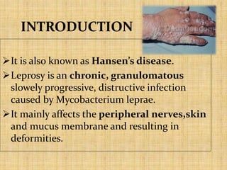
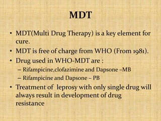
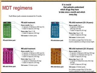
LEPROSY
(Hansen’s Disease)
INDEX
 Introduction
 History of leprosy
 Epidemiology
 Aetiology
 Classification
 Pathogenesis
 Transmission
 Incubation period
 Clinical features
 Treatment
 Reactions of leprosy
 MDT
 Management of leprosy
INTRODUCTION
It is also known as Hansen’s disease.
Leprosy is an chronic, granulomatous
slowely progressive, distructive infection
caused by Mycobacterium leprae.
It mainly affects the peripheral nerves,skin
and mucus membrane and resulting in
deformities.
HISTORY
• Gerhard henrik armauer hansen was a
physician who idetified the Mycobacterium
leprae as the cause of leprosy in 1873.
• India is considered the point of origin of
leprosy with skeletal evidence of the disease
dating to 2000 B.C.
• The disease is thought to have spread through
trade and war to other parts of Asia, the
Middle East, North Africa, and later Europe
and the Americas.
• In ancient Indian society, individuals suffering
from leprosy were alienated because the
disease was chronic, contagious, resulted in
disfigurement, had no cure at the time, and
was associated with sin.
Hansen (1841-1912)
EPIDEMIOLOGY
• According to the official reports recieved from 138
countries from all WHO regions,global registered
prevelance of leprosy at the end of 2015,1,76, 176
cases (0.2 cases per 10,000 poeple).
• Leprosy currently affects approximately a quarter of
a million people throughout the world, with majority
of these cases being reported from India.
• Still, 1.2 to 1.3 hundred thousand new cases of
leprosy reported every year, 58.8% of the total
number of new cases reported every year.
ETIOLOGY
 It is caused by the Mycobacterium leprae.
 M.leprae is also known as the Hansen’s
bacillus spirily.
 It is obligate intracellular paracite.
 Gram +ve bacteria.
 Shape – slender,lightly curved
 Size – 1-8 micro(long)
0.3-1 micro(wide)
 Weakly acid fast bacteria
 Can not be cultured
Ridley and Jopling’s classification :
Depends on clinico pathological spectrum of disease , determined by the
immune resistance of the host.
a. Tuberculoid leprosy (TT) – Show maximum immune response.
b. Borderline tuberculoid (BT) – Immune response falls between the BB
and BT.
c. Borderline leprosy (BB) – Immune response falls between BT and BL.
d. Borderline lapromatous (BL) – Immune response falls between BB
and LL.
e. Lapromatous leprosy (LL) – Least immune response.
WHO Classification :
1.Paucibacillary leprosy : All cases of tuberculoid and some
cases of borderline type.
2.Multibacillary leprosy : All cases of lepromatous leprosy
and some cases of borderline type.
PATHOGENESIS OF LEPROSY
CLINICAL FEATURES
TUBERCULOID LEPROSY
• Skin lesions :
• Number : single or few
• Site : usually face,trunk and
extremities
• Types : localised,hypopigmented,red
or pale in colour
• Numbness and loss of temperature in skin
• Amputation of fingers or toes
• Paralysis of eyelids,keratinitis and corneal
ulcerations
LEPROMATOUS LEPROSY
• Lesions of skin :
Number : multiple
site : common at cooler areas of skin ( ear
lobes,wrist, elbows , knees ,feet ) than
warmer areas ( e.g. groin ,axilla).
Type : macular, papular, or nodular,
symmetric,hypoaesthetic
LEONINE FACIES - When the nodular lesions of
face and earlobes get coalesce to
produce a lion like face.
• Loss of eye brows
• Pendulous ear lobes
• Claw hand
• Saddle nose
• Particularly the ulnar and paroneal nearves
are invaded by the mycobacterium.
• Blindness : Anterior chamber of the eye is
damaged
• Chronic nasal discharge
MODE OF TRANSMISSION
• Transmission by inhalation
• Droplet infecton (most common)
• Transmission by contact
• Skin to skin contact with infectious person
• Contact with soil or fomites
• Other routes
• Insects vectors e.g. Mosquito, Bed bugs
• Tatting needles
• Breast feeding and transplacental infection do not occure.
INCUBATION PERIOD :
• Tuberculoid Leprosy : 2-5 yr
• Lepromatous Leprosy : 8-12 yr
INVESTIGATION
• Sensory testing
• Peripheral nerve examination
• Demonstration of acid fast bacilli
» Skin smears - slit / scrape method
» Nasal swab - Ziehl-Neelsen stain method
• Molecular method : Polymerase chain reaction (PCR).
• LEPROMIN TEST : It is not a diagnostic test .
• Method : Lepromin ( the antigen extract of M. Leprae) is injected
intradermally
• Reaction : Fernandez reaction : Early 9 24-48 hr ) and positive reaction
Mistuda reaction : Delayed granulomatous reaction ( 3-5
weeks)
• Uses : 1. Classification of leprosy
2. Evaluation of cellmediated immunity status in patient
3. Know the prognosis
Reactions in leprosy
The immunity in leprosy may change spontaneously or following
treatment.
• Type I reaction: – Borderline leprosy is the most unstable form of
leprosy where immune status may shift up or down. These are
called as type I reaction, which may be of two types :
 Upgrading reactions : If immunity improves, the disease
may shift towards tuberculoid leprosy.
 Downgrading reaction: If the immunity decreases, the
disease moves towards lepromatous leprosy.
• Type II reaction (erythema nodosum leprosum) – It occurs in
mostly in lepromatous leprosy, particularly when on treatment.
– Clinical features: 1) Tender red plaque or nodules and
2) fever, malaise and arthralgia(joint pain)
Treatment of leprosy
• Dapsone (DDS) : Diamino diphenyl sulfone
• Clofazimine
• Rifampicin
• Ofloxacin
• Minocycline
Treatment of immunologic
reactions
Type I reactions
• Clofazimine 200mg daily
• Corticosteroids
(Prednisolone)
• Rest
Type II reactions
• Corticosteroids
– Prednisolone
– Thalidomide – 100 -300 mg/day
** avoided during pregnancy
and lactation
• Rest
MDT
• MDT(Multi Drug Therapy) is a key element for
cure.
• MDT is free of charge from WHO (From 1981).
• Drug used in WHO-MDT are :
– Rifampicine,clofazimine and Dapsone –MB
– Rifampicine and Dapsone – PB
• Treatment of leprosy with only single drug will
always result in development of drug
resistance
Management of leprosy
• In 2016 WHO has launched a new global
leprosy strategy – The Global Leprosy Strategy
2016-2020 accelerating towards a leprosy free
world.
• India is currently running a leprosy eradication
National Leprosy Eradication
Programme(NLEP) since 1983. c cf
Every last
Sunday of
january....
30thjan,20
leprosy final.pptx

leprosy final.pptx

  • 1.
  • 2.
    INDEX  Introduction  Historyof leprosy  Epidemiology  Aetiology  Classification  Pathogenesis  Transmission  Incubation period  Clinical features  Treatment  Reactions of leprosy  MDT  Management of leprosy
  • 3.
    INTRODUCTION It is alsoknown as Hansen’s disease. Leprosy is an chronic, granulomatous slowely progressive, distructive infection caused by Mycobacterium leprae. It mainly affects the peripheral nerves,skin and mucus membrane and resulting in deformities.
  • 4.
    HISTORY • Gerhard henrikarmauer hansen was a physician who idetified the Mycobacterium leprae as the cause of leprosy in 1873. • India is considered the point of origin of leprosy with skeletal evidence of the disease dating to 2000 B.C. • The disease is thought to have spread through trade and war to other parts of Asia, the Middle East, North Africa, and later Europe and the Americas. • In ancient Indian society, individuals suffering from leprosy were alienated because the disease was chronic, contagious, resulted in disfigurement, had no cure at the time, and was associated with sin. Hansen (1841-1912)
  • 5.
    EPIDEMIOLOGY • According tothe official reports recieved from 138 countries from all WHO regions,global registered prevelance of leprosy at the end of 2015,1,76, 176 cases (0.2 cases per 10,000 poeple). • Leprosy currently affects approximately a quarter of a million people throughout the world, with majority of these cases being reported from India. • Still, 1.2 to 1.3 hundred thousand new cases of leprosy reported every year, 58.8% of the total number of new cases reported every year.
  • 8.
    ETIOLOGY  It iscaused by the Mycobacterium leprae.  M.leprae is also known as the Hansen’s bacillus spirily.  It is obligate intracellular paracite.  Gram +ve bacteria.  Shape – slender,lightly curved  Size – 1-8 micro(long) 0.3-1 micro(wide)  Weakly acid fast bacteria  Can not be cultured
  • 10.
    Ridley and Jopling’sclassification : Depends on clinico pathological spectrum of disease , determined by the immune resistance of the host. a. Tuberculoid leprosy (TT) – Show maximum immune response. b. Borderline tuberculoid (BT) – Immune response falls between the BB and BT. c. Borderline leprosy (BB) – Immune response falls between BT and BL. d. Borderline lapromatous (BL) – Immune response falls between BB and LL. e. Lapromatous leprosy (LL) – Least immune response. WHO Classification : 1.Paucibacillary leprosy : All cases of tuberculoid and some cases of borderline type. 2.Multibacillary leprosy : All cases of lepromatous leprosy and some cases of borderline type.
  • 12.
  • 13.
    CLINICAL FEATURES TUBERCULOID LEPROSY •Skin lesions : • Number : single or few • Site : usually face,trunk and extremities • Types : localised,hypopigmented,red or pale in colour • Numbness and loss of temperature in skin • Amputation of fingers or toes • Paralysis of eyelids,keratinitis and corneal ulcerations
  • 14.
    LEPROMATOUS LEPROSY • Lesionsof skin : Number : multiple site : common at cooler areas of skin ( ear lobes,wrist, elbows , knees ,feet ) than warmer areas ( e.g. groin ,axilla). Type : macular, papular, or nodular, symmetric,hypoaesthetic LEONINE FACIES - When the nodular lesions of face and earlobes get coalesce to produce a lion like face. • Loss of eye brows • Pendulous ear lobes • Claw hand • Saddle nose • Particularly the ulnar and paroneal nearves are invaded by the mycobacterium. • Blindness : Anterior chamber of the eye is damaged • Chronic nasal discharge
  • 16.
    MODE OF TRANSMISSION •Transmission by inhalation • Droplet infecton (most common) • Transmission by contact • Skin to skin contact with infectious person • Contact with soil or fomites • Other routes • Insects vectors e.g. Mosquito, Bed bugs • Tatting needles • Breast feeding and transplacental infection do not occure. INCUBATION PERIOD : • Tuberculoid Leprosy : 2-5 yr • Lepromatous Leprosy : 8-12 yr
  • 17.
    INVESTIGATION • Sensory testing •Peripheral nerve examination • Demonstration of acid fast bacilli » Skin smears - slit / scrape method » Nasal swab - Ziehl-Neelsen stain method • Molecular method : Polymerase chain reaction (PCR). • LEPROMIN TEST : It is not a diagnostic test . • Method : Lepromin ( the antigen extract of M. Leprae) is injected intradermally • Reaction : Fernandez reaction : Early 9 24-48 hr ) and positive reaction Mistuda reaction : Delayed granulomatous reaction ( 3-5 weeks) • Uses : 1. Classification of leprosy 2. Evaluation of cellmediated immunity status in patient 3. Know the prognosis
  • 18.
    Reactions in leprosy Theimmunity in leprosy may change spontaneously or following treatment. • Type I reaction: – Borderline leprosy is the most unstable form of leprosy where immune status may shift up or down. These are called as type I reaction, which may be of two types :  Upgrading reactions : If immunity improves, the disease may shift towards tuberculoid leprosy.  Downgrading reaction: If the immunity decreases, the disease moves towards lepromatous leprosy. • Type II reaction (erythema nodosum leprosum) – It occurs in mostly in lepromatous leprosy, particularly when on treatment. – Clinical features: 1) Tender red plaque or nodules and 2) fever, malaise and arthralgia(joint pain)
  • 19.
    Treatment of leprosy •Dapsone (DDS) : Diamino diphenyl sulfone • Clofazimine • Rifampicin • Ofloxacin • Minocycline
  • 20.
    Treatment of immunologic reactions TypeI reactions • Clofazimine 200mg daily • Corticosteroids (Prednisolone) • Rest Type II reactions • Corticosteroids – Prednisolone – Thalidomide – 100 -300 mg/day ** avoided during pregnancy and lactation • Rest
  • 21.
    MDT • MDT(Multi DrugTherapy) is a key element for cure. • MDT is free of charge from WHO (From 1981). • Drug used in WHO-MDT are : – Rifampicine,clofazimine and Dapsone –MB – Rifampicine and Dapsone – PB • Treatment of leprosy with only single drug will always result in development of drug resistance
  • 23.
    Management of leprosy •In 2016 WHO has launched a new global leprosy strategy – The Global Leprosy Strategy 2016-2020 accelerating towards a leprosy free world. • India is currently running a leprosy eradication National Leprosy Eradication Programme(NLEP) since 1983. c cf
  • 24.