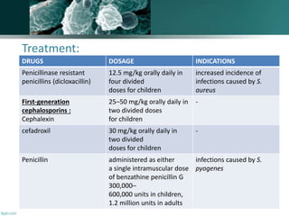
By,
M.Logeshwary
(PharmD III year)
IMPETIGO
• Impetigo is a superficial skin infection that is seen
most commonly in children and is transmitted easily
from person to person.
• Based on clinical presentations:
DEINITION:
TYPES
BULLOUS
NON-
BULLOUS
• There are two ways an initial infection can occur:
• primary impetigo - is when the bacteria invades the
skin through a cut , insect bite, or other injury, and
• secondary impetigo - is where the bacteria invades
the skin because the skin barrier has been disrupted
by another skin infection, such as scabies or eczema.
Epidemiology:
• The bullous form most frequently affects neonates and
accounts for approximately 10% of all cases of impetigo
• Based on data from studies published since 2000 from
low and low-middle income countries, we estimate the
global population of children suffering from impetigo at
any one time to be in excess of 162 million,
predominantly in tropical, resource-poor contexts.
Impetigo is an under-recognised disease and in
conjunction with scabies, comprises a major childhood
dermatological condition with potential lifelong
consequences if untreated.
Occurence
• On exposed skin mainly on face.
• most common during hot, humid weather,
which facilitates microbial colonization of
the skin.
• Minor trauma, such as scratches or insect
bites, then allows entry of organisms into
the superficial layers of skin, and infection
ensues
• Caused by S. pyogenes
• But S. aureus either alone or in combination with S.
pyogenes has emerged more recently as the principal cause
of impetigo
• The bullous form is caused by strains of S. aureus capable of
producing exfoliative toxins
Causes
BULLOUS IMPETIGO (BLISTERS)
• This form is caused by staph bacteria that produce a
toxin that causes a break between the top layer
(epidermis) and the lower levels of skin forming a
blister. (The medical term for blister is bulla.) Blisters
can appear in various skin areas, especially the
buttocks, though these blisters are fragile and often
break and leave red, raw skin with a ragged edge. No
prior trauma is needed for these blisters to appear.
NON -BULLOUS IMPETIGO
This is the common form, caused by both staph and
strep bacteria. It appears as small blisters or scabs,
which then form yellow or honey-colored crusts.
These often start around the nose and on the face,
but they also may affect the arms and legs. At times ,
there may be swollen glands nearby.
Pathophysiology:
• Bullous impetigo is caused by staphylococci producing exfoliative
toxin that contains serine proteases acting on desmoglein , a
structurally critical peptide bond in a molecule that holds
epidermal cells together. This process allows Staphylococcus
aureus to spread under the stratum corneum in the space
formed by the toxin, causing the epidermis to split just below
the stratum granulosum. Large blisters then form in the
epidermis with neutrophil .
• In bullous impetigo, the bullae rupture quickly, causing
superficial erosion and a yellow crust,
• while in non-bullous impetigo, Streptococcus typically produces
a thick-walled pustule with an erythematous base. Histology of
non-bullous established lesions shows a thick surface crust
composed of serum and neutrophils in various stages of
breakdown with parakeratotic material
Symptoms:
• Pruritus(severe itching) is common, and scratching of
the lesions may further spread infection through
excoriation of the skin.
• Other systemic signs of infection are minimal.
• Weakness, fever, and diarrhea sometimes are seen
with bullous impetigo.
Signs:
• Non bullous impetigo manifests initially as small, fluid
filled vesicles.
• These lesions rapidly develop into pus-filled blisters that
rupture readily.
• Purulent discharge from the lesions dries to form golden-
yellow crusts that are characteristic of impetigo.
• In the bullous form of impetigo, the lesions begin as
vesicles and turn into bullae containing clear yellow
fluid.
• Bullae soon rupture, forming thin, light brown crusts.
• Regional lymph nodes may be enlarged.
IS IMPETIGO CONTAGIOUS?
• Impetigo is contagious, mostly from direct contact
with someone who has it.
• Can be transmitted through:
1. towels,
2. toys,
3. clothing or
4. household items
DIAGNOSIS
• Doctors generally diagnose impetigo by looking at the
distinctive sores.
• Sometimes culture test are done rarely to identify the type of
bacteria causing lesions.
• A complete blood count is often performed because
leukocytosis is common.
TREATMENT
• Impetigo is not serious, may go away and dry up on its
own, and is easy to treat.
• Mild cases can be handled by gentle cleansing, removing
crusts, and applying the prescription-strength antibiotic
ointment mupirocin ( Bactroban).
• More severe or widespread cases, especially of bullous
impetigo, may require oral antibiotic medication for
impetigo.
• impetigo may resolve spontaneously, antimicrobial
treatment is indicated to relieve symptoms, prevent
formation of new lesions, and prevent complications,
such as cellulitis.
Treatment:
DRUGS DOSAGE INDICATIONS
Penicillinase resistant
penicillins (dicloxacillin)
12.5 mg/kg orally daily in
four divided
doses for children
increased incidence of
infections caused by S.
aureus
First-generation
cephalosporins :
Cephalexin
25–50 mg/kg orally daily in
two divided doses
for children
-
cefadroxil 30 mg/kg orally daily in
two divided
doses for children
-
Penicillin administered as either
a single intramuscular dose
of benzathine penicillin G
300,000–
600,000 units in children,
1.2 million units in adults
infections caused by S.
pyogenes
TREATMENT
clindamycin adults
150–300 mg orally every 6
to 8 hours; children 10–30
mg/kg per
day in three to four divided
doses
The duration of therapy is
7 to
10 days.
Penicillin-allergic patients
can be treated
Topical antibiotics, such as
mupirocin and bacitracin
- used to treat non-bullous
impetigo.
Mupirocin ointment applied
three times daily for 7 days
as effective as
erythromycin.
• With proper treatment, healing of skin lesions
generally is rapid and occurs without residual
scarring.
• Removal of crusts by soaking in soap and warm
water also may be helpful in providing symptomatic
relief
EVALUATION OF THERAPEUTIC OUTCOMES
• Clinical response should be seen within 7 days of
initiating antimicrobial therapy for impetigo.
• Treatment failures could be due to noncompliance or
antimicrobial resistance.
• A follow-up culture of exudates should be collected
for culture and sensitivity, with treatment modified
accordingly.
Case study
• OB, a 3-year-old boy, is brought to the clinic with a facial rash. According to OB's
mother, the rash started 4 days ago as little red bumps below his nose. The rash
has spread around his mouth and chin. The rash also has changed in appearance to
flat, reddened areas with fluid-filled pustules. On physical examination, the
pediatrician finds OB to be a content and alert child in no acute distress. His vital
signs are stable and within normal limits. The pediatrician notes that some of the
pustules have ruptured, leaving weepy, red lesions and honey-colored crusts. The
affected area is not excessively warm or swollen.
• The pediatrician suspects that OB has impetigo. He explains to the mother that
impetigo is a contagious condition that requires treatment with antibiotics. He
knows that the most common pathogen causing impetigo is ( ? ),
with ( ? )coinfection. The pediatrician is aware that impetigo was
traditionally treated with penicillin, but resistance has limited the usefulness of
this antibiotic. Instead he hopes to use an antibiotic that effectively will cover
staphylococci and streptococci.
• As the pediatrician checks the supplies of medications available in the clinic, the
mother comments that OB will not take any medications by mouth. She asks
whether there are any medications that can be applied to the rash, rather than
given by mouth.
• Are there any topical options available to treat OB's impetigo?
• Whether it is bullous or non-bullous impetigo?
• Because many cases of impetigo involve coinfection with
streptococci, antibiotic selection must consider covering for
both organisms. Antimicrobial agents that will cover for both
organisms include dicloxcillin, cephalexin, erythromycin, and
amoxicillin/clavulanate. Since OB will not take oral antibiotics,
mupirocin ointment is another option. Mupirocin should be
used only for mild cases, however.
• The pediatrician should advise the mother about the
importance of not spreading the infection to the rest of the
family (or even to other parts of OB's body). The most
important measure of prevention is frequent hand washing.
OB also should be reminded not to touch the rash.
• Non-bullous impetigo.
Impetigo

Impetigo

  • 1.
  • 2.
    • Impetigo isa superficial skin infection that is seen most commonly in children and is transmitted easily from person to person. • Based on clinical presentations: DEINITION: TYPES BULLOUS NON- BULLOUS
  • 3.
    • There aretwo ways an initial infection can occur: • primary impetigo - is when the bacteria invades the skin through a cut , insect bite, or other injury, and • secondary impetigo - is where the bacteria invades the skin because the skin barrier has been disrupted by another skin infection, such as scabies or eczema.
  • 4.
    Epidemiology: • The bullousform most frequently affects neonates and accounts for approximately 10% of all cases of impetigo • Based on data from studies published since 2000 from low and low-middle income countries, we estimate the global population of children suffering from impetigo at any one time to be in excess of 162 million, predominantly in tropical, resource-poor contexts. Impetigo is an under-recognised disease and in conjunction with scabies, comprises a major childhood dermatological condition with potential lifelong consequences if untreated.
  • 5.
    Occurence • On exposedskin mainly on face. • most common during hot, humid weather, which facilitates microbial colonization of the skin. • Minor trauma, such as scratches or insect bites, then allows entry of organisms into the superficial layers of skin, and infection ensues
  • 6.
    • Caused byS. pyogenes • But S. aureus either alone or in combination with S. pyogenes has emerged more recently as the principal cause of impetigo • The bullous form is caused by strains of S. aureus capable of producing exfoliative toxins Causes
  • 7.
    BULLOUS IMPETIGO (BLISTERS) •This form is caused by staph bacteria that produce a toxin that causes a break between the top layer (epidermis) and the lower levels of skin forming a blister. (The medical term for blister is bulla.) Blisters can appear in various skin areas, especially the buttocks, though these blisters are fragile and often break and leave red, raw skin with a ragged edge. No prior trauma is needed for these blisters to appear.
  • 8.
    NON -BULLOUS IMPETIGO Thisis the common form, caused by both staph and strep bacteria. It appears as small blisters or scabs, which then form yellow or honey-colored crusts. These often start around the nose and on the face, but they also may affect the arms and legs. At times , there may be swollen glands nearby.
  • 10.
    Pathophysiology: • Bullous impetigois caused by staphylococci producing exfoliative toxin that contains serine proteases acting on desmoglein , a structurally critical peptide bond in a molecule that holds epidermal cells together. This process allows Staphylococcus aureus to spread under the stratum corneum in the space formed by the toxin, causing the epidermis to split just below the stratum granulosum. Large blisters then form in the epidermis with neutrophil . • In bullous impetigo, the bullae rupture quickly, causing superficial erosion and a yellow crust, • while in non-bullous impetigo, Streptococcus typically produces a thick-walled pustule with an erythematous base. Histology of non-bullous established lesions shows a thick surface crust composed of serum and neutrophils in various stages of breakdown with parakeratotic material
  • 13.
    Symptoms: • Pruritus(severe itching)is common, and scratching of the lesions may further spread infection through excoriation of the skin. • Other systemic signs of infection are minimal. • Weakness, fever, and diarrhea sometimes are seen with bullous impetigo.
  • 14.
    Signs: • Non bullousimpetigo manifests initially as small, fluid filled vesicles. • These lesions rapidly develop into pus-filled blisters that rupture readily. • Purulent discharge from the lesions dries to form golden- yellow crusts that are characteristic of impetigo. • In the bullous form of impetigo, the lesions begin as vesicles and turn into bullae containing clear yellow fluid. • Bullae soon rupture, forming thin, light brown crusts. • Regional lymph nodes may be enlarged.
  • 15.
    IS IMPETIGO CONTAGIOUS? •Impetigo is contagious, mostly from direct contact with someone who has it. • Can be transmitted through: 1. towels, 2. toys, 3. clothing or 4. household items
  • 16.
    DIAGNOSIS • Doctors generallydiagnose impetigo by looking at the distinctive sores. • Sometimes culture test are done rarely to identify the type of bacteria causing lesions. • A complete blood count is often performed because leukocytosis is common.
  • 17.
    TREATMENT • Impetigo isnot serious, may go away and dry up on its own, and is easy to treat. • Mild cases can be handled by gentle cleansing, removing crusts, and applying the prescription-strength antibiotic ointment mupirocin ( Bactroban). • More severe or widespread cases, especially of bullous impetigo, may require oral antibiotic medication for impetigo. • impetigo may resolve spontaneously, antimicrobial treatment is indicated to relieve symptoms, prevent formation of new lesions, and prevent complications, such as cellulitis.
  • 18.
    Treatment: DRUGS DOSAGE INDICATIONS Penicillinaseresistant penicillins (dicloxacillin) 12.5 mg/kg orally daily in four divided doses for children increased incidence of infections caused by S. aureus First-generation cephalosporins : Cephalexin 25–50 mg/kg orally daily in two divided doses for children - cefadroxil 30 mg/kg orally daily in two divided doses for children - Penicillin administered as either a single intramuscular dose of benzathine penicillin G 300,000– 600,000 units in children, 1.2 million units in adults infections caused by S. pyogenes
  • 19.
    TREATMENT clindamycin adults 150–300 mgorally every 6 to 8 hours; children 10–30 mg/kg per day in three to four divided doses The duration of therapy is 7 to 10 days. Penicillin-allergic patients can be treated Topical antibiotics, such as mupirocin and bacitracin - used to treat non-bullous impetigo. Mupirocin ointment applied three times daily for 7 days as effective as erythromycin.
  • 20.
    • With propertreatment, healing of skin lesions generally is rapid and occurs without residual scarring. • Removal of crusts by soaking in soap and warm water also may be helpful in providing symptomatic relief
  • 21.
    EVALUATION OF THERAPEUTICOUTCOMES • Clinical response should be seen within 7 days of initiating antimicrobial therapy for impetigo. • Treatment failures could be due to noncompliance or antimicrobial resistance. • A follow-up culture of exudates should be collected for culture and sensitivity, with treatment modified accordingly.
  • 22.
    Case study • OB,a 3-year-old boy, is brought to the clinic with a facial rash. According to OB's mother, the rash started 4 days ago as little red bumps below his nose. The rash has spread around his mouth and chin. The rash also has changed in appearance to flat, reddened areas with fluid-filled pustules. On physical examination, the pediatrician finds OB to be a content and alert child in no acute distress. His vital signs are stable and within normal limits. The pediatrician notes that some of the pustules have ruptured, leaving weepy, red lesions and honey-colored crusts. The affected area is not excessively warm or swollen. • The pediatrician suspects that OB has impetigo. He explains to the mother that impetigo is a contagious condition that requires treatment with antibiotics. He knows that the most common pathogen causing impetigo is ( ? ), with ( ? )coinfection. The pediatrician is aware that impetigo was traditionally treated with penicillin, but resistance has limited the usefulness of this antibiotic. Instead he hopes to use an antibiotic that effectively will cover staphylococci and streptococci. • As the pediatrician checks the supplies of medications available in the clinic, the mother comments that OB will not take any medications by mouth. She asks whether there are any medications that can be applied to the rash, rather than given by mouth. • Are there any topical options available to treat OB's impetigo? • Whether it is bullous or non-bullous impetigo?
  • 23.
    • Because manycases of impetigo involve coinfection with streptococci, antibiotic selection must consider covering for both organisms. Antimicrobial agents that will cover for both organisms include dicloxcillin, cephalexin, erythromycin, and amoxicillin/clavulanate. Since OB will not take oral antibiotics, mupirocin ointment is another option. Mupirocin should be used only for mild cases, however. • The pediatrician should advise the mother about the importance of not spreading the infection to the rest of the family (or even to other parts of OB's body). The most important measure of prevention is frequent hand washing. OB also should be reminded not to touch the rash. • Non-bullous impetigo.