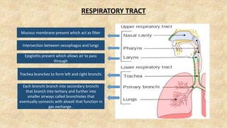
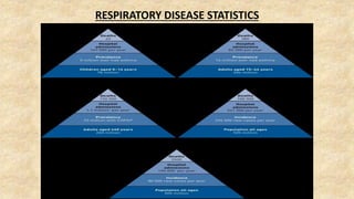
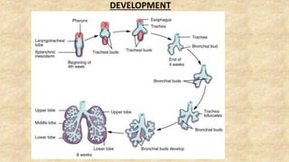
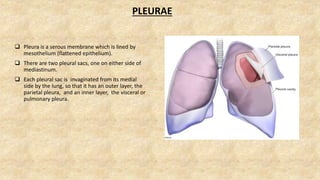
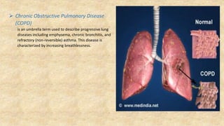
SlideShare a Scribd company logo
RESPIRATORY TRACT
Mucous membrane present which act as filter
Intersection between oesophagus and lungs
Epiglottis present which allows air to pass
through
Trachea branches to form left and right bronchi.
Each bronchi branch into secondary bronchi
that branch into tertiary and further into
smaller airways called bronchioles that
eventually connects with alveoli that function in
gas exchange .
RESPIRATORY DISEASE STATISTICS
DEVELOPMENT OF RESPIRATORY SYSTEM
DEVELOPMENT
PLEURAE
 Pleura is a serous membrane which is lined by
mesothelium (flattened epithelium).
 There are two pleural sacs, one on either side of
mediastinum.
 Each pleural sac is invaginated from its medial
side by the lung, so that it has an outer layer, the
parietal pleura, and an inner layer, the visceral or
pulmonary pleura.
RECESS OF PLEURA
There are two recesses of parietal pleura, which act as “reserve spaces
” for the lung to expand during deep inspiration.
 Costomediastinal recess
Lies anteriorly, behind the sternum and Costal cartilages,
between the Costal and mediastinal pleurae, particularly in
relation to the cardiac notch of the left lung.
 Costodiaphragmatic / Costovertebral recess
Lies inferiorly between the Costal and diaphragmatic
pleurae. Vertically, it measures about 5 cm, and extends from
the eighth to tenth ribs along the midaxillary line.
HISTOLOGY
Intrapulmonary Bronchus
Terminal Bronchiole
Respiratory Bronchiole
Alveoli
Features
of
Lungs
Shape - Conical
Texture - Spongy
Color -
In young ones – Brown or Grey
In Adults – Mottled Black
Weight –
Right Lung – 445 grams
Left Lung – 395 grams
Position –
On either side of mediastinum within thoracic
cavity
Lung
structure
Apex
Three
surfaces
Costal surface
Medial surface
Diaphragmatic
surface
Three
borders
Anterior border
Posterior border
Inferior border
Base
 Apex
 Blunt superior end of the lung.
 Projects upwards ,above the level of the first rib.
 Reaches nearly 2.5 cm above the medial one-third of the
clavicle, just medial to the supraclavicular fossa.
 Covered by cervical pleura, the Supraplueral membrane.
 Base
 Semilunar and concave.
 On inferior surface of lung and rests on the diaphragm..
 Separates the right lung from right lobe of liver, and left lung
from the left lobe of liver ,the fundus of stomach, and the
spleen.
 Three borders
Anterior border
 Shorter.
 Formed by convergence of mediastinal and
Costal surfaces.
 On the left lung , it is marked by a deep notch
created by the apex of the heart. It is known as
Cardiac notch.
Posterior border
 Thick and ill defined, Smooth and rounded.
 formed by Costal and medial surfaces meeting
posteriorly.
Inferior border
 Separates the base from the Costal and medial
surface.
 Three surfaces
Costal surface
 Large ,convex ,smooth.
 It is related to the Costal pleura ,which separates it from
ribs and intercostal muscles.
Medial surface
 It is divided into Posterior/Vertebral part and an
Anterior Mediastinal part.
 The vertebral part is related to the vertebral bodies,
intervertebral discs, the posterior intercostal vessels and
splanchnic nerves .
 The mediastinal part is related to the mediastinal
septum, and shows a cardiac impression, the hilum and
a number of other impressions which differ on the two
sides.
Diaphragmatic surface
 It lies on the top of diaphragm.
 Fissures and lobes of the lungs –
 The right lung is divided into 3 lobes (upper, middle, lower) by two
fissures (oblique and horizontal).
 The left lung is divided into two lobes by the oblique fissure.
 The presence of oblique fissure of each lung allows a more uniform
expansion of the whole lung.
 Root of the lung
It is short, broad pedicle which connects the medial surface of the lung to
the mediastinum.It is formed by structures which either enter or come
out of the lung at the hilum.
Contents
1. Principal bronchus on the left side, and eparterial and hypaterial
bronchi on the right side.
2. One pulmonary artery.
3. Two pulmonary veins, superior and inferior.
4. Bronchial arteries, one on the right side and two on the left side.
5. Bronchial veins.
6. Anterior and posterior pulmonary plexuses of nerves.
7. Lymphatic of the lung.
8. Brochopulmonary lymph nodes.
9. Areolar tissue .
 Blood supply
 The lungs have a dual blood supply provided by
a bronchial and a pulmonary circulation.
 The bronchial arteries supply nutrition to the bronchial and
to the pulmonary tissue.
On the right side
One bronchial artery (From the third right posterior
intercostal artery).
On the left side
Two bronchial arteries (From the descending thoracic
aorta, the upper opposite fifth thoracic vertebra and
the lower just below the left bronchus).
 The pulmonary circulation includes two pulmonary arteries
and four pulmonary veins.
 Venous Drainage
 The two bronchial veins on each side carries the venous
blood from first and second divisions of bronchi.
 The right bronchial veins drain into azygous vein.
 The left bronchial veins drain into hemiazygous vein.
 The greater Part of venous blood from the lungs is drained
by the pulmonary veins.
 Lymphatic Drainage
There are two sets of lymphatic which drain into Broncho
pulmonary nodes.
1) Superficial vessels drain the peripheral lung tissue.
2) Deep lymphatics drain the bronchial tree ,pulmonary
vessels and the connective tissue septa.
 Nervous supply
The nerves of the lungs are derived from the pulmonary
plexuses. They feature sympathetic, parasympathetic and
visceral afferent fibers .
 Parasympathetic
- Derived from the vagus nerve.
 Sympathetic
- Derived from the sympathetic trunks.(second to fifth
sympathetic ganglia).
 Visceral afferent
- Conduct pain impulses to the sensory ganglion of the
vagus nerve.
 Broncho pulmonary
segments
 There are 10 segments on the right
side and 10 on the left side.
 There are well -defined anatomical
,functional and surgical sectors of
the lung.
 Each segment is pyramidal in shape
and each segment has a segmental
bronchus, segmental artery,
autonomic nerves and lymph
vessels.
CLINICAL ANATOMY
 Pleuritis or pleurisy
This is the inflammation of the pleura. Acute pleuritis is marked by sharp, stabbing pain, especially on exertion.
 Aspiration of foreign bodies
Since the right bronchus is wider and shorter and runs more vertically than the left bronchus, foreign bodies are
more likely to enter and lodge in it or in one of its branches.
 Bronchoscopy
When examining with a bronchoscope, one
observes a keel like ridge called the carina. It is a
cartilaginous projection of the last tracheal ring.
Morphological changes in the carina are
important diagnostic signs to bronchoscopists in
assisting with the differential diagnosis of
respiratory disease.
 Bronchial asthma
Bronchial asthma is an increasingly common condition of
the lungs in which widespread narrowing of the airways is
present.
 Asthma is caused by varying degrees by contraction of the
smooth muscle, edema of the mucosa, and mucus in the
lumen of the bronchi and bronchioles.
 In asthma, there is difficult expiration because the
bronchioles which are opened during inspiration also have
to remain open during expiration if they are to permit a
rapid outflow of air owing to elastic recoil of the lung
tissues.
 Mycobacterium tuberculosis
Man is the main host.
 The microbes cause pulmonary tuberculosis and are spread
either by droplet infection from an individual with active
tuberculosis, or in dust contaminated by infected sputum.
 Pneumonia
 This occurs when protective processes fail to prevent inhaled or blood
born microbes reaching and colonizing the lungs.
 Types- lobar pneumonia and bronchopneumonia
Lobar pneumonia
This is the infection of one or more lobes by Streptococcus pneumoniae .
Bronchopneumonia
Infection is spread from the bronchi to terminal bronchioles and alveoli.
 Floatation of the lungs
Fresh healthy lungs always contain some air. Diseased lungs filled with fluid, fetal lungs, and lungs from a still born
infant will not float.
 Bronchial carcinoma
Primary bronchial carcinoma is a common form of
malignancy. The tumour usually develops in a main
bronchus, forming a large friable mass that project into the
lumen sometimes causing obstruction.
 Emphysema is a long-term, progressive disease of the
lungs that primarily causes shortness of breath due to over-
inflation of the alveoli (air sacs in the lung). In people with
emphysema, the lung tissue involved in exchange of gases
(oxygen and carbon dioxide) is impaired or destroyed.
 Pneumoconiosis
This is a group of lung diseases caused by inhaling organic or inorganic atmospheric pollutants. Some of them are-
 Coal workers pneumoconiosis- soft bituminous coal
 Silicosis- dust containing silicon compounds
 Asbestosis- asbestos
 Pleural mesothelioma- asbestos
 Byssinosis- cotton fibres
 Extrinsic allergic alveolitis
This is a group of conditions caused by inhaling materials contaminated by moulds and fungi.
 Farmers lung- mouldy hay
 Bagassosis- mouldy sugar waste
 Bird handler’s lung- mould in bird droppings
 Malt worker’s lung- mouldy barley
 Chronic Obstructive Pulmonary Disease
(COPD)
is an umbrella term used to describe progressive lung
diseases including emphysema, chronic bronchitis, and
refractory (non-reversible) asthma. This disease is
characterized by increasing breathlessness.
Respiratory system and lungs.pptx

More Related Content

What's hot

The pleurae
The pleuraeThe pleurae
The pleurae
Idris Siddiqui
 
Blood supply of head & neck. Arterial & venous anastomoses
Blood supply of head & neck. Arterial & venous anastomosesBlood supply of head & neck. Arterial & venous anastomoses
Blood supply of head & neck. Arterial & venous anastomoses
Eneutron
 
Thorax
ThoraxThorax
Blood supplyand lymphatic drainage to oral cavity
Blood supplyand lymphatic drainage to oral cavityBlood supplyand lymphatic drainage to oral cavity
Blood supplyand lymphatic drainage to oral cavity
Aishwarya Hajare
 
5th week -_respiratory_system_ii
5th week -_respiratory_system_ii5th week -_respiratory_system_ii
5th week -_respiratory_system_iiMUBOSScz
 
The Respiratory System
The Respiratory SystemThe Respiratory System
The Respiratory System
raj kumar
 
Anatomy of lungs, pleura and diaphragm
Anatomy  of lungs, pleura and diaphragmAnatomy  of lungs, pleura and diaphragm
Anatomy of lungs, pleura and diaphragm
Dr. Ashish kumar
 
Lung anatomy.
Lung anatomy.Lung anatomy.
Lung anatomy.
siti hamidah
 
E lecture respiratory system
E lecture respiratory systemE lecture respiratory system
E lecture respiratory system
Nisar Arain
 
Anatomy of trachea & lungs
Anatomy of trachea & lungsAnatomy of trachea & lungs
Anatomy of trachea & lungs
AnuSebastian18
 
Trachea + Lungs
Trachea + LungsTrachea + Lungs
Trachea + Lungsmsu
 
Lecture 3 lungs & pleura
Lecture 3 lungs & pleuraLecture 3 lungs & pleura
Lecture 3 lungs & pleura
Dr. Noura El Tahawy
 
Surgery 5th year, 1st lecture (Dr. Ahmed Al-Azzawi)
Surgery 5th year, 1st lecture (Dr. Ahmed Al-Azzawi)Surgery 5th year, 1st lecture (Dr. Ahmed Al-Azzawi)
Surgery 5th year, 1st lecture (Dr. Ahmed Al-Azzawi)
College of Medicine, Sulaymaniyah
 
Arterial supply of face
Arterial  supply of faceArterial  supply of face
Arterial supply of face
Dr. Aparna Ray
 
4+-+Thoracic+Cavity+Grays.ppt
4+-+Thoracic+Cavity+Grays.ppt4+-+Thoracic+Cavity+Grays.ppt
4+-+Thoracic+Cavity+Grays.pptempite
 
Anatomy lungs
Anatomy lungsAnatomy lungs
Anatomy lungs
surajitkundu
 
Arterial supply of head & neck
Arterial supply of head & neckArterial supply of head & neck
Arterial supply of head & neck
shanender sambyal
 
Anatomy of bronchial tree, lungs and pleura
Anatomy of bronchial tree, lungs and pleuraAnatomy of bronchial tree, lungs and pleura
Anatomy of bronchial tree, lungs and pleura
Dr.Apurva Patel
 
Arteria venous and lymphatic drinage of head and neck basics
Arteria venous and lymphatic drinage of head and neck basicsArteria venous and lymphatic drinage of head and neck basics
Arteria venous and lymphatic drinage of head and neck basics
Manoj Kumar
 

What's hot (20)

The pleurae
The pleuraeThe pleurae
The pleurae
 
Blood supply of head & neck. Arterial & venous anastomoses
Blood supply of head & neck. Arterial & venous anastomosesBlood supply of head & neck. Arterial & venous anastomoses
Blood supply of head & neck. Arterial & venous anastomoses
 
Thorax
ThoraxThorax
Thorax
 
Blood supplyand lymphatic drainage to oral cavity
Blood supplyand lymphatic drainage to oral cavityBlood supplyand lymphatic drainage to oral cavity
Blood supplyand lymphatic drainage to oral cavity
 
5th week -_respiratory_system_ii
5th week -_respiratory_system_ii5th week -_respiratory_system_ii
5th week -_respiratory_system_ii
 
The Respiratory System
The Respiratory SystemThe Respiratory System
The Respiratory System
 
Anatomy of lungs, pleura and diaphragm
Anatomy  of lungs, pleura and diaphragmAnatomy  of lungs, pleura and diaphragm
Anatomy of lungs, pleura and diaphragm
 
Lung anatomy.
Lung anatomy.Lung anatomy.
Lung anatomy.
 
E lecture respiratory system
E lecture respiratory systemE lecture respiratory system
E lecture respiratory system
 
Arteries of head and neck
Arteries of head and neckArteries of head and neck
Arteries of head and neck
 
Anatomy of trachea & lungs
Anatomy of trachea & lungsAnatomy of trachea & lungs
Anatomy of trachea & lungs
 
Trachea + Lungs
Trachea + LungsTrachea + Lungs
Trachea + Lungs
 
Lecture 3 lungs & pleura
Lecture 3 lungs & pleuraLecture 3 lungs & pleura
Lecture 3 lungs & pleura
 
Surgery 5th year, 1st lecture (Dr. Ahmed Al-Azzawi)
Surgery 5th year, 1st lecture (Dr. Ahmed Al-Azzawi)Surgery 5th year, 1st lecture (Dr. Ahmed Al-Azzawi)
Surgery 5th year, 1st lecture (Dr. Ahmed Al-Azzawi)
 
Arterial supply of face
Arterial  supply of faceArterial  supply of face
Arterial supply of face
 
4+-+Thoracic+Cavity+Grays.ppt
4+-+Thoracic+Cavity+Grays.ppt4+-+Thoracic+Cavity+Grays.ppt
4+-+Thoracic+Cavity+Grays.ppt
 
Anatomy lungs
Anatomy lungsAnatomy lungs
Anatomy lungs
 
Arterial supply of head & neck
Arterial supply of head & neckArterial supply of head & neck
Arterial supply of head & neck
 
Anatomy of bronchial tree, lungs and pleura
Anatomy of bronchial tree, lungs and pleuraAnatomy of bronchial tree, lungs and pleura
Anatomy of bronchial tree, lungs and pleura
 
Arteria venous and lymphatic drinage of head and neck basics
Arteria venous and lymphatic drinage of head and neck basicsArteria venous and lymphatic drinage of head and neck basics
Arteria venous and lymphatic drinage of head and neck basics
 

Similar to Respiratory system and lungs.pptx

ASTHMA.docx
ASTHMA.docxASTHMA.docx
ASTHMA.docx
Anusha Amit
 
LUNGS ANATOMY
LUNGS ANATOMYLUNGS ANATOMY
LUNGS ANATOMY
Muhammad Bilal
 
Lungs,Pleura & Tracheo-bronchial Tree
Lungs,Pleura & Tracheo-bronchial TreeLungs,Pleura & Tracheo-bronchial Tree
Lungs,Pleura & Tracheo-bronchial Tree
Prabhakar Yadav
 
Pleura$lung ppt.pptx assignment of anatomy
Pleura$lung ppt.pptx assignment of anatomyPleura$lung ppt.pptx assignment of anatomy
Pleura$lung ppt.pptx assignment of anatomy
AmanuelIbrahim
 
Lec.8 lungs pt&rc
Lec.8 lungs pt&rcLec.8 lungs pt&rc
Lec.8 lungs pt&rc
Dr Motawei
 
Anatomy Of The Lungs.pptx
Anatomy Of The Lungs.pptxAnatomy Of The Lungs.pptx
Anatomy Of The Lungs.pptx
Saurabh Puri
 
Respiratory system
Respiratory systemRespiratory system
Respiratory system
ajith joseph
 
Anatomy Of Lungs.pptx
Anatomy Of Lungs.pptxAnatomy Of Lungs.pptx
Anatomy Of Lungs.pptx
DipeshGamare
 
Anatomy and physiology of lung
Anatomy and physiology of lungAnatomy and physiology of lung
Anatomy and physiology of lung
jithahari
 
Basic anatomy of lung .ppt
Basic anatomy of lung  .pptBasic anatomy of lung  .ppt
Basic anatomy of lung .ppt
DrSreelakshmiSuresh
 
gross anatomy of resp. system.pptx
gross anatomy of resp. system.pptxgross anatomy of resp. system.pptx
gross anatomy of resp. system.pptx
Shabana Ali
 
Respiratory system.ppsx
Respiratory system.ppsxRespiratory system.ppsx
Respiratory system.ppsx
lumaGhaziALzamel
 
Lecture (4) Anatomy of Lung & Pleura.pdf
Lecture (4) Anatomy of Lung & Pleura.pdfLecture (4) Anatomy of Lung & Pleura.pdf
Lecture (4) Anatomy of Lung & Pleura.pdf
AmanuelIbrahim
 
Respiratory system, physiology of respiratory system and neural control
Respiratory system, physiology of respiratory system and neural control Respiratory system, physiology of respiratory system and neural control
Respiratory system, physiology of respiratory system and neural control
sunil JMI
 
Respiratory System - Human Anatomy and Physiology bPharm
Respiratory System - Human Anatomy and Physiology bPharmRespiratory System - Human Anatomy and Physiology bPharm
Respiratory System - Human Anatomy and Physiology bPharm
Pankaj Saha
 
Lungs & Pleura.pptx
Lungs & Pleura.pptxLungs & Pleura.pptx
Lungs & Pleura.pptx
AKASHKUMARKAMLESHPRA
 
Pleura & lungs 1 l4 dr kandil
Pleura & lungs 1   l4  dr kandilPleura & lungs 1   l4  dr kandil
Pleura & lungs 1 l4 dr kandil
mostafa soliman
 
Mediastinum & pleurae
Mediastinum & pleurae Mediastinum & pleurae
Gross anatomy and development of respiratory system
Gross anatomy and development of respiratory systemGross anatomy and development of respiratory system
Gross anatomy and development of respiratory system
Dr Laxman Khanal
 
Anatomy & physiology of respiratory system
Anatomy & physiology of respiratory systemAnatomy & physiology of respiratory system
Anatomy & physiology of respiratory system
Zeeshan Ahmad Awan
 

Similar to Respiratory system and lungs.pptx (20)

ASTHMA.docx
ASTHMA.docxASTHMA.docx
ASTHMA.docx
 
LUNGS ANATOMY
LUNGS ANATOMYLUNGS ANATOMY
LUNGS ANATOMY
 
Lungs,Pleura & Tracheo-bronchial Tree
Lungs,Pleura & Tracheo-bronchial TreeLungs,Pleura & Tracheo-bronchial Tree
Lungs,Pleura & Tracheo-bronchial Tree
 
Pleura$lung ppt.pptx assignment of anatomy
Pleura$lung ppt.pptx assignment of anatomyPleura$lung ppt.pptx assignment of anatomy
Pleura$lung ppt.pptx assignment of anatomy
 
Lec.8 lungs pt&rc
Lec.8 lungs pt&rcLec.8 lungs pt&rc
Lec.8 lungs pt&rc
 
Anatomy Of The Lungs.pptx
Anatomy Of The Lungs.pptxAnatomy Of The Lungs.pptx
Anatomy Of The Lungs.pptx
 
Respiratory system
Respiratory systemRespiratory system
Respiratory system
 
Anatomy Of Lungs.pptx
Anatomy Of Lungs.pptxAnatomy Of Lungs.pptx
Anatomy Of Lungs.pptx
 
Anatomy and physiology of lung
Anatomy and physiology of lungAnatomy and physiology of lung
Anatomy and physiology of lung
 
Basic anatomy of lung .ppt
Basic anatomy of lung  .pptBasic anatomy of lung  .ppt
Basic anatomy of lung .ppt
 
gross anatomy of resp. system.pptx
gross anatomy of resp. system.pptxgross anatomy of resp. system.pptx
gross anatomy of resp. system.pptx
 
Respiratory system.ppsx
Respiratory system.ppsxRespiratory system.ppsx
Respiratory system.ppsx
 
Lecture (4) Anatomy of Lung & Pleura.pdf
Lecture (4) Anatomy of Lung & Pleura.pdfLecture (4) Anatomy of Lung & Pleura.pdf
Lecture (4) Anatomy of Lung & Pleura.pdf
 
Respiratory system, physiology of respiratory system and neural control
Respiratory system, physiology of respiratory system and neural control Respiratory system, physiology of respiratory system and neural control
Respiratory system, physiology of respiratory system and neural control
 
Respiratory System - Human Anatomy and Physiology bPharm
Respiratory System - Human Anatomy and Physiology bPharmRespiratory System - Human Anatomy and Physiology bPharm
Respiratory System - Human Anatomy and Physiology bPharm
 
Lungs & Pleura.pptx
Lungs & Pleura.pptxLungs & Pleura.pptx
Lungs & Pleura.pptx
 
Pleura & lungs 1 l4 dr kandil
Pleura & lungs 1   l4  dr kandilPleura & lungs 1   l4  dr kandil
Pleura & lungs 1 l4 dr kandil
 
Mediastinum & pleurae
Mediastinum & pleurae Mediastinum & pleurae
Mediastinum & pleurae
 
Gross anatomy and development of respiratory system
Gross anatomy and development of respiratory systemGross anatomy and development of respiratory system
Gross anatomy and development of respiratory system
 
Anatomy & physiology of respiratory system
Anatomy & physiology of respiratory systemAnatomy & physiology of respiratory system
Anatomy & physiology of respiratory system
 

More from Shubham Shukla

tuberculosis-150808111627-lva1-app6891.pdf
tuberculosis-150808111627-lva1-app6891.pdftuberculosis-150808111627-lva1-app6891.pdf
tuberculosis-150808111627-lva1-app6891.pdf
Shubham Shukla
 
Autonomic nervous system.pptx
Autonomic nervous system.pptxAutonomic nervous system.pptx
Autonomic nervous system.pptx
Shubham Shukla
 
LIVER [Autosaved](1).pptx
LIVER [Autosaved](1).pptxLIVER [Autosaved](1).pptx
LIVER [Autosaved](1).pptx
Shubham Shukla
 
#Tests for detection of Vish-PPT Final#.pptx
#Tests for detection of Vish-PPT Final#.pptx#Tests for detection of Vish-PPT Final#.pptx
#Tests for detection of Vish-PPT Final#.pptx
Shubham Shukla
 
Environmental toxicology.pptx
Environmental toxicology.pptxEnvironmental toxicology.pptx
Environmental toxicology.pptx
Shubham Shukla
 
SHUBHAM SHUKLA ROLL NO - 47 SNAKE BITE.pptx
SHUBHAM SHUKLA ROLL NO - 47 SNAKE BITE.pptxSHUBHAM SHUKLA ROLL NO - 47 SNAKE BITE.pptx
SHUBHAM SHUKLA ROLL NO - 47 SNAKE BITE.pptx
Shubham Shukla
 
Ulcerative collitis.pptx
Ulcerative collitis.pptxUlcerative collitis.pptx
Ulcerative collitis.pptx
Shubham Shukla
 
Rheumatoid Arthritis.pptx
Rheumatoid Arthritis.pptxRheumatoid Arthritis.pptx
Rheumatoid Arthritis.pptx
Shubham Shukla
 
obesity .ppt
obesity .pptobesity .ppt
obesity .ppt
Shubham Shukla
 
Symptoms Signs and Investigations in Cardiovascular Disease.pptx
Symptoms Signs and Investigations in Cardiovascular Disease.pptxSymptoms Signs and Investigations in Cardiovascular Disease.pptx
Symptoms Signs and Investigations in Cardiovascular Disease.pptx
Shubham Shukla
 
रोग निदान परीक्षा विज्ञानीय.pptx
रोग निदान परीक्षा विज्ञानीय.pptxरोग निदान परीक्षा विज्ञानीय.pptx
रोग निदान परीक्षा विज्ञानीय.pptx
Shubham Shukla
 
sciatica final.pptx
sciatica final.pptxsciatica final.pptx
sciatica final.pptx
Shubham Shukla
 
rheumatic fever.ppsx
rheumatic fever.ppsxrheumatic fever.ppsx
rheumatic fever.ppsx
Shubham Shukla
 
गृहणी रोग.pptx
गृहणी रोग.pptxगृहणी रोग.pptx
गृहणी रोग.pptx
Shubham Shukla
 
UTI.pptx
UTI.pptxUTI.pptx
UTI.pptx
Shubham Shukla
 
Skin diseases.pptx
Skin diseases.pptxSkin diseases.pptx
Skin diseases.pptx
Shubham Shukla
 
Prameh presentation.pptx
Prameh presentation.pptxPrameh presentation.pptx
Prameh presentation.pptx
Shubham Shukla
 
THYROID'.pptx
THYROID'.pptxTHYROID'.pptx
THYROID'.pptx
Shubham Shukla
 
OSTEOPOROSIS.pptx
OSTEOPOROSIS.pptxOSTEOPOROSIS.pptx
OSTEOPOROSIS.pptx
Shubham Shukla
 
Pneumonia.pptx
Pneumonia.pptxPneumonia.pptx
Pneumonia.pptx
Shubham Shukla
 

More from Shubham Shukla (20)

tuberculosis-150808111627-lva1-app6891.pdf
tuberculosis-150808111627-lva1-app6891.pdftuberculosis-150808111627-lva1-app6891.pdf
tuberculosis-150808111627-lva1-app6891.pdf
 
Autonomic nervous system.pptx
Autonomic nervous system.pptxAutonomic nervous system.pptx
Autonomic nervous system.pptx
 
LIVER [Autosaved](1).pptx
LIVER [Autosaved](1).pptxLIVER [Autosaved](1).pptx
LIVER [Autosaved](1).pptx
 
#Tests for detection of Vish-PPT Final#.pptx
#Tests for detection of Vish-PPT Final#.pptx#Tests for detection of Vish-PPT Final#.pptx
#Tests for detection of Vish-PPT Final#.pptx
 
Environmental toxicology.pptx
Environmental toxicology.pptxEnvironmental toxicology.pptx
Environmental toxicology.pptx
 
SHUBHAM SHUKLA ROLL NO - 47 SNAKE BITE.pptx
SHUBHAM SHUKLA ROLL NO - 47 SNAKE BITE.pptxSHUBHAM SHUKLA ROLL NO - 47 SNAKE BITE.pptx
SHUBHAM SHUKLA ROLL NO - 47 SNAKE BITE.pptx
 
Ulcerative collitis.pptx
Ulcerative collitis.pptxUlcerative collitis.pptx
Ulcerative collitis.pptx
 
Rheumatoid Arthritis.pptx
Rheumatoid Arthritis.pptxRheumatoid Arthritis.pptx
Rheumatoid Arthritis.pptx
 
obesity .ppt
obesity .pptobesity .ppt
obesity .ppt
 
Symptoms Signs and Investigations in Cardiovascular Disease.pptx
Symptoms Signs and Investigations in Cardiovascular Disease.pptxSymptoms Signs and Investigations in Cardiovascular Disease.pptx
Symptoms Signs and Investigations in Cardiovascular Disease.pptx
 
रोग निदान परीक्षा विज्ञानीय.pptx
रोग निदान परीक्षा विज्ञानीय.pptxरोग निदान परीक्षा विज्ञानीय.pptx
रोग निदान परीक्षा विज्ञानीय.pptx
 
sciatica final.pptx
sciatica final.pptxsciatica final.pptx
sciatica final.pptx
 
rheumatic fever.ppsx
rheumatic fever.ppsxrheumatic fever.ppsx
rheumatic fever.ppsx
 
गृहणी रोग.pptx
गृहणी रोग.pptxगृहणी रोग.pptx
गृहणी रोग.pptx
 
UTI.pptx
UTI.pptxUTI.pptx
UTI.pptx
 
Skin diseases.pptx
Skin diseases.pptxSkin diseases.pptx
Skin diseases.pptx
 
Prameh presentation.pptx
Prameh presentation.pptxPrameh presentation.pptx
Prameh presentation.pptx
 
THYROID'.pptx
THYROID'.pptxTHYROID'.pptx
THYROID'.pptx
 
OSTEOPOROSIS.pptx
OSTEOPOROSIS.pptxOSTEOPOROSIS.pptx
OSTEOPOROSIS.pptx
 
Pneumonia.pptx
Pneumonia.pptxPneumonia.pptx
Pneumonia.pptx
 

Recently uploaded

Are There Any Natural Remedies To Treat Syphilis.pdf
Are There Any Natural Remedies To Treat Syphilis.pdfAre There Any Natural Remedies To Treat Syphilis.pdf
Are There Any Natural Remedies To Treat Syphilis.pdf
Little Cross Family Clinic
 
Report Back from SGO 2024: What’s the Latest in Cervical Cancer?
Report Back from SGO 2024: What’s the Latest in Cervical Cancer?Report Back from SGO 2024: What’s the Latest in Cervical Cancer?
Report Back from SGO 2024: What’s the Latest in Cervical Cancer?
bkling
 
Phone Us ❤85270-49040❤ #ℂall #gIRLS In Surat By Surat @ℂall @Girls Hotel With...
Phone Us ❤85270-49040❤ #ℂall #gIRLS In Surat By Surat @ℂall @Girls Hotel With...Phone Us ❤85270-49040❤ #ℂall #gIRLS In Surat By Surat @ℂall @Girls Hotel With...
Phone Us ❤85270-49040❤ #ℂall #gIRLS In Surat By Surat @ℂall @Girls Hotel With...
Savita Shen $i11
 
Charaka Samhita Sutra sthana Chapter 15 Upakalpaniyaadhyaya
Charaka Samhita Sutra sthana Chapter 15 UpakalpaniyaadhyayaCharaka Samhita Sutra sthana Chapter 15 Upakalpaniyaadhyaya
Charaka Samhita Sutra sthana Chapter 15 Upakalpaniyaadhyaya
Dr KHALID B.M
 
24 Upakrama.pptx class ppt useful in all
24 Upakrama.pptx class ppt useful in all24 Upakrama.pptx class ppt useful in all
24 Upakrama.pptx class ppt useful in all
DrSathishMS1
 
heat stroke and heat exhaustion in children
heat stroke and heat exhaustion in childrenheat stroke and heat exhaustion in children
heat stroke and heat exhaustion in children
SumeraAhmad5
 
Alcohol_Dr. Jeenal Mistry MD Pharmacology.pdf
Alcohol_Dr. Jeenal Mistry MD Pharmacology.pdfAlcohol_Dr. Jeenal Mistry MD Pharmacology.pdf
Alcohol_Dr. Jeenal Mistry MD Pharmacology.pdf
Dr Jeenal Mistry
 
How to Give Better Lectures: Some Tips for Doctors
How to Give Better Lectures: Some Tips for DoctorsHow to Give Better Lectures: Some Tips for Doctors
How to Give Better Lectures: Some Tips for Doctors
LanceCatedral
 
basicmodesofventilation2022-220313203758.pdf
basicmodesofventilation2022-220313203758.pdfbasicmodesofventilation2022-220313203758.pdf
basicmodesofventilation2022-220313203758.pdf
aljamhori teaching hospital
 
Superficial & Deep Fascia of the NECK.pptx
Superficial & Deep Fascia of the NECK.pptxSuperficial & Deep Fascia of the NECK.pptx
Superficial & Deep Fascia of the NECK.pptx
Dr. Rabia Inam Gandapore
 
The Normal Electrocardiogram - Part I of II
The Normal Electrocardiogram - Part I of IIThe Normal Electrocardiogram - Part I of II
The Normal Electrocardiogram - Part I of II
MedicoseAcademics
 
New Directions in Targeted Therapeutic Approaches for Older Adults With Mantl...
New Directions in Targeted Therapeutic Approaches for Older Adults With Mantl...New Directions in Targeted Therapeutic Approaches for Older Adults With Mantl...
New Directions in Targeted Therapeutic Approaches for Older Adults With Mantl...
i3 Health
 
POST OPERATIVE OLIGURIA and its management
POST OPERATIVE OLIGURIA and its managementPOST OPERATIVE OLIGURIA and its management
POST OPERATIVE OLIGURIA and its management
touseefaziz1
 
ARTHROLOGY PPT NCISM SYLLABUS AYURVEDA STUDENTS
ARTHROLOGY PPT NCISM SYLLABUS AYURVEDA STUDENTSARTHROLOGY PPT NCISM SYLLABUS AYURVEDA STUDENTS
ARTHROLOGY PPT NCISM SYLLABUS AYURVEDA STUDENTS
Dr. Vinay Pareek
 
Non-respiratory Functions of the Lungs.pdf
Non-respiratory Functions of the Lungs.pdfNon-respiratory Functions of the Lungs.pdf
Non-respiratory Functions of the Lungs.pdf
MedicoseAcademics
 
Factory Supply Best Quality Pmk Oil CAS 28578–16–7 PMK Powder in Stock
Factory Supply Best Quality Pmk Oil CAS 28578–16–7 PMK Powder in StockFactory Supply Best Quality Pmk Oil CAS 28578–16–7 PMK Powder in Stock
Factory Supply Best Quality Pmk Oil CAS 28578–16–7 PMK Powder in Stock
rebeccabio
 
Pulmonary Thromboembolism - etilogy, types, medical- Surgical and nursing man...
Pulmonary Thromboembolism - etilogy, types, medical- Surgical and nursing man...Pulmonary Thromboembolism - etilogy, types, medical- Surgical and nursing man...
Pulmonary Thromboembolism - etilogy, types, medical- Surgical and nursing man...
VarunMahajani
 
BRACHYTHERAPY OVERVIEW AND APPLICATORS
BRACHYTHERAPY OVERVIEW  AND  APPLICATORSBRACHYTHERAPY OVERVIEW  AND  APPLICATORS
BRACHYTHERAPY OVERVIEW AND APPLICATORS
Krishan Murari
 
Ozempic: Preoperative Management of Patients on GLP-1 Receptor Agonists
Ozempic: Preoperative Management of Patients on GLP-1 Receptor Agonists  Ozempic: Preoperative Management of Patients on GLP-1 Receptor Agonists
Ozempic: Preoperative Management of Patients on GLP-1 Receptor Agonists
Saeid Safari
 
MANAGEMENT OF ATRIOVENTRICULAR CONDUCTION BLOCK.pdf
MANAGEMENT OF ATRIOVENTRICULAR CONDUCTION BLOCK.pdfMANAGEMENT OF ATRIOVENTRICULAR CONDUCTION BLOCK.pdf
MANAGEMENT OF ATRIOVENTRICULAR CONDUCTION BLOCK.pdf
Jim Jacob Roy
 

Recently uploaded (20)

Are There Any Natural Remedies To Treat Syphilis.pdf
Are There Any Natural Remedies To Treat Syphilis.pdfAre There Any Natural Remedies To Treat Syphilis.pdf
Are There Any Natural Remedies To Treat Syphilis.pdf
 
Report Back from SGO 2024: What’s the Latest in Cervical Cancer?
Report Back from SGO 2024: What’s the Latest in Cervical Cancer?Report Back from SGO 2024: What’s the Latest in Cervical Cancer?
Report Back from SGO 2024: What’s the Latest in Cervical Cancer?
 
Phone Us ❤85270-49040❤ #ℂall #gIRLS In Surat By Surat @ℂall @Girls Hotel With...
Phone Us ❤85270-49040❤ #ℂall #gIRLS In Surat By Surat @ℂall @Girls Hotel With...Phone Us ❤85270-49040❤ #ℂall #gIRLS In Surat By Surat @ℂall @Girls Hotel With...
Phone Us ❤85270-49040❤ #ℂall #gIRLS In Surat By Surat @ℂall @Girls Hotel With...
 
Charaka Samhita Sutra sthana Chapter 15 Upakalpaniyaadhyaya
Charaka Samhita Sutra sthana Chapter 15 UpakalpaniyaadhyayaCharaka Samhita Sutra sthana Chapter 15 Upakalpaniyaadhyaya
Charaka Samhita Sutra sthana Chapter 15 Upakalpaniyaadhyaya
 
24 Upakrama.pptx class ppt useful in all
24 Upakrama.pptx class ppt useful in all24 Upakrama.pptx class ppt useful in all
24 Upakrama.pptx class ppt useful in all
 
heat stroke and heat exhaustion in children
heat stroke and heat exhaustion in childrenheat stroke and heat exhaustion in children
heat stroke and heat exhaustion in children
 
Alcohol_Dr. Jeenal Mistry MD Pharmacology.pdf
Alcohol_Dr. Jeenal Mistry MD Pharmacology.pdfAlcohol_Dr. Jeenal Mistry MD Pharmacology.pdf
Alcohol_Dr. Jeenal Mistry MD Pharmacology.pdf
 
How to Give Better Lectures: Some Tips for Doctors
How to Give Better Lectures: Some Tips for DoctorsHow to Give Better Lectures: Some Tips for Doctors
How to Give Better Lectures: Some Tips for Doctors
 
basicmodesofventilation2022-220313203758.pdf
basicmodesofventilation2022-220313203758.pdfbasicmodesofventilation2022-220313203758.pdf
basicmodesofventilation2022-220313203758.pdf
 
Superficial & Deep Fascia of the NECK.pptx
Superficial & Deep Fascia of the NECK.pptxSuperficial & Deep Fascia of the NECK.pptx
Superficial & Deep Fascia of the NECK.pptx
 
The Normal Electrocardiogram - Part I of II
The Normal Electrocardiogram - Part I of IIThe Normal Electrocardiogram - Part I of II
The Normal Electrocardiogram - Part I of II
 
New Directions in Targeted Therapeutic Approaches for Older Adults With Mantl...
New Directions in Targeted Therapeutic Approaches for Older Adults With Mantl...New Directions in Targeted Therapeutic Approaches for Older Adults With Mantl...
New Directions in Targeted Therapeutic Approaches for Older Adults With Mantl...
 
POST OPERATIVE OLIGURIA and its management
POST OPERATIVE OLIGURIA and its managementPOST OPERATIVE OLIGURIA and its management
POST OPERATIVE OLIGURIA and its management
 
ARTHROLOGY PPT NCISM SYLLABUS AYURVEDA STUDENTS
ARTHROLOGY PPT NCISM SYLLABUS AYURVEDA STUDENTSARTHROLOGY PPT NCISM SYLLABUS AYURVEDA STUDENTS
ARTHROLOGY PPT NCISM SYLLABUS AYURVEDA STUDENTS
 
Non-respiratory Functions of the Lungs.pdf
Non-respiratory Functions of the Lungs.pdfNon-respiratory Functions of the Lungs.pdf
Non-respiratory Functions of the Lungs.pdf
 
Factory Supply Best Quality Pmk Oil CAS 28578–16–7 PMK Powder in Stock
Factory Supply Best Quality Pmk Oil CAS 28578–16–7 PMK Powder in StockFactory Supply Best Quality Pmk Oil CAS 28578–16–7 PMK Powder in Stock
Factory Supply Best Quality Pmk Oil CAS 28578–16–7 PMK Powder in Stock
 
Pulmonary Thromboembolism - etilogy, types, medical- Surgical and nursing man...
Pulmonary Thromboembolism - etilogy, types, medical- Surgical and nursing man...Pulmonary Thromboembolism - etilogy, types, medical- Surgical and nursing man...
Pulmonary Thromboembolism - etilogy, types, medical- Surgical and nursing man...
 
BRACHYTHERAPY OVERVIEW AND APPLICATORS
BRACHYTHERAPY OVERVIEW  AND  APPLICATORSBRACHYTHERAPY OVERVIEW  AND  APPLICATORS
BRACHYTHERAPY OVERVIEW AND APPLICATORS
 
Ozempic: Preoperative Management of Patients on GLP-1 Receptor Agonists
Ozempic: Preoperative Management of Patients on GLP-1 Receptor Agonists  Ozempic: Preoperative Management of Patients on GLP-1 Receptor Agonists
Ozempic: Preoperative Management of Patients on GLP-1 Receptor Agonists
 
MANAGEMENT OF ATRIOVENTRICULAR CONDUCTION BLOCK.pdf
MANAGEMENT OF ATRIOVENTRICULAR CONDUCTION BLOCK.pdfMANAGEMENT OF ATRIOVENTRICULAR CONDUCTION BLOCK.pdf
MANAGEMENT OF ATRIOVENTRICULAR CONDUCTION BLOCK.pdf
 

Respiratory system and lungs.pptx

  • 1. RESPIRATORY TRACT Mucous membrane present which act as filter Intersection between oesophagus and lungs Epiglottis present which allows air to pass through Trachea branches to form left and right bronchi. Each bronchi branch into secondary bronchi that branch into tertiary and further into smaller airways called bronchioles that eventually connects with alveoli that function in gas exchange .
  • 3. DEVELOPMENT OF RESPIRATORY SYSTEM DEVELOPMENT
  • 4. PLEURAE  Pleura is a serous membrane which is lined by mesothelium (flattened epithelium).  There are two pleural sacs, one on either side of mediastinum.  Each pleural sac is invaginated from its medial side by the lung, so that it has an outer layer, the parietal pleura, and an inner layer, the visceral or pulmonary pleura.
  • 5. RECESS OF PLEURA There are two recesses of parietal pleura, which act as “reserve spaces ” for the lung to expand during deep inspiration.  Costomediastinal recess Lies anteriorly, behind the sternum and Costal cartilages, between the Costal and mediastinal pleurae, particularly in relation to the cardiac notch of the left lung.  Costodiaphragmatic / Costovertebral recess Lies inferiorly between the Costal and diaphragmatic pleurae. Vertically, it measures about 5 cm, and extends from the eighth to tenth ribs along the midaxillary line.
  • 7. Features of Lungs Shape - Conical Texture - Spongy Color - In young ones – Brown or Grey In Adults – Mottled Black Weight – Right Lung – 445 grams Left Lung – 395 grams Position – On either side of mediastinum within thoracic cavity
  • 9.  Apex  Blunt superior end of the lung.  Projects upwards ,above the level of the first rib.  Reaches nearly 2.5 cm above the medial one-third of the clavicle, just medial to the supraclavicular fossa.  Covered by cervical pleura, the Supraplueral membrane.  Base  Semilunar and concave.  On inferior surface of lung and rests on the diaphragm..  Separates the right lung from right lobe of liver, and left lung from the left lobe of liver ,the fundus of stomach, and the spleen.
  • 10.  Three borders Anterior border  Shorter.  Formed by convergence of mediastinal and Costal surfaces.  On the left lung , it is marked by a deep notch created by the apex of the heart. It is known as Cardiac notch. Posterior border  Thick and ill defined, Smooth and rounded.  formed by Costal and medial surfaces meeting posteriorly. Inferior border  Separates the base from the Costal and medial surface.  Three surfaces Costal surface  Large ,convex ,smooth.  It is related to the Costal pleura ,which separates it from ribs and intercostal muscles. Medial surface  It is divided into Posterior/Vertebral part and an Anterior Mediastinal part.  The vertebral part is related to the vertebral bodies, intervertebral discs, the posterior intercostal vessels and splanchnic nerves .  The mediastinal part is related to the mediastinal septum, and shows a cardiac impression, the hilum and a number of other impressions which differ on the two sides. Diaphragmatic surface  It lies on the top of diaphragm.
  • 11.  Fissures and lobes of the lungs –  The right lung is divided into 3 lobes (upper, middle, lower) by two fissures (oblique and horizontal).  The left lung is divided into two lobes by the oblique fissure.  The presence of oblique fissure of each lung allows a more uniform expansion of the whole lung.  Root of the lung It is short, broad pedicle which connects the medial surface of the lung to the mediastinum.It is formed by structures which either enter or come out of the lung at the hilum. Contents 1. Principal bronchus on the left side, and eparterial and hypaterial bronchi on the right side. 2. One pulmonary artery. 3. Two pulmonary veins, superior and inferior. 4. Bronchial arteries, one on the right side and two on the left side. 5. Bronchial veins. 6. Anterior and posterior pulmonary plexuses of nerves. 7. Lymphatic of the lung. 8. Brochopulmonary lymph nodes. 9. Areolar tissue .
  • 12.
  • 13.
  • 14.  Blood supply  The lungs have a dual blood supply provided by a bronchial and a pulmonary circulation.  The bronchial arteries supply nutrition to the bronchial and to the pulmonary tissue. On the right side One bronchial artery (From the third right posterior intercostal artery). On the left side Two bronchial arteries (From the descending thoracic aorta, the upper opposite fifth thoracic vertebra and the lower just below the left bronchus).  The pulmonary circulation includes two pulmonary arteries and four pulmonary veins.  Venous Drainage  The two bronchial veins on each side carries the venous blood from first and second divisions of bronchi.  The right bronchial veins drain into azygous vein.  The left bronchial veins drain into hemiazygous vein.  The greater Part of venous blood from the lungs is drained by the pulmonary veins.
  • 15.  Lymphatic Drainage There are two sets of lymphatic which drain into Broncho pulmonary nodes. 1) Superficial vessels drain the peripheral lung tissue. 2) Deep lymphatics drain the bronchial tree ,pulmonary vessels and the connective tissue septa.  Nervous supply The nerves of the lungs are derived from the pulmonary plexuses. They feature sympathetic, parasympathetic and visceral afferent fibers .  Parasympathetic - Derived from the vagus nerve.  Sympathetic - Derived from the sympathetic trunks.(second to fifth sympathetic ganglia).  Visceral afferent - Conduct pain impulses to the sensory ganglion of the vagus nerve.
  • 16.  Broncho pulmonary segments  There are 10 segments on the right side and 10 on the left side.  There are well -defined anatomical ,functional and surgical sectors of the lung.  Each segment is pyramidal in shape and each segment has a segmental bronchus, segmental artery, autonomic nerves and lymph vessels.
  • 17. CLINICAL ANATOMY  Pleuritis or pleurisy This is the inflammation of the pleura. Acute pleuritis is marked by sharp, stabbing pain, especially on exertion.  Aspiration of foreign bodies Since the right bronchus is wider and shorter and runs more vertically than the left bronchus, foreign bodies are more likely to enter and lodge in it or in one of its branches.  Bronchoscopy When examining with a bronchoscope, one observes a keel like ridge called the carina. It is a cartilaginous projection of the last tracheal ring. Morphological changes in the carina are important diagnostic signs to bronchoscopists in assisting with the differential diagnosis of respiratory disease.
  • 18.  Bronchial asthma Bronchial asthma is an increasingly common condition of the lungs in which widespread narrowing of the airways is present.  Asthma is caused by varying degrees by contraction of the smooth muscle, edema of the mucosa, and mucus in the lumen of the bronchi and bronchioles.  In asthma, there is difficult expiration because the bronchioles which are opened during inspiration also have to remain open during expiration if they are to permit a rapid outflow of air owing to elastic recoil of the lung tissues.  Mycobacterium tuberculosis Man is the main host.  The microbes cause pulmonary tuberculosis and are spread either by droplet infection from an individual with active tuberculosis, or in dust contaminated by infected sputum.
  • 19.  Pneumonia  This occurs when protective processes fail to prevent inhaled or blood born microbes reaching and colonizing the lungs.  Types- lobar pneumonia and bronchopneumonia Lobar pneumonia This is the infection of one or more lobes by Streptococcus pneumoniae . Bronchopneumonia Infection is spread from the bronchi to terminal bronchioles and alveoli.  Floatation of the lungs Fresh healthy lungs always contain some air. Diseased lungs filled with fluid, fetal lungs, and lungs from a still born infant will not float.
  • 20.  Bronchial carcinoma Primary bronchial carcinoma is a common form of malignancy. The tumour usually develops in a main bronchus, forming a large friable mass that project into the lumen sometimes causing obstruction.  Emphysema is a long-term, progressive disease of the lungs that primarily causes shortness of breath due to over- inflation of the alveoli (air sacs in the lung). In people with emphysema, the lung tissue involved in exchange of gases (oxygen and carbon dioxide) is impaired or destroyed.
  • 21.  Pneumoconiosis This is a group of lung diseases caused by inhaling organic or inorganic atmospheric pollutants. Some of them are-  Coal workers pneumoconiosis- soft bituminous coal  Silicosis- dust containing silicon compounds  Asbestosis- asbestos  Pleural mesothelioma- asbestos  Byssinosis- cotton fibres  Extrinsic allergic alveolitis This is a group of conditions caused by inhaling materials contaminated by moulds and fungi.  Farmers lung- mouldy hay  Bagassosis- mouldy sugar waste  Bird handler’s lung- mould in bird droppings  Malt worker’s lung- mouldy barley
  • 22.  Chronic Obstructive Pulmonary Disease (COPD) is an umbrella term used to describe progressive lung diseases including emphysema, chronic bronchitis, and refractory (non-reversible) asthma. This disease is characterized by increasing breathlessness.