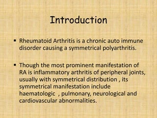
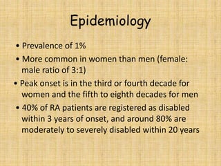
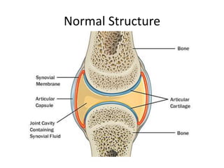
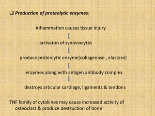
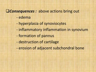
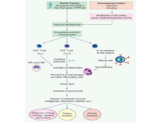
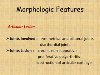
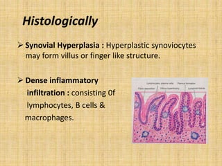
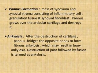
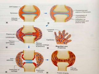
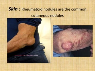
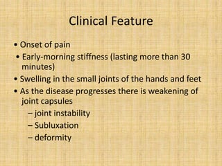
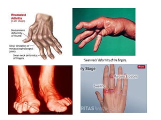
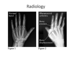
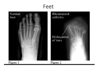
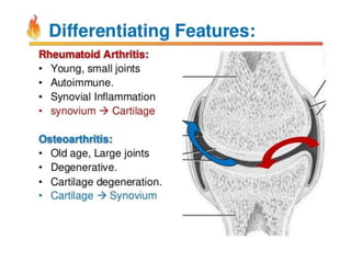
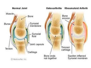
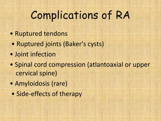
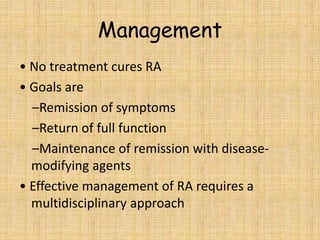
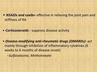
Presented By-
Paridhi Jain
Batch- 2017-18
Index
 Introduction
 Epidemiology
 Normal Structure
 Etiology
 Pathogenesis
 Morphology & Clinical Manifestation
- Articular
- Extra articular
 Variants
 Score
 Laboratory Investigation
 Differential Diagnosis
 Management
Introduction
 Rheumatoid Arthritis is a chronic auto immune
disorder causing a symmetrical polyarthritis.
 Though the most prominent manifestation of
RA is inflammatory arthritis of peripheral joints,
usually with symmetrical distribution , its
symmetrical manifestation include
haematologic , pulmonary, neurological and
cardiovascular abnormalities.
Epidemiology
• Prevalence of 1%
• More common in women than men (female:
male ratio of 3:1)
• Peak onset is in the third or fourth decade for
women and the fifth to eighth decades for men
• 40% of RA patients are registered as disabled
within 3 years of onset, and around 80% are
moderately to severely disabled within 20 years
Normal Structure
Etiology
Causes of RA remains unknown. Following three
factors play important role:
• Genetic Factor : Genetic susceptibility is major factor
in pathogenesis of RA
HLA gene : HLA-DRB1
Non- HLA gene: Polymorphism in PTPN22
• Environmental arthritogen : Smoking & Microbial
agents
• Immunological Derangement
Pathogenesis
 Break down of self tolerance : RA is initiated in genetically susceptible
individual by an exposure to arthitogenc agent which results in break down of
self tolerance.
 Cytokine- mediated inflammation:
loss of self tolerance
activation of CD4+ cells( TH1 &TH17 and interferon)
recruit macrophages & other inflammatory cell
tissue injury
activation of synovial cells( synoviocytes)
 Activation of B lymphocytes : produce 2 types
of antibodies
- Anti – CCPs : antibodies against cyclic citrulinated peptides
(CCPs).
Antibodies against CCPs form immune complex and deposit in
various tissues.
-Rheumatoid Factor : RF is serum immunoglobin M (IgM) auto
antibody that binds to Fc portion of own/self IgG.
Rheumatoid factor form immune complex with self IgG & can
produce damage to joints and other tissues.
 Production of proteolytic enzymes:
inflammation causes tissue injury
activaton of synoviocytes
produce proteolytic enzyme(collagenase , elastase)
enzymes along with antigen antibody complex
destroys articular cartilage, ligaments & tendons
TNF family of cytokines may cause increased activity of
osteoclast & produce destruction of bone
Consequences : above actions bring out
- edema
- hyperplasia of synoviocytes
- inflammatory inflammation in synovium
- formation of pannus
- destruction of cartilage
- erosion of adjacent subchondral bone
Morphologic Features
Articular Lesion
Joints Involved : -symmetrical and bilateral joints
- diarthordial joints
Joints Lesion : - chronic non supprative
proliferative polyarthritis
-destruction of articular cartilage
Histologically
 Synovial Hyperplasia : Hyperplastic synoviocytes
may form villus or finger like structure.
 Dense inflammatory
infiltration : consisting 0f
lymphocytes, B cells &
macrophages.
 Pannus Formation : mass of synovium and
synovial stroma consisting of inflammatory cell ,
granulation tissue & synovial fibroblast . Pannus
grows over the articular cartilage and destroys
it.
Ankylosis : After the destruction of cartilage ,
pannus bridges the opposite bones to form
fibrous ankylosis , which may result in bony
ankylosis. Destruction of joint followed by fusion
is termed as ankylosis.
Skin : Rheumatoid nodules are the common
cutaneous nodules
Clinical Feature
• Onset of pain
• Early-morning stiffness (lasting more than 30
minutes)
• Swelling in the small joints of the hands and feet
• As the disease progresses there is weakening of
joint capsules
– joint instability
– Subluxation
– deformity
Hammer toe
Non-articular manifestations of
RA
•Systemic – Fever, Fatigue, Weight loss
•Eyes- Scleritis, Scleromalacia perforans
(perforation of the eye)
•Neurological- Carpal tunnel syndrome,
Atlantoaxial subluxation, Cord compression
•Hematological- Lymphadenopathy, Felty’s
syndrome (rheumatoid arthritis, splenomegaly,
neutropenia), Anaemia (chronic disease,
NSAIDinduced, gastrointestinal blood loss,
haemolysis, hypersplenism), Thrombocytosis
• Pulmonary - Pleural effusion, Lung fibrosis,
Rheumatoid nodules, Rheumatoid
pneumoconiosis
• Heart and peripheral vessels – Pericarditis,
Pericardial effusion, Raynaud’s syndrome
• Vasculitis - Leg ulcers, Nail fold infarcts,
Gangrene of fingers and toes
• Kidneys - Amyloidosis causes the nephrotic
syndrome and renal failure
Variants of RA
 Juvenile RA : Found in adolescent patient.
 Felty’s syndrome : Consist of polyarticular RA
associated with splenomeagly & hypersplenism.
 Ankylosing Spondylitis or Rheumatoid spondylitis :
Rheumatoid involvement of spine particularly sacro
iliac joint. This condition has a strong HLA-B27
association & may have association with other
inflammatory disorder.
*Criteria for Diagnosis of Rheumatoid
Arthritis2,7
Criterion Score
• Joints affected
1 large joint 0
2–10 large joints 1
1–3 small joints 2
4 -10 small joints 5
• Serology
Negative RF and ACPA 0
Low positive RF or ACPA 2
High positive RF or ACPA 3
• Duration of symptoms
< 6 wks 0
> 6 wks 1
• Acute phase reactants
Normal CRP and ESR 0
Abnormal CRP or ESR 1
Patients with a score ≥ 6 are considered to have definite RA.
*European League Against Rheumatism/American College of Rheumatology 2010 Criteria.
Investigation
•Blood count- usually a normochromic, normocytic
anemia, ESR and CRP are raised
•Serum autoantibodies - Anti-CCP has high
specificity (90%) and, Rheumatoid factor is
positive in 70% of cases sensitivity (80%) for RA.
•X-ray- joint narrowing, erosions at the joint
margins
•Synovial fluid - high neutrophil count in
uncomplicated disease
Complications of RA
• Ruptured tendons
• Ruptured joints (Baker's cysts)
• Joint infection
• Spinal cord compression (atlantoaxial or upper
cervical spine)
• Amyloidosis (rare)
• Side-effects of therapy
Management
• No treatment cures RA
• Goals are
–Remission of symptoms
–Return of full function
–Maintenance of remission with disease-
modifying agents
• Effective management of RA requires a
multidisciplinary approach
• NSAIDs and coxibs- effective in relieving the joint pain and
stiffness of RA
• Corticosteroids - suppress disease activity
• Disease-modifying anti-rheumatic drugs (DMARDs)- act
mainly through inhibition of inflammatory cytokines (6
weeks to 6 months of disease onset)
–Sulfasalazine, Methotrexate
Rheumatoid Arthritis.pptx

Rheumatoid Arthritis.pptx

  • 1.
  • 2.
    Index  Introduction  Epidemiology Normal Structure  Etiology  Pathogenesis  Morphology & Clinical Manifestation - Articular - Extra articular  Variants  Score  Laboratory Investigation  Differential Diagnosis  Management
  • 3.
    Introduction  Rheumatoid Arthritisis a chronic auto immune disorder causing a symmetrical polyarthritis.  Though the most prominent manifestation of RA is inflammatory arthritis of peripheral joints, usually with symmetrical distribution , its symmetrical manifestation include haematologic , pulmonary, neurological and cardiovascular abnormalities.
  • 4.
    Epidemiology • Prevalence of1% • More common in women than men (female: male ratio of 3:1) • Peak onset is in the third or fourth decade for women and the fifth to eighth decades for men • 40% of RA patients are registered as disabled within 3 years of onset, and around 80% are moderately to severely disabled within 20 years
  • 5.
  • 6.
    Etiology Causes of RAremains unknown. Following three factors play important role: • Genetic Factor : Genetic susceptibility is major factor in pathogenesis of RA HLA gene : HLA-DRB1 Non- HLA gene: Polymorphism in PTPN22 • Environmental arthritogen : Smoking & Microbial agents • Immunological Derangement
  • 7.
    Pathogenesis  Break downof self tolerance : RA is initiated in genetically susceptible individual by an exposure to arthitogenc agent which results in break down of self tolerance.  Cytokine- mediated inflammation: loss of self tolerance activation of CD4+ cells( TH1 &TH17 and interferon) recruit macrophages & other inflammatory cell tissue injury activation of synovial cells( synoviocytes)
  • 8.
     Activation ofB lymphocytes : produce 2 types of antibodies - Anti – CCPs : antibodies against cyclic citrulinated peptides (CCPs). Antibodies against CCPs form immune complex and deposit in various tissues. -Rheumatoid Factor : RF is serum immunoglobin M (IgM) auto antibody that binds to Fc portion of own/self IgG. Rheumatoid factor form immune complex with self IgG & can produce damage to joints and other tissues.
  • 9.
     Production ofproteolytic enzymes: inflammation causes tissue injury activaton of synoviocytes produce proteolytic enzyme(collagenase , elastase) enzymes along with antigen antibody complex destroys articular cartilage, ligaments & tendons TNF family of cytokines may cause increased activity of osteoclast & produce destruction of bone
  • 10.
    Consequences : aboveactions bring out - edema - hyperplasia of synoviocytes - inflammatory inflammation in synovium - formation of pannus - destruction of cartilage - erosion of adjacent subchondral bone
  • 12.
    Morphologic Features Articular Lesion JointsInvolved : -symmetrical and bilateral joints - diarthordial joints Joints Lesion : - chronic non supprative proliferative polyarthritis -destruction of articular cartilage
  • 13.
    Histologically  Synovial Hyperplasia: Hyperplastic synoviocytes may form villus or finger like structure.  Dense inflammatory infiltration : consisting 0f lymphocytes, B cells & macrophages.
  • 14.
     Pannus Formation: mass of synovium and synovial stroma consisting of inflammatory cell , granulation tissue & synovial fibroblast . Pannus grows over the articular cartilage and destroys it. Ankylosis : After the destruction of cartilage , pannus bridges the opposite bones to form fibrous ankylosis , which may result in bony ankylosis. Destruction of joint followed by fusion is termed as ankylosis.
  • 16.
    Skin : Rheumatoidnodules are the common cutaneous nodules
  • 17.
    Clinical Feature • Onsetof pain • Early-morning stiffness (lasting more than 30 minutes) • Swelling in the small joints of the hands and feet • As the disease progresses there is weakening of joint capsules – joint instability – Subluxation – deformity
  • 18.
  • 19.
    Non-articular manifestations of RA •Systemic– Fever, Fatigue, Weight loss •Eyes- Scleritis, Scleromalacia perforans (perforation of the eye) •Neurological- Carpal tunnel syndrome, Atlantoaxial subluxation, Cord compression •Hematological- Lymphadenopathy, Felty’s syndrome (rheumatoid arthritis, splenomegaly, neutropenia), Anaemia (chronic disease, NSAIDinduced, gastrointestinal blood loss, haemolysis, hypersplenism), Thrombocytosis
  • 20.
    • Pulmonary -Pleural effusion, Lung fibrosis, Rheumatoid nodules, Rheumatoid pneumoconiosis • Heart and peripheral vessels – Pericarditis, Pericardial effusion, Raynaud’s syndrome • Vasculitis - Leg ulcers, Nail fold infarcts, Gangrene of fingers and toes • Kidneys - Amyloidosis causes the nephrotic syndrome and renal failure
  • 21.
    Variants of RA Juvenile RA : Found in adolescent patient.  Felty’s syndrome : Consist of polyarticular RA associated with splenomeagly & hypersplenism.  Ankylosing Spondylitis or Rheumatoid spondylitis : Rheumatoid involvement of spine particularly sacro iliac joint. This condition has a strong HLA-B27 association & may have association with other inflammatory disorder.
  • 22.
    *Criteria for Diagnosisof Rheumatoid Arthritis2,7 Criterion Score • Joints affected 1 large joint 0 2–10 large joints 1 1–3 small joints 2 4 -10 small joints 5 • Serology Negative RF and ACPA 0 Low positive RF or ACPA 2 High positive RF or ACPA 3
  • 23.
    • Duration ofsymptoms < 6 wks 0 > 6 wks 1 • Acute phase reactants Normal CRP and ESR 0 Abnormal CRP or ESR 1 Patients with a score ≥ 6 are considered to have definite RA. *European League Against Rheumatism/American College of Rheumatology 2010 Criteria.
  • 24.
    Investigation •Blood count- usuallya normochromic, normocytic anemia, ESR and CRP are raised •Serum autoantibodies - Anti-CCP has high specificity (90%) and, Rheumatoid factor is positive in 70% of cases sensitivity (80%) for RA. •X-ray- joint narrowing, erosions at the joint margins •Synovial fluid - high neutrophil count in uncomplicated disease
  • 29.
    Complications of RA •Ruptured tendons • Ruptured joints (Baker's cysts) • Joint infection • Spinal cord compression (atlantoaxial or upper cervical spine) • Amyloidosis (rare) • Side-effects of therapy
  • 30.
    Management • No treatmentcures RA • Goals are –Remission of symptoms –Return of full function –Maintenance of remission with disease- modifying agents • Effective management of RA requires a multidisciplinary approach
  • 31.
    • NSAIDs andcoxibs- effective in relieving the joint pain and stiffness of RA • Corticosteroids - suppress disease activity • Disease-modifying anti-rheumatic drugs (DMARDs)- act mainly through inhibition of inflammatory cytokines (6 weeks to 6 months of disease onset) –Sulfasalazine, Methotrexate