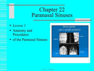
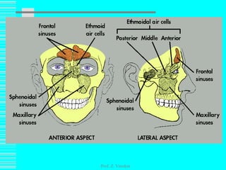
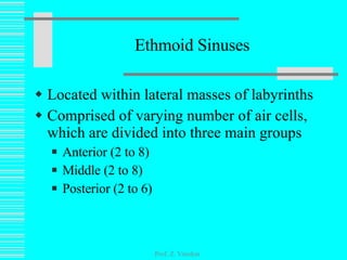
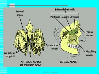
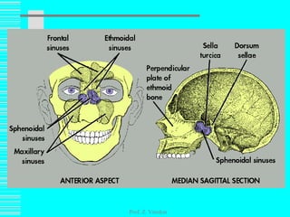
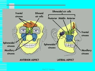
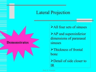
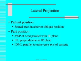
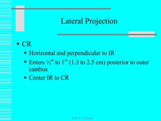
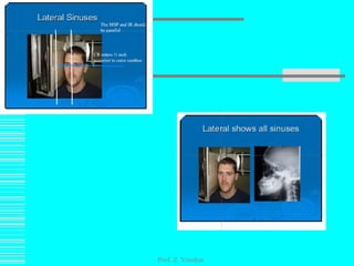
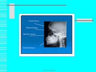
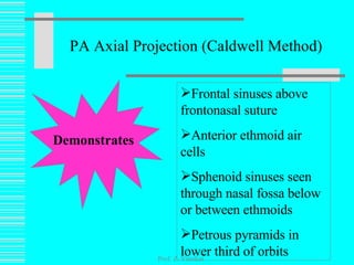
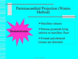
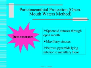
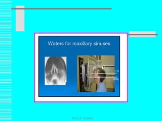
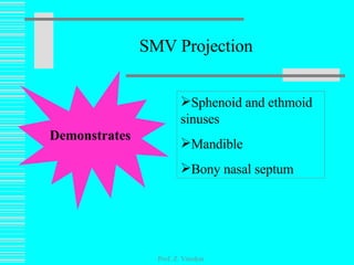
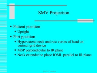
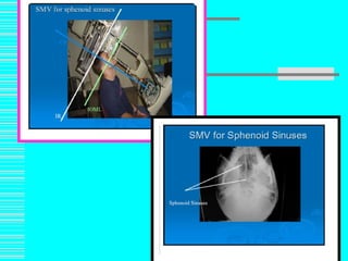
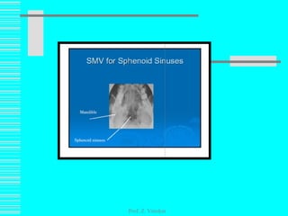
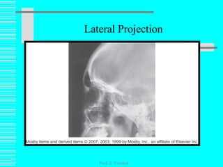
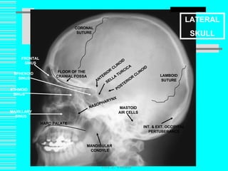
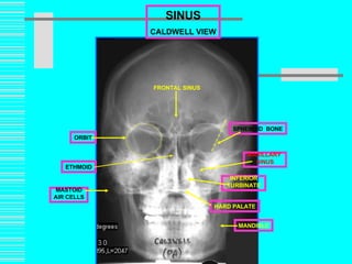
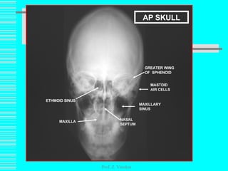
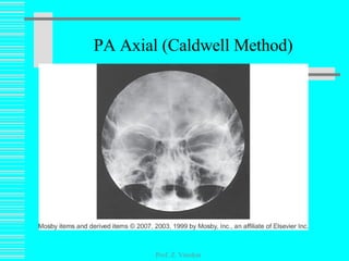
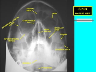
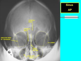
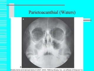
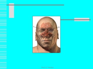
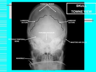
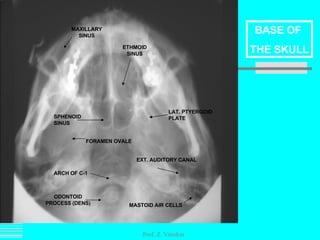
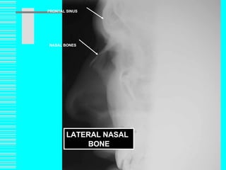
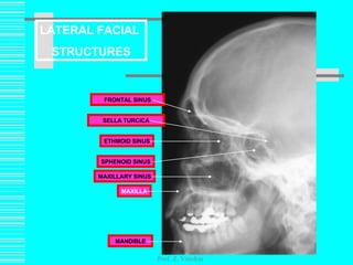
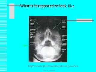
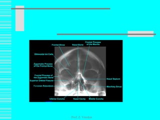
Chapter 22 Paranasal Sinuses Lesson 1 Anatomy and Procedures of the Paranasal Sinuses Prof. Z. Vinokur
Paranasal Sinuses Defined as the air-filled cavities located in the frontal, ethmoid, and sphenoid bones of the cranium, as well as the maxillae of the face So named because of their formation from the nasal mucosa and their continued communication with the nasal fossas Prof. Z. Vinokur
Functions Serve as resonating chamber for the voice Decrease weight of skull Aid in warming and moisturizing inhaled air Act as shock absorbers in trauma Possibly control the immune system Prof. Z. Vinokur
Development Begins in fetal life Maxillary sinuses are usually the only ones developed enough to be demonstrated radiographically at birth By 6 to 7 years of age, frontal and sphenoid are distinguishable from ethmoids Ethmoids do not fully develop until 17 to 18 years of age Prof. Z. Vinokur
Maxillary Sinuses Largest and most symmetric Paired (one in each facial bone)  Vary in size and shape but are roughly pyramidal in shape Prof. Z. Vinokur
Frontal Sinuses Second largest Paired Located between vertical plates of frontal bone Vary greatly in size and shape Occasionally absent Rarely symmetric Prof. Z. Vinokur
Prof. Z. Vinokur
Ethmoid Sinuses Located within lateral masses of labyrinths Comprised of varying number of air cells, which are divided into three main groups Anterior (2 to 8) Middle (2 to 8) Posterior (2 to 6) Prof. Z. Vinokur
Prof. Z. Vinokur
Sphenoid Sinuses Normally paired Occupy body of sphenoid bone Often only one sinus develops, but never more than two Vary in size and shape Usually asymmetric Located below sella and extend between dorsum and posterior ethmoids Prof. Z. Vinokur
Prof. Z. Vinokur
Prof. Z. Vinokur
Essential Projections Lateral PA axial (Caldwell method) Parietoacanthial (Waters method) Parietoacanthial (Waters) open-mouth modification Submentovertical Prof. Z. Vinokur
Technical Considerations Density most critical to demonstrate pathology Upright position necessary to demonstrate fluid levels CR should remain horizontal Shielding must be used on pregnant patients and pediatric patients Proper collimation critical in reducing scatter radiation and patient dose Prof. Z. Vinokur
Lateral Projection Prof. Z. Vinokur Demonstrates All four sets of sinuses AP and superoinferior dimensions of paranasal sinuses Thickness of frontal bone Detail of side closer to IR
Lateral Projection Patient position Seated erect in anterior oblique position Part position MSP of head parallel with IR plane IPL perpendicular to IR plane IOML parallel to transverse axis of cassette Prof. Z. Vinokur
Lateral Projection CR Horizontal and perpendicular to IR Enters ½   to 1   (1.3 to 2.5 cm) posterior to outer canthus Center IR to CR Prof. Z. Vinokur
Prof. Z. Vinokur
Prof. Z. Vinokur
PA Axial Projection (Caldwell Method) Prof. Z. Vinokur Demonstrates Frontal sinuses above frontonasal suture Anterior ethmoid air cells  Sphenoid sinuses seen through nasal fossa below or between ethmoids Petrous pyramids in lower third of orbits
PA Axial (Caldwell Method) Angled Grid Technique Patient position Seated upright facing Bucky  MSP centered to midline Prof. Z. Vinokur
PA Axial (Caldwell Method) Patient position Tilt vertical Bucky down 15 degrees Rest patient’s forehead and nose on device Center nasion to IR MSP and OML perpendicular to IR plane Prof. Z. Vinokur
Prof. Z. Vinokur
PA Axial (Caldwell Method) Vertical Grid Technique Position patient’s OML at 15-degree   angle to horizontal CR Center nasion to IR MSP perpendicular Radiolucent support may be needed at forehead This method is not preferred due to increased OID, which results in decreased resolution Prof. Z. Vinokur
PA Axial (Caldwell Method) CR Horizontal Exits nasion Center IR and CR Prof. Z. Vinokur
Parietoacanthial Projection (Waters Method) Prof. Z. Vinokur Demonstrates Maxillary sinuses Petrous pyramids lying inferior to maxillary floor Frontal and ethmoid sinuses are distorted
Patient position Seated erect facing Bucky  MSP centered to midline Part position Hyperextend neck to place OML at 37-degree angle from IR plane MSP and MML perpendicular to IR plane Parietoacanthial Projection (Waters Method) Prof. Z. Vinokur
CR Horizontal Exits acanthion Parietoacanthial Projection (Waters Method) Prof. Z. Vinokur
Parietoacanthial Projection (Open-Mouth Waters Method) Prof. Z. Vinokur Demonstrates Sphenoid sinuses through open mouth Maxillary sinuses Petrous pyramids lying inferior to maxillary floor
Patient position Seated erect facing Bucky MSP centered to midline Part position Hyperextend neck to place OML at 37-degree angle from IR plane MSP and MML perpendicular to IR plane Open mouth wide while holding position Parietoacanthial Projection (Open-Mouth Waters Method) Prof. Z. Vinokur
CR Horizontal Exits acanthion Parietoacanthial Projection (Open-Mouth Waters Method) Prof. Z. Vinokur
Prof. Z. Vinokur
Prof. Z. Vinokur
Prof. Z. Vinokur
SMV Projection Prof. Z. Vinokur Demonstrates Sphenoid and ethmoid sinuses Mandible Bony nasal septum
SMV Projection Patient position Upright Part position  Hyperextend neck and rest vertex of head on vertical grid device MSP perpendicular to IR plane Neck extended to place IOML parallel to IR plane Prof. Z. Vinokur
SMV Projection CR Horizontal and perpendicular to IOML Enters MSP ¾   (1.9 cm) anterior to level of EAM Prof. Z. Vinokur
Prof. Z. Vinokur
Prof. Z. Vinokur
Image Critique Criteria for Essential Projection Slide
Lateral Projection All four sinus groups Sphenoid of primary importance No rotation of sella turcica Superimposed orbital roofs Superimposed mandibular rami Clearly visible sinuses Close beam restriction Clearly visible air-fluid levels, if present Prof. Z. Vinokur
Lateral Projection Prof. Z. Vinokur
Prof. Z. Vinokur L LATERAL SKULL CORONAL SUTURE LAMBOID SUTURE INT. & EXT. OCCIPITAL PERTUBERANCE MASTOID AIR CELLS HARD PALATE NASOPHARYNX ANTERIOR CLINOID POSTERIOR CLINOID MANDIBULAR  CONDYLE FLOOR OF THE CRANIAL FOSSA SELLA TURCICA FRONTAL SINUS MAXILLARY SINUS SPHENOID  SINUS ETHMOID SINUS
PA Axial (Caldwell Method) No rotation Equal distance between lateral border of skull and lateral border of orbits Petrous ridges symmetric Petrous ridge lying in lower third of orbit Frontal sinuses above frontonasal suture and anterior ethmoidal air cells above petrous ridges Prof. Z. Vinokur
PA Axial (Caldwell Method) Frontal and anterior ethmoidal air cells Clearly visible air-fluid levels, if present Close beam restriction Prof. Z. Vinokur
Prof. Z. Vinokur SINUS CALDWELL VIEW FRONTAL SINUS SPHENOID   BONE MAXILLARY  SINUS INFERIOR TURBINATE MANDIBLE HARD PALATE MASTOID AIR CELLS ORBIT ETHMOID
Prof. Z. Vinokur ETHMOID SINUS NASAL SEPTUM MASTOID AIR CELLS MAXILLARY SINUS MAXILLA GREATER WING OF  SPHENOID AP SKULL
PA Axial (Caldwell Method) Prof. Z. Vinokur
Petrous pyramids immediately inferior to floor of maxillary sinuses No rotation Equal distance between lateral border of skull and lateral border of orbit Orbits and maxillary sinuses symmetric Maxillary sinuses Close beam restriction Clearly visible air-fluid levels, if present Parietoacanthial Projection (Waters Method) Prof. Z. Vinokur
Prof. Z. Vinokur Sinus WATERS VIEW MANDIBLE FRONTAL  SINUS ETHMOID SINUS MAXILLARY SINUS ZYGOMA ZYGOMATIC ARCH INFERIOR ORBITAL MARGIN ORBIT NASAL SEPTUM ZYGOMATICAL –FRONTAL SUTURE ODONTOID
Prof. Z. Vinokur Sinus  AP FRONTAL  SINUS ETHIMOID  SINUS ORBIT NASAL SEPTUM GREATER WING  OF  SPHENOID MAXILLARY  SINUS
Parietoacanthial (Waters) Prof. Z. Vinokur
Petrous pyramids immediately inferior to floor of maxillary sinuses No rotation Equal distance between lateral border of skull and lateral border of orbit Orbits and maxillary sinuses symmetric Maxillary sinuses Parietoacanthial Projection (Open-Mouth Waters Method) Prof. Z. Vinokur
Close beam restriction Clearly visible air-fluid levels, if present Sphenoid sinuses projected through open mouth Parietoacanthial Projection (Open-Mouth Waters Method) Prof. Z. Vinokur
Parietoacanthial (Waters) Prof. Z. Vinokur
SMV Projection No tilt Equal distance from lateral border of skull to mandibular condyles on both sides Anterior frontal bone superimposed by mental protuberance Indicates IOML is parallel (full extension) Mandibular condyles anterior to petrous pyramids Clearly visible air-fluid levels, if present Prof. Z. Vinokur
SMV Projection Prof. Z. Vinokur
Prof. Z. Vinokur
Prof. Z. Vinokur SKULL TOWNE VIEW MANDIBLE MASTOID AIR CELLS PETROUS TEMPORAL  BONE FORAMEN MAGNUM PARIETAL BONES LAMBDOID  SUTURE LAMBDOID  SUTURE
Prof. Z. Vinokur ODONTOID PROCESS (DENS) ETHMOID SINUS SPHENOID SINUS MAXILLARY SINUS LAT. PTYERGOID PLATE FORAMEN OVALE ARCH OF C-1 EXT. AUDITORY CANAL MASTOID AIR CELLS BASE OF  THE SKULL
Prof. Z. Vinokur FRONTAL SINUS LATERAL NASAL BONE NASAL BONES
Prof. Z. Vinokur FRONTAL SINUS MAXILLARY SINUS MANDIBLE MAXILLA ETHMOID SINUS SELLA   TURCICA SPHENOID SINUS LATERAL FACIAL  STRUCTURES
Prof. Z. Vinokur AIRWAY EPIGLOTTIS HYOID BONE CALCIFIED TRACHEAL CARTILAGE RINGS THYROID  CARTILAGE CRICOID  CARTILAGE
Prof. Z. Vinokur ARTERIOGRAM  CAROTID SYPHON OCCIPITAL ARTERY INTERNAL CAROTID ARTERY EXTERNAL CAROTID  ARTERY COMMON CAROTID ARTERY MAXILLARY ARTERY MIDDLE CEREBRAL ARTERY ANTERIOR CEREBRAL ARTERY FACIAL ARTERY
What is it supposed to look like Prof. Z. Vinokur http://www.jeffersonhospital.org/webcast/sinus/index.html
Prof. Z. Vinokur
links Prof. Z. Vinokur http://classes.kumc.edu/som/radanatomy/results2.asp?RegionID=1&PathologyID=2

Lec1222509slideshare

  • 1.
    Chapter 22 ParanasalSinuses Lesson 1 Anatomy and Procedures of the Paranasal Sinuses Prof. Z. Vinokur
  • 2.
    Paranasal Sinuses Definedas the air-filled cavities located in the frontal, ethmoid, and sphenoid bones of the cranium, as well as the maxillae of the face So named because of their formation from the nasal mucosa and their continued communication with the nasal fossas Prof. Z. Vinokur
  • 3.
    Functions Serve asresonating chamber for the voice Decrease weight of skull Aid in warming and moisturizing inhaled air Act as shock absorbers in trauma Possibly control the immune system Prof. Z. Vinokur
  • 4.
    Development Begins infetal life Maxillary sinuses are usually the only ones developed enough to be demonstrated radiographically at birth By 6 to 7 years of age, frontal and sphenoid are distinguishable from ethmoids Ethmoids do not fully develop until 17 to 18 years of age Prof. Z. Vinokur
  • 5.
    Maxillary Sinuses Largestand most symmetric Paired (one in each facial bone) Vary in size and shape but are roughly pyramidal in shape Prof. Z. Vinokur
  • 6.
    Frontal Sinuses Secondlargest Paired Located between vertical plates of frontal bone Vary greatly in size and shape Occasionally absent Rarely symmetric Prof. Z. Vinokur
  • 7.
  • 8.
    Ethmoid Sinuses Locatedwithin lateral masses of labyrinths Comprised of varying number of air cells, which are divided into three main groups Anterior (2 to 8) Middle (2 to 8) Posterior (2 to 6) Prof. Z. Vinokur
  • 9.
  • 10.
    Sphenoid Sinuses Normallypaired Occupy body of sphenoid bone Often only one sinus develops, but never more than two Vary in size and shape Usually asymmetric Located below sella and extend between dorsum and posterior ethmoids Prof. Z. Vinokur
  • 11.
  • 12.
  • 13.
    Essential Projections LateralPA axial (Caldwell method) Parietoacanthial (Waters method) Parietoacanthial (Waters) open-mouth modification Submentovertical Prof. Z. Vinokur
  • 14.
    Technical Considerations Densitymost critical to demonstrate pathology Upright position necessary to demonstrate fluid levels CR should remain horizontal Shielding must be used on pregnant patients and pediatric patients Proper collimation critical in reducing scatter radiation and patient dose Prof. Z. Vinokur
  • 15.
    Lateral Projection Prof.Z. Vinokur Demonstrates All four sets of sinuses AP and superoinferior dimensions of paranasal sinuses Thickness of frontal bone Detail of side closer to IR
  • 16.
    Lateral Projection Patientposition Seated erect in anterior oblique position Part position MSP of head parallel with IR plane IPL perpendicular to IR plane IOML parallel to transverse axis of cassette Prof. Z. Vinokur
  • 17.
    Lateral Projection CRHorizontal and perpendicular to IR Enters ½  to 1  (1.3 to 2.5 cm) posterior to outer canthus Center IR to CR Prof. Z. Vinokur
  • 18.
  • 19.
  • 20.
    PA Axial Projection(Caldwell Method) Prof. Z. Vinokur Demonstrates Frontal sinuses above frontonasal suture Anterior ethmoid air cells Sphenoid sinuses seen through nasal fossa below or between ethmoids Petrous pyramids in lower third of orbits
  • 21.
    PA Axial (CaldwellMethod) Angled Grid Technique Patient position Seated upright facing Bucky MSP centered to midline Prof. Z. Vinokur
  • 22.
    PA Axial (CaldwellMethod) Patient position Tilt vertical Bucky down 15 degrees Rest patient’s forehead and nose on device Center nasion to IR MSP and OML perpendicular to IR plane Prof. Z. Vinokur
  • 23.
  • 24.
    PA Axial (CaldwellMethod) Vertical Grid Technique Position patient’s OML at 15-degree angle to horizontal CR Center nasion to IR MSP perpendicular Radiolucent support may be needed at forehead This method is not preferred due to increased OID, which results in decreased resolution Prof. Z. Vinokur
  • 25.
    PA Axial (CaldwellMethod) CR Horizontal Exits nasion Center IR and CR Prof. Z. Vinokur
  • 26.
    Parietoacanthial Projection (WatersMethod) Prof. Z. Vinokur Demonstrates Maxillary sinuses Petrous pyramids lying inferior to maxillary floor Frontal and ethmoid sinuses are distorted
  • 27.
    Patient position Seatederect facing Bucky MSP centered to midline Part position Hyperextend neck to place OML at 37-degree angle from IR plane MSP and MML perpendicular to IR plane Parietoacanthial Projection (Waters Method) Prof. Z. Vinokur
  • 28.
    CR Horizontal Exitsacanthion Parietoacanthial Projection (Waters Method) Prof. Z. Vinokur
  • 29.
    Parietoacanthial Projection (Open-MouthWaters Method) Prof. Z. Vinokur Demonstrates Sphenoid sinuses through open mouth Maxillary sinuses Petrous pyramids lying inferior to maxillary floor
  • 30.
    Patient position Seatederect facing Bucky MSP centered to midline Part position Hyperextend neck to place OML at 37-degree angle from IR plane MSP and MML perpendicular to IR plane Open mouth wide while holding position Parietoacanthial Projection (Open-Mouth Waters Method) Prof. Z. Vinokur
  • 31.
    CR Horizontal Exitsacanthion Parietoacanthial Projection (Open-Mouth Waters Method) Prof. Z. Vinokur
  • 32.
  • 33.
  • 34.
  • 35.
    SMV Projection Prof.Z. Vinokur Demonstrates Sphenoid and ethmoid sinuses Mandible Bony nasal septum
  • 36.
    SMV Projection Patientposition Upright Part position Hyperextend neck and rest vertex of head on vertical grid device MSP perpendicular to IR plane Neck extended to place IOML parallel to IR plane Prof. Z. Vinokur
  • 37.
    SMV Projection CRHorizontal and perpendicular to IOML Enters MSP ¾  (1.9 cm) anterior to level of EAM Prof. Z. Vinokur
  • 38.
  • 39.
  • 40.
    Image Critique Criteriafor Essential Projection Slide
  • 41.
    Lateral Projection Allfour sinus groups Sphenoid of primary importance No rotation of sella turcica Superimposed orbital roofs Superimposed mandibular rami Clearly visible sinuses Close beam restriction Clearly visible air-fluid levels, if present Prof. Z. Vinokur
  • 42.
  • 43.
    Prof. Z. VinokurL LATERAL SKULL CORONAL SUTURE LAMBOID SUTURE INT. & EXT. OCCIPITAL PERTUBERANCE MASTOID AIR CELLS HARD PALATE NASOPHARYNX ANTERIOR CLINOID POSTERIOR CLINOID MANDIBULAR CONDYLE FLOOR OF THE CRANIAL FOSSA SELLA TURCICA FRONTAL SINUS MAXILLARY SINUS SPHENOID SINUS ETHMOID SINUS
  • 44.
    PA Axial (CaldwellMethod) No rotation Equal distance between lateral border of skull and lateral border of orbits Petrous ridges symmetric Petrous ridge lying in lower third of orbit Frontal sinuses above frontonasal suture and anterior ethmoidal air cells above petrous ridges Prof. Z. Vinokur
  • 45.
    PA Axial (CaldwellMethod) Frontal and anterior ethmoidal air cells Clearly visible air-fluid levels, if present Close beam restriction Prof. Z. Vinokur
  • 46.
    Prof. Z. VinokurSINUS CALDWELL VIEW FRONTAL SINUS SPHENOID BONE MAXILLARY SINUS INFERIOR TURBINATE MANDIBLE HARD PALATE MASTOID AIR CELLS ORBIT ETHMOID
  • 47.
    Prof. Z. VinokurETHMOID SINUS NASAL SEPTUM MASTOID AIR CELLS MAXILLARY SINUS MAXILLA GREATER WING OF SPHENOID AP SKULL
  • 48.
    PA Axial (CaldwellMethod) Prof. Z. Vinokur
  • 49.
    Petrous pyramids immediatelyinferior to floor of maxillary sinuses No rotation Equal distance between lateral border of skull and lateral border of orbit Orbits and maxillary sinuses symmetric Maxillary sinuses Close beam restriction Clearly visible air-fluid levels, if present Parietoacanthial Projection (Waters Method) Prof. Z. Vinokur
  • 50.
    Prof. Z. VinokurSinus WATERS VIEW MANDIBLE FRONTAL SINUS ETHMOID SINUS MAXILLARY SINUS ZYGOMA ZYGOMATIC ARCH INFERIOR ORBITAL MARGIN ORBIT NASAL SEPTUM ZYGOMATICAL –FRONTAL SUTURE ODONTOID
  • 51.
    Prof. Z. VinokurSinus AP FRONTAL SINUS ETHIMOID SINUS ORBIT NASAL SEPTUM GREATER WING OF SPHENOID MAXILLARY SINUS
  • 52.
  • 53.
    Petrous pyramids immediatelyinferior to floor of maxillary sinuses No rotation Equal distance between lateral border of skull and lateral border of orbit Orbits and maxillary sinuses symmetric Maxillary sinuses Parietoacanthial Projection (Open-Mouth Waters Method) Prof. Z. Vinokur
  • 54.
    Close beam restrictionClearly visible air-fluid levels, if present Sphenoid sinuses projected through open mouth Parietoacanthial Projection (Open-Mouth Waters Method) Prof. Z. Vinokur
  • 55.
  • 56.
    SMV Projection Notilt Equal distance from lateral border of skull to mandibular condyles on both sides Anterior frontal bone superimposed by mental protuberance Indicates IOML is parallel (full extension) Mandibular condyles anterior to petrous pyramids Clearly visible air-fluid levels, if present Prof. Z. Vinokur
  • 57.
  • 58.
  • 59.
    Prof. Z. VinokurSKULL TOWNE VIEW MANDIBLE MASTOID AIR CELLS PETROUS TEMPORAL BONE FORAMEN MAGNUM PARIETAL BONES LAMBDOID SUTURE LAMBDOID SUTURE
  • 60.
    Prof. Z. VinokurSKULL TOWNE VIEW MANDIBLE MASTOID AIR CELLS PETROUS TEMPORAL BONE FORAMEN MAGNUM PARIETAL BONES LAMBDOID SUTURE LAMBDOID SUTURE
  • 61.
    Prof. Z. VinokurODONTOID PROCESS (DENS) ETHMOID SINUS SPHENOID SINUS MAXILLARY SINUS LAT. PTYERGOID PLATE FORAMEN OVALE ARCH OF C-1 EXT. AUDITORY CANAL MASTOID AIR CELLS BASE OF THE SKULL
  • 62.
    Prof. Z. VinokurFRONTAL SINUS LATERAL NASAL BONE NASAL BONES
  • 63.
    Prof. Z. VinokurFRONTAL SINUS MAXILLARY SINUS MANDIBLE MAXILLA ETHMOID SINUS SELLA TURCICA SPHENOID SINUS LATERAL FACIAL STRUCTURES
  • 64.
    Prof. Z. VinokurAIRWAY EPIGLOTTIS HYOID BONE CALCIFIED TRACHEAL CARTILAGE RINGS THYROID CARTILAGE CRICOID CARTILAGE
  • 65.
    Prof. Z. VinokurARTERIOGRAM CAROTID SYPHON OCCIPITAL ARTERY INTERNAL CAROTID ARTERY EXTERNAL CAROTID ARTERY COMMON CAROTID ARTERY MAXILLARY ARTERY MIDDLE CEREBRAL ARTERY ANTERIOR CEREBRAL ARTERY FACIAL ARTERY
  • 66.
    What is itsupposed to look like Prof. Z. Vinokur http://www.jeffersonhospital.org/webcast/sinus/index.html
  • 67.
  • 68.
    links Prof. Z.Vinokur http://classes.kumc.edu/som/radanatomy/results2.asp?RegionID=1&PathologyID=2