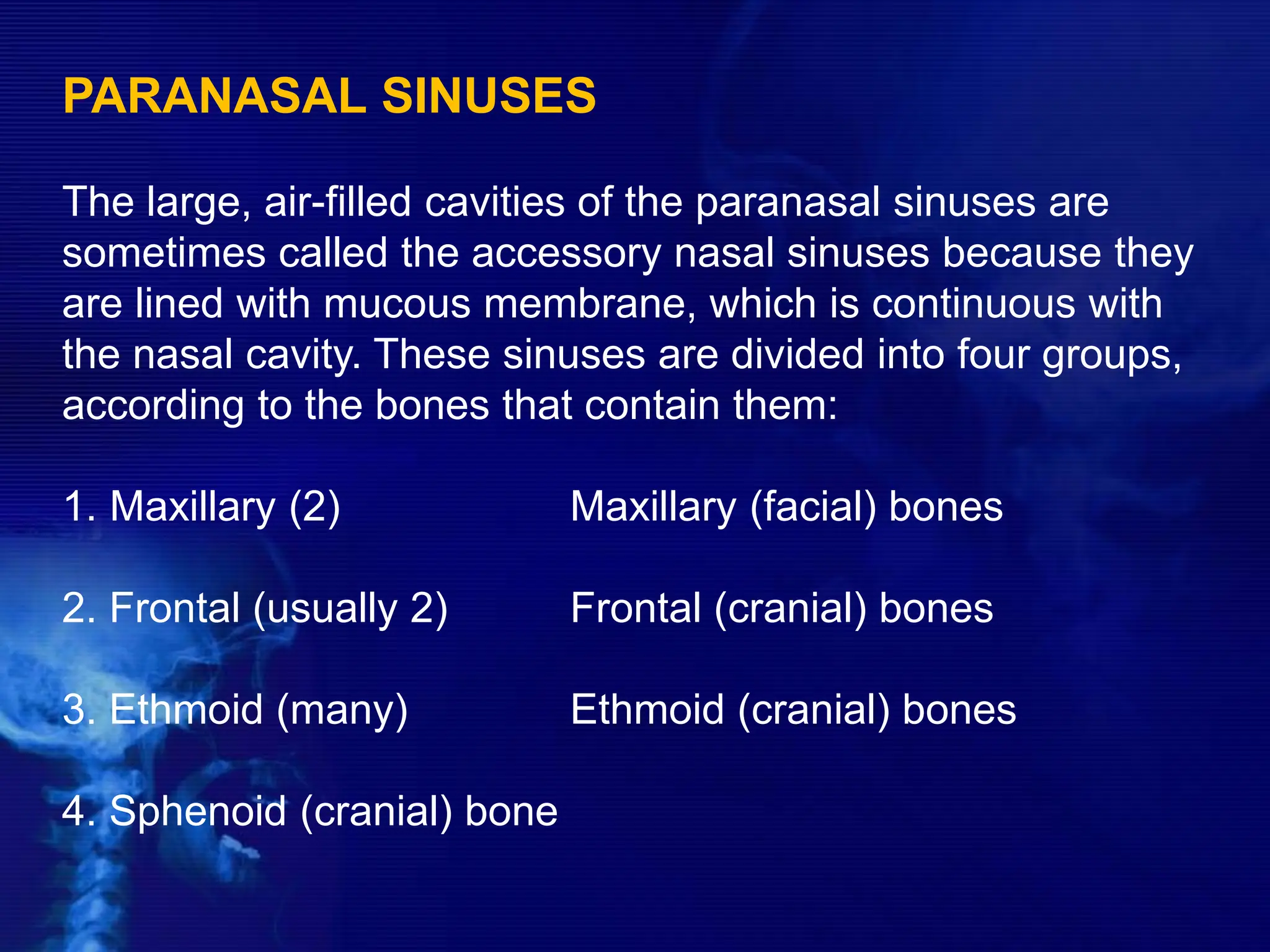
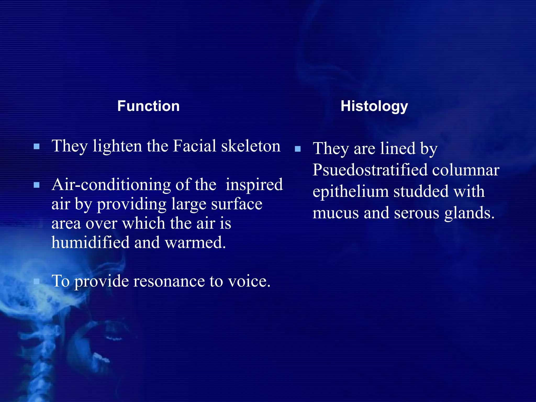
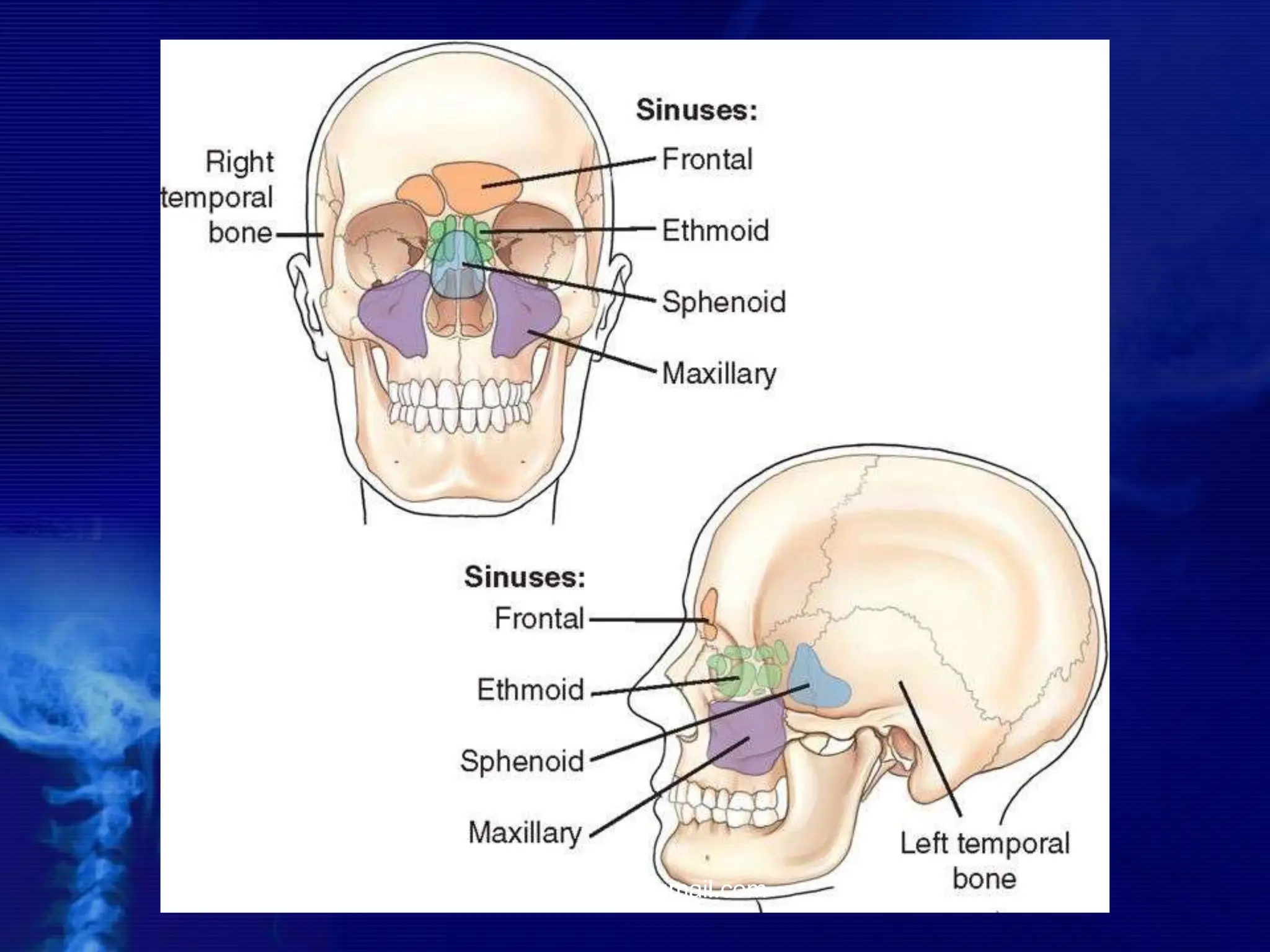
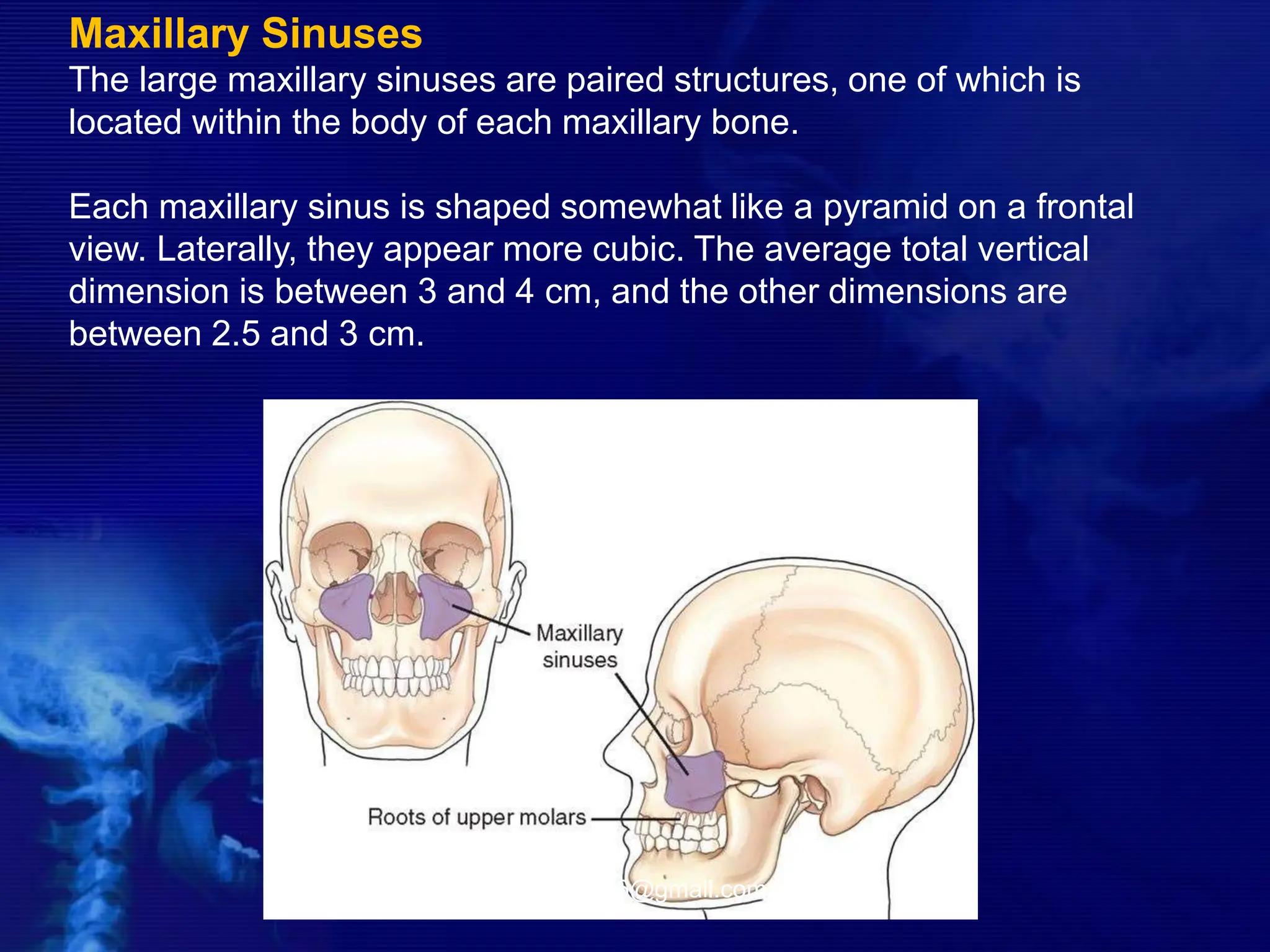
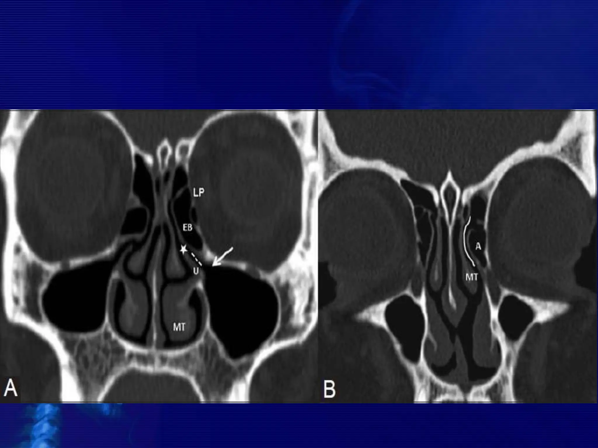
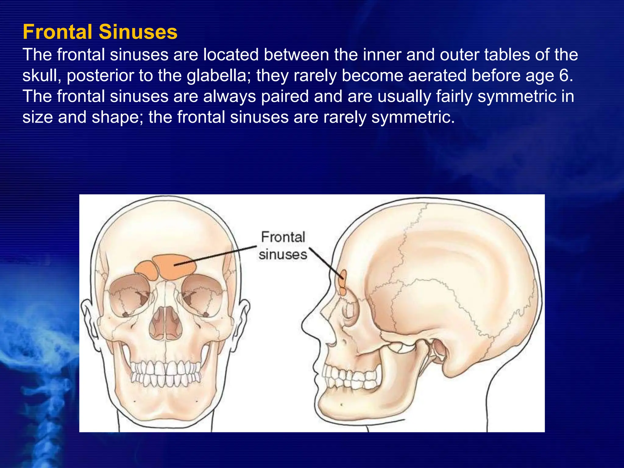
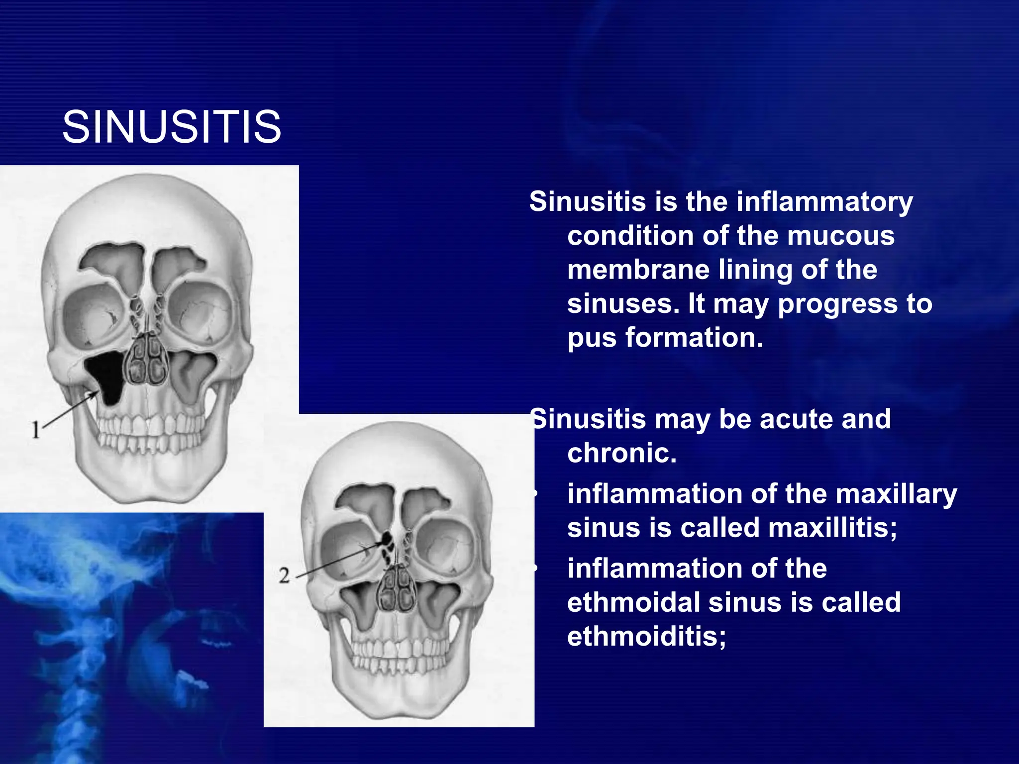
PARANASAL SINUSES-
ANATOMY & PATHOLOGY
PRESENTER:
DR ASHOK SHARMA
JUNIOR RESIDENT II
GUIDE:
DR P.K. LAMGHARE SIR
& DR MOHIT PATIL SIR
PARANASAL SINUSES
The large, air-filled cavities of the paranasal sinuses are
sometimes called the accessory nasal sinuses because they
are lined with mucous membrane, which is continuous with
the nasal cavity. These sinuses are divided into four groups,
according to the bones that contain them:
1. Maxillary (2) Maxillary (facial) bones
2. Frontal (usually 2) Frontal (cranial) bones
3. Ethmoid (many) Ethmoid (cranial) bones
4. Sphenoid (cranial) bone
 They lighten the Facial skeleton
 Air-conditioning of the inspired
air by providing large surface
area over which the air is
humidified and warmed.
 To provide resonance to voice.
 They are lined by
Psuedostratified columnar
epithelium studded with
mucus and serous glands.
Function Histology
-drdhiru456@gmail.com
Maxillary Sinuses
The large maxillary sinuses are paired structures, one of which is
located within the body of each maxillary bone.
Each maxillary sinus is shaped somewhat like a pyramid on a frontal
view. Laterally, they appear more cubic. The average total vertical
dimension is between 3 and 4 cm, and the other dimensions are
between 2.5 and 3 cm.
-drdhiru456@gmail.com
Osteomeatal unit
• The osteomeatal unit (OMU) includes the (1) maxillary
sinus ostium, (2) ethmoid infundibulum, (3) anterior
ethmoid air cells, and (4) frontal recess.
• The OMU is the key factor in the pathogenesis of chronic
sinusitis.
Frontal Sinuses
The frontal sinuses are located between the inner and outer tables of the
skull, posterior to the glabella; they rarely become aerated before age 6.
The frontal sinuses are always paired and are usually fairly symmetric in
size and shape; the frontal sinuses are rarely symmetric.
Ethmoid Sinuses
The ethmoid sinuses are contained within the lateral masses or
labyrinths of the ethmoid bone. These air cells are grouped into anterior,
middle, and posterior collections, but they all intercommunicate.
When viewed from the side, the anterior ethmoid sinuses appear to fill
the orbits. This occurs because portions of the ethmoid sinuses are
contained in the lateral masses of the ethmoid bone, which helps to form
the medial wall of each orbit.
-drdhiru456@gmail.com
Sphenoid Sinuses
The sphenoid sinuses lie in the body of the sphenoid bone directly below
the sella turcica. The body of the sphenoid that contains these sinuses is
cubic and frequently is divided by a thin septum to form two cavities. This
septum may be incomplete or absent entirely, however, resulting in only
one cavity.
The Lateral Wall of Nasal Cavity
Marked by 3
projections:
• Superior concha
• Middle concha
• Inferior concha
• The space below
each concha is
called a meatus.
The Lateral Wall of Nasal Cavity
1. Inferior meatus:
nasolacrimal duct
2. Middle meatus:
• Maxillary sinus
• Frontal sinus
• Anterior ethmoid
sinuses
3. Superior meatus:
posterior ethmoid
sinuses
4. Sphenoethmoidal
recess: sphenoid
sinus
LATERAL VIEW
Lateral side of the skull lies against
the film and x-ray beam is projected
perpendicular from the other side.
Center CR to a point midway between
outer canthus and EAM.
LATERAL POSITION—RIGHT OR LEFT LATERAL: SINUSES
Respiration
Suspend respiration during exposure.
Structures Shown: • All four paranasal sinus groups are
shown.
STRUCTURES SEEN -
 ANTERIOR AND POSTERIOR EXTENT
OF SPHENOID, FRONTAL AND
MAXILLARY SINUSES
 SELLA TURCICA
 ETHMOID SINUSES
 CONDYLE AND NECK OF MANDIBLE
CALDWELL VIEW
• A/K/A OCCIPITOFRONTAL VIEW OR
NOSE FOREHEAD POSITION
Part Position
 Place patient's nose and forehead against upright table
with neck extended to elevate the OML 15° from horizontal. A
radiolucent support between forehead and upright Bucky or
table may be used to maintain this position. CR remains
horizontal. (alternate method if Bucky can be tilted 15°.)
 Center X-RAY to CR and to nasion, ensuring no rotation.
 Align CR horizontal, parallel to floor.
POSITION: SINUSES Caldwell Method
.
Structures Shown: • Frontal sinuses projected above the
frontonasal suture. • Anterior ethmoid air cells visualized lateral to
each nasal bone, directly below the frontal sinuses.
STRUCTURES SEEN
1. FRONTAL SINUSES (SEEN BEST)
2. ETHMOID SINUSES
3. MAXILLARY SINUSES
4. FRONTAL PROCESS OF ZYGOMA AND
ZYGOMATIC PROCESS OF FRONTAL
BONE
5. SUPERIOR MARGIN OF ORBIT AND
LAMINA PAPYRACEA
6. SUPERIOR ORBITAL FISSURE
WATER’S VIEW
 OCCIPITOMENTAL VIEW OR NOSE
CHIN POSITION
 IT IS TAKEN IN SUCH A WAY THAT
NOSE AND CHIN OF THE PATIENT
TOUCH THE FILM WHILE X-RAY BEAM
IS PROJECTED FROM BEHIND.
Part Position
• Extend neck, placing chin and nose against table/film.
• Adjust head until MML is perpendicular to film; OML will form
a 37° angle with the plane of the film.
• Ensure that no rotation or tilt exists.
• Center film to CR and to acanthion.
PARIETOACANTHIAL PROJECTION: SINUSES Waters
Method
Structures Shown: • Maxillary sinuses with the inferior
aspect visualized free from superimposing alveolar
processes and petrous ridges, the inferior orbital rim, and
an oblique view of the frontal sinuses
STRUCTURES SEEN
• Maxillary sinuses (seen best)
• Frontal sinuses
• Sphenoid sinuses (if the film is taken with
open mouth)
• Zygoma
• Zygomatic arch
• Nasal bone
• Frontal process of maxilla
SUBMENTOVERTICAL (BASAL)
VIEW
 THE VIEW IS TAKEN WITH VERTEX NEAR THE
FILM AND X-RAY BEAM PROJECTED AT RIGHT
ANGLES TO THE FILM FROM THE SUBMENTAL
AREA.
Part Position
• Raise chin, hyperextend neck if possible until
OML is parallel to table/film.
• Head rests on vertex of skull.
• Ensure no rotation or tilt.
SUBMENTOVERTEX (SMV) PROJECTION: SINUSES
STRUCTURES SEEN
• Sphenoid, posterior Ethmoid and Maxillarry sinuses
(seen best in that order)
• Mandible
Structures Shown: • Sphenoid sinuses,
ethmoid sinuses, nasal fossae, and maxillary
sinuses.
PARIETOACANTHIAL TRANSORAL PROJECTION: SINUSES
Open Mouth Waters Method
Structures Shown: • Maxillary sinuses with the inferior aspect visualized, free from
superimposing alveolar processes and petrous ridges, the inferior orbital rim, an
oblique view of the frontal sinuses, and the sphenoid sinuses visualized through
the open mouth.
Advantages of x-ray imaging in rhinology
include:
1. Cost effectiveness of the investigation
2. Easy availability
3. Currently available digital x-ray imaging
techniques provide better soft tissue and
bone resolution when compared to
conventional x-rays.
Disadvantages of conventional radiographs:
1. Plain radiographs have a false positive
rate of 4% .
2. Plain radiographs have false negative rate
of more than 30%
CT NOSE
AND PNS
BASIC CONCEPTS
• CT scans typically obtained for visualizing the
paranasal sinus should include coronal and axial (3-
mm) cross
sections.
 Soft tissue and bony windows facilitate evaluation of
disease processes and the bony architecture.
 The use of intravenous contrast material just prior
to scanning can help define soft tissue lesions and
delineate vascularized structures, such as vascular
tumors.
 Contrast-enhanced CT is particularly useful in
evaluating neoplastic, chronic, and inflammatory
processes.
 The CT scan is the GOLD STANDARD
investigation in all preoperative cases as it
gives detailed bony anatomy of the area
and serves as a ‘road map’ for the
operating surgeon.
 CT scans are best done after a course of
antibiotics, so that acute inflammation is not
mistaken for chronic mucosal disease.
CROSS SECTIONAL ANATOMY –
AXIALS
CORONAL ANATOMY
SAGITTAL ANATOMY
PATHOLOGY OF THE
PARANASAL SINUSES
SINUSITIS
Sinusitis is the inflammatory
condition of the mucous
membrane lining of the
sinuses. It may progress to
pus formation.
Sinusitis may be acute and
chronic.
• inflammation of the maxillary
sinus is called maxillitis;
• inflammation of the
ethmoidal sinus is called
ethmoiditis;
SINUSITIS
• inflammation of the
frontal sinus is called
frontal sinusitis;
• inflammation of the
sphenoid sinus is
called sphenoiditis;
SINUSITIS
• hemisinusitis – the
involvement all
sinuses on one side
into inflammation
process;
• pansinusitis – all
sinuses are involved;
• polisinusitis –
several sinuses, but
not all, are involved
SINUSITIS
Sinusitis may divided into:
• Rhinogenous – infection spreads from
the nasal cavity. It is the most common
way for infection and such sinusitis is
the complication of the flu.
• Odontogenic – infection spreads from
upper teeth. This way is typical only for
maxillary sinus. The pathologic process
may spreads from 4,5,6 cheek-teeth
apex to the inferior wall of the maxillary
sinus
• Traumatic
• Hematogenic
• Allergic.
SINUSITIS
ACUTE sinusitis are
divided into:
• Catarrhal.
• Purulent.
• Necrotic.
CHRONIC:
• Purulent
• Polyps of sinuses
• mixed
SINUSITIS SYMPTOMS
Common symptoms:
• rise in temperature
• bad appetite
• sleep disturbances
• changes of the blood
(leukocytosis)
Local symptoms:
• Pain located in the affected
sinus.
• Nasal obstruction.
• Purulent discharge from the
nose.
• Oedema of facial tissues.
• Watering.
• Smell disturbances
(hyposmia).
ONE-SIDED MAXILLARY
SINUSITIS
ONE-SIDED
ETHMOIDITIS
ONE-SIDED
SPHENOIDITIS
FUNGAL SUNUSITIS
OSTEOMA
Mucocele
Fibrous dysplasia
INVERTED PAPILLOMA
JUVENILE NASOPHARYNGEAL ANGIOFIBROMA
ESTHESIONEUROBLASTOMA
-drdhiru456@gmail.com
ADENOCYSTIC CA
AMELOBLASTOMA
THANK YOU

an overview of the paranasal sinuses.ppt

  • 1.
    PARANASAL SINUSES- ANATOMY &PATHOLOGY PRESENTER: DR ASHOK SHARMA JUNIOR RESIDENT II GUIDE: DR P.K. LAMGHARE SIR & DR MOHIT PATIL SIR
  • 2.
    PARANASAL SINUSES The large,air-filled cavities of the paranasal sinuses are sometimes called the accessory nasal sinuses because they are lined with mucous membrane, which is continuous with the nasal cavity. These sinuses are divided into four groups, according to the bones that contain them: 1. Maxillary (2) Maxillary (facial) bones 2. Frontal (usually 2) Frontal (cranial) bones 3. Ethmoid (many) Ethmoid (cranial) bones 4. Sphenoid (cranial) bone
  • 3.
     They lightenthe Facial skeleton  Air-conditioning of the inspired air by providing large surface area over which the air is humidified and warmed.  To provide resonance to voice.  They are lined by Psuedostratified columnar epithelium studded with mucus and serous glands. Function Histology
  • 4.
  • 5.
    Maxillary Sinuses The largemaxillary sinuses are paired structures, one of which is located within the body of each maxillary bone. Each maxillary sinus is shaped somewhat like a pyramid on a frontal view. Laterally, they appear more cubic. The average total vertical dimension is between 3 and 4 cm, and the other dimensions are between 2.5 and 3 cm. -drdhiru456@gmail.com
  • 6.
    Osteomeatal unit • Theosteomeatal unit (OMU) includes the (1) maxillary sinus ostium, (2) ethmoid infundibulum, (3) anterior ethmoid air cells, and (4) frontal recess. • The OMU is the key factor in the pathogenesis of chronic sinusitis.
  • 8.
    Frontal Sinuses The frontalsinuses are located between the inner and outer tables of the skull, posterior to the glabella; they rarely become aerated before age 6. The frontal sinuses are always paired and are usually fairly symmetric in size and shape; the frontal sinuses are rarely symmetric.
  • 9.
    Ethmoid Sinuses The ethmoidsinuses are contained within the lateral masses or labyrinths of the ethmoid bone. These air cells are grouped into anterior, middle, and posterior collections, but they all intercommunicate. When viewed from the side, the anterior ethmoid sinuses appear to fill the orbits. This occurs because portions of the ethmoid sinuses are contained in the lateral masses of the ethmoid bone, which helps to form the medial wall of each orbit. -drdhiru456@gmail.com
  • 10.
    Sphenoid Sinuses The sphenoidsinuses lie in the body of the sphenoid bone directly below the sella turcica. The body of the sphenoid that contains these sinuses is cubic and frequently is divided by a thin septum to form two cavities. This septum may be incomplete or absent entirely, however, resulting in only one cavity.
  • 11.
    The Lateral Wallof Nasal Cavity Marked by 3 projections: • Superior concha • Middle concha • Inferior concha • The space below each concha is called a meatus.
  • 12.
    The Lateral Wallof Nasal Cavity 1. Inferior meatus: nasolacrimal duct 2. Middle meatus: • Maxillary sinus • Frontal sinus • Anterior ethmoid sinuses 3. Superior meatus: posterior ethmoid sinuses 4. Sphenoethmoidal recess: sphenoid sinus
  • 13.
    LATERAL VIEW Lateral sideof the skull lies against the film and x-ray beam is projected perpendicular from the other side. Center CR to a point midway between outer canthus and EAM.
  • 14.
    LATERAL POSITION—RIGHT ORLEFT LATERAL: SINUSES Respiration Suspend respiration during exposure.
  • 15.
    Structures Shown: •All four paranasal sinus groups are shown.
  • 16.
    STRUCTURES SEEN - ANTERIOR AND POSTERIOR EXTENT OF SPHENOID, FRONTAL AND MAXILLARY SINUSES  SELLA TURCICA  ETHMOID SINUSES  CONDYLE AND NECK OF MANDIBLE
  • 17.
    CALDWELL VIEW • A/K/AOCCIPITOFRONTAL VIEW OR NOSE FOREHEAD POSITION
  • 18.
    Part Position  Placepatient's nose and forehead against upright table with neck extended to elevate the OML 15° from horizontal. A radiolucent support between forehead and upright Bucky or table may be used to maintain this position. CR remains horizontal. (alternate method if Bucky can be tilted 15°.)  Center X-RAY to CR and to nasion, ensuring no rotation.  Align CR horizontal, parallel to floor.
  • 19.
  • 20.
    Structures Shown: •Frontal sinuses projected above the frontonasal suture. • Anterior ethmoid air cells visualized lateral to each nasal bone, directly below the frontal sinuses.
  • 21.
    STRUCTURES SEEN 1. FRONTALSINUSES (SEEN BEST) 2. ETHMOID SINUSES 3. MAXILLARY SINUSES 4. FRONTAL PROCESS OF ZYGOMA AND ZYGOMATIC PROCESS OF FRONTAL BONE 5. SUPERIOR MARGIN OF ORBIT AND LAMINA PAPYRACEA 6. SUPERIOR ORBITAL FISSURE
  • 22.
    WATER’S VIEW  OCCIPITOMENTALVIEW OR NOSE CHIN POSITION  IT IS TAKEN IN SUCH A WAY THAT NOSE AND CHIN OF THE PATIENT TOUCH THE FILM WHILE X-RAY BEAM IS PROJECTED FROM BEHIND.
  • 23.
    Part Position • Extendneck, placing chin and nose against table/film. • Adjust head until MML is perpendicular to film; OML will form a 37° angle with the plane of the film. • Ensure that no rotation or tilt exists. • Center film to CR and to acanthion.
  • 24.
  • 25.
    Structures Shown: •Maxillary sinuses with the inferior aspect visualized free from superimposing alveolar processes and petrous ridges, the inferior orbital rim, and an oblique view of the frontal sinuses
  • 26.
    STRUCTURES SEEN • Maxillarysinuses (seen best) • Frontal sinuses • Sphenoid sinuses (if the film is taken with open mouth) • Zygoma • Zygomatic arch • Nasal bone • Frontal process of maxilla
  • 27.
    SUBMENTOVERTICAL (BASAL) VIEW  THEVIEW IS TAKEN WITH VERTEX NEAR THE FILM AND X-RAY BEAM PROJECTED AT RIGHT ANGLES TO THE FILM FROM THE SUBMENTAL AREA.
  • 28.
    Part Position • Raisechin, hyperextend neck if possible until OML is parallel to table/film. • Head rests on vertex of skull. • Ensure no rotation or tilt.
  • 29.
  • 30.
    STRUCTURES SEEN • Sphenoid,posterior Ethmoid and Maxillarry sinuses (seen best in that order) • Mandible
  • 31.
    Structures Shown: •Sphenoid sinuses, ethmoid sinuses, nasal fossae, and maxillary sinuses.
  • 32.
    PARIETOACANTHIAL TRANSORAL PROJECTION:SINUSES Open Mouth Waters Method
  • 33.
    Structures Shown: •Maxillary sinuses with the inferior aspect visualized, free from superimposing alveolar processes and petrous ridges, the inferior orbital rim, an oblique view of the frontal sinuses, and the sphenoid sinuses visualized through the open mouth.
  • 34.
    Advantages of x-rayimaging in rhinology include: 1. Cost effectiveness of the investigation 2. Easy availability 3. Currently available digital x-ray imaging techniques provide better soft tissue and bone resolution when compared to conventional x-rays.
  • 35.
    Disadvantages of conventionalradiographs: 1. Plain radiographs have a false positive rate of 4% . 2. Plain radiographs have false negative rate of more than 30%
  • 36.
  • 37.
    BASIC CONCEPTS • CTscans typically obtained for visualizing the paranasal sinus should include coronal and axial (3- mm) cross sections.  Soft tissue and bony windows facilitate evaluation of disease processes and the bony architecture.  The use of intravenous contrast material just prior to scanning can help define soft tissue lesions and delineate vascularized structures, such as vascular tumors.  Contrast-enhanced CT is particularly useful in evaluating neoplastic, chronic, and inflammatory processes.
  • 38.
     The CTscan is the GOLD STANDARD investigation in all preoperative cases as it gives detailed bony anatomy of the area and serves as a ‘road map’ for the operating surgeon.  CT scans are best done after a course of antibiotics, so that acute inflammation is not mistaken for chronic mucosal disease.
  • 39.
  • 48.
  • 56.
  • 60.
  • 61.
    SINUSITIS Sinusitis is theinflammatory condition of the mucous membrane lining of the sinuses. It may progress to pus formation. Sinusitis may be acute and chronic. • inflammation of the maxillary sinus is called maxillitis; • inflammation of the ethmoidal sinus is called ethmoiditis;
  • 62.
    SINUSITIS • inflammation ofthe frontal sinus is called frontal sinusitis; • inflammation of the sphenoid sinus is called sphenoiditis;
  • 63.
    SINUSITIS • hemisinusitis –the involvement all sinuses on one side into inflammation process; • pansinusitis – all sinuses are involved; • polisinusitis – several sinuses, but not all, are involved
  • 64.
    SINUSITIS Sinusitis may dividedinto: • Rhinogenous – infection spreads from the nasal cavity. It is the most common way for infection and such sinusitis is the complication of the flu. • Odontogenic – infection spreads from upper teeth. This way is typical only for maxillary sinus. The pathologic process may spreads from 4,5,6 cheek-teeth apex to the inferior wall of the maxillary sinus • Traumatic • Hematogenic • Allergic.
  • 65.
    SINUSITIS ACUTE sinusitis are dividedinto: • Catarrhal. • Purulent. • Necrotic. CHRONIC: • Purulent • Polyps of sinuses • mixed
  • 66.
    SINUSITIS SYMPTOMS Common symptoms: •rise in temperature • bad appetite • sleep disturbances • changes of the blood (leukocytosis) Local symptoms: • Pain located in the affected sinus. • Nasal obstruction. • Purulent discharge from the nose. • Oedema of facial tissues. • Watering. • Smell disturbances (hyposmia).
  • 67.
  • 68.
  • 69.
  • 71.
  • 76.
  • 77.
  • 78.
  • 79.
  • 80.
  • 81.
  • 82.
  • 83.
  • 84.

Editor's Notes

  • #8 Figure 1 (A, B): (A) Coronal CT scan shows the osteomeatal complex which comprises of - infundibulum (dotted line), hiatus semilunaris (asterisk), maxillary ostium (arrow) and Ethmoidal bulla (EB). MT-middle turbinate, LP-lamina papyracea. (B) Coronal CT images reveals a prominent agger nasi cell (A) inferior and lateral to the nasofrontal recess (solid curved line). The medial relationship of the recess is formed by the middle turbinate (MT)
  • #40 Intersinus septum.