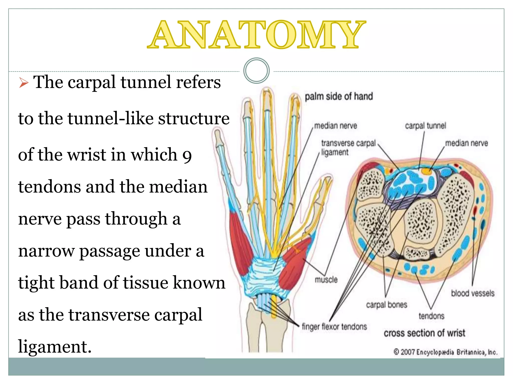
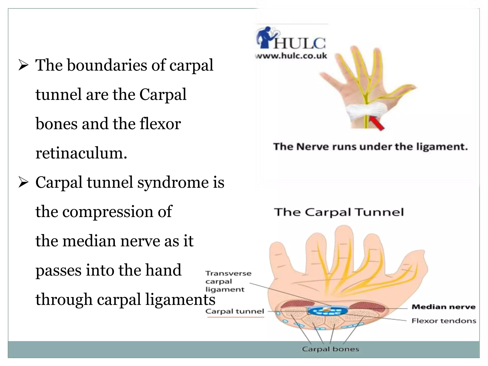
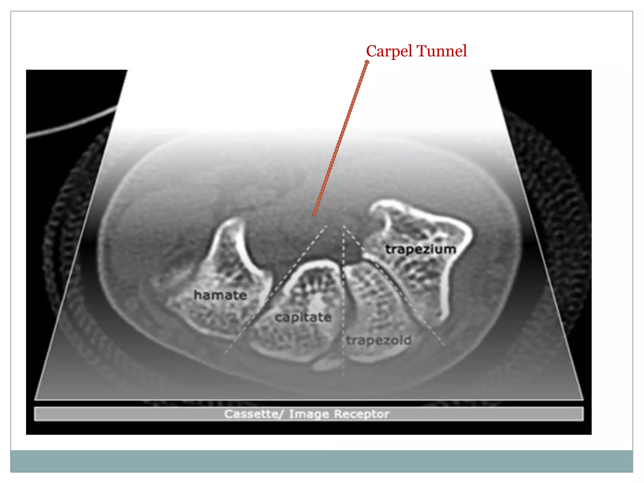
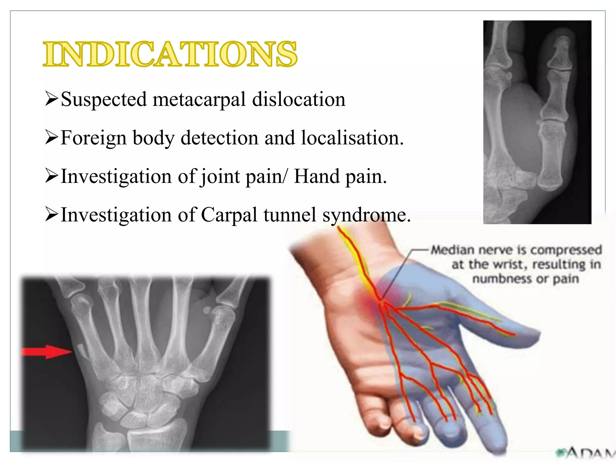
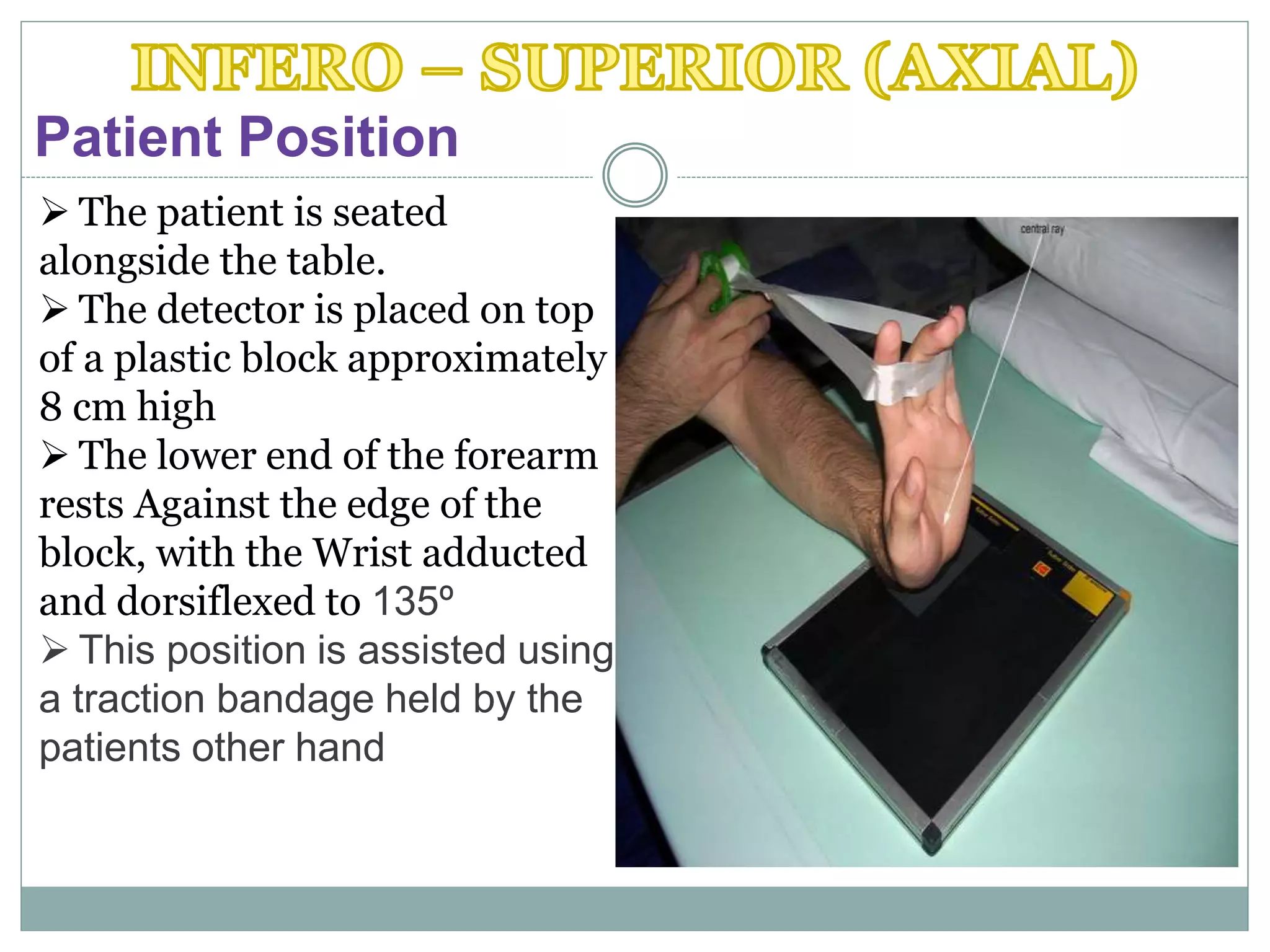
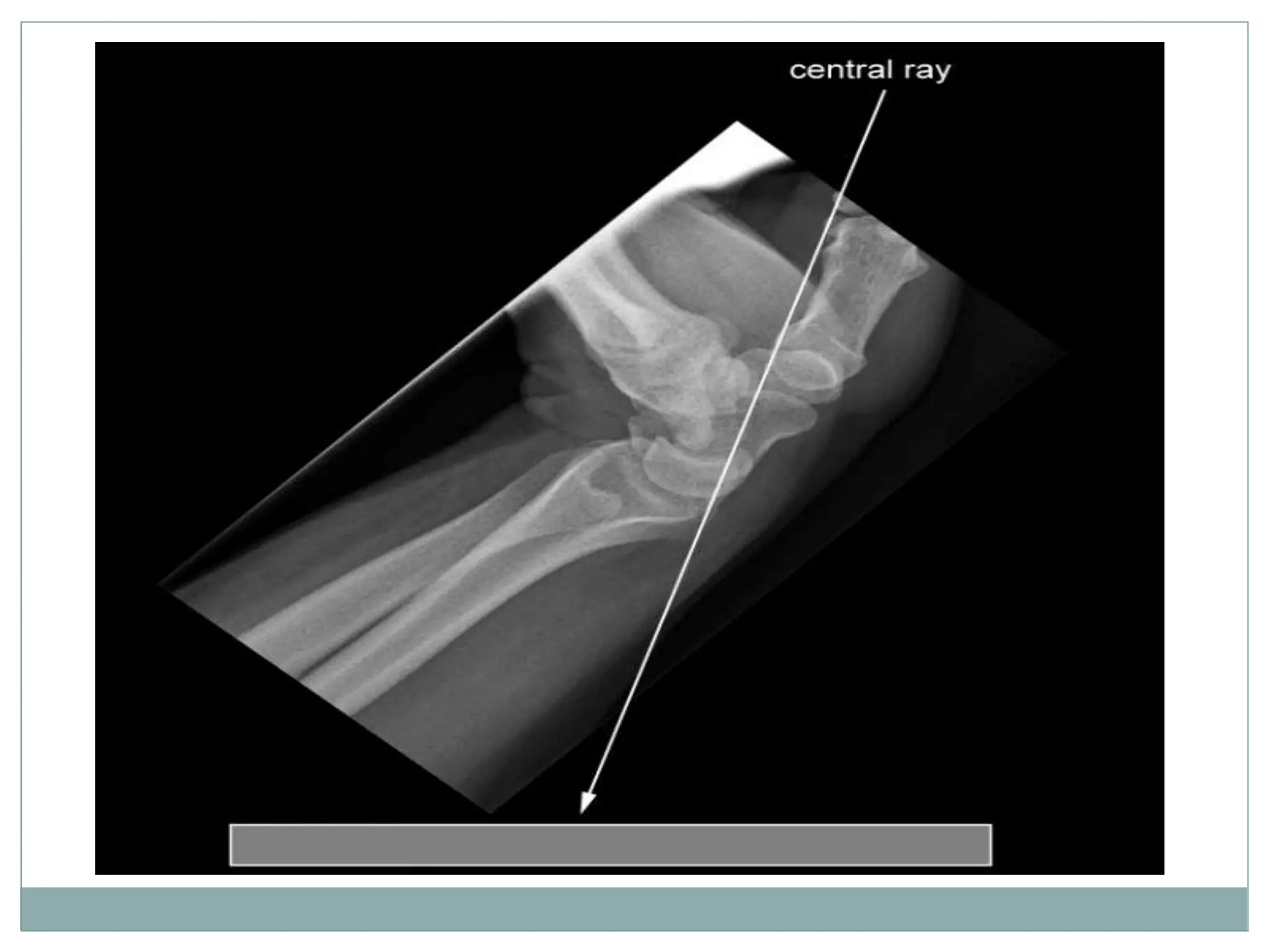
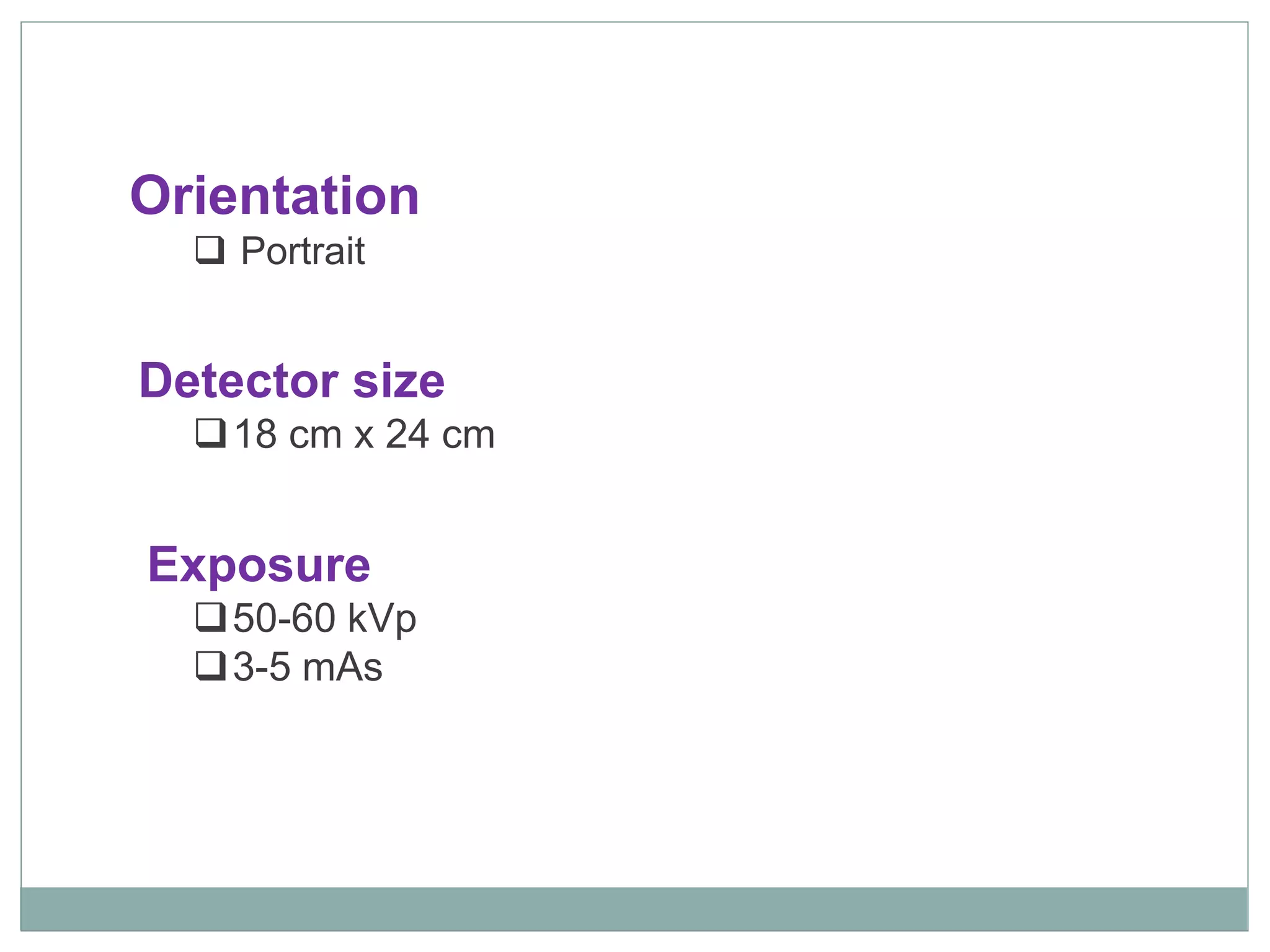
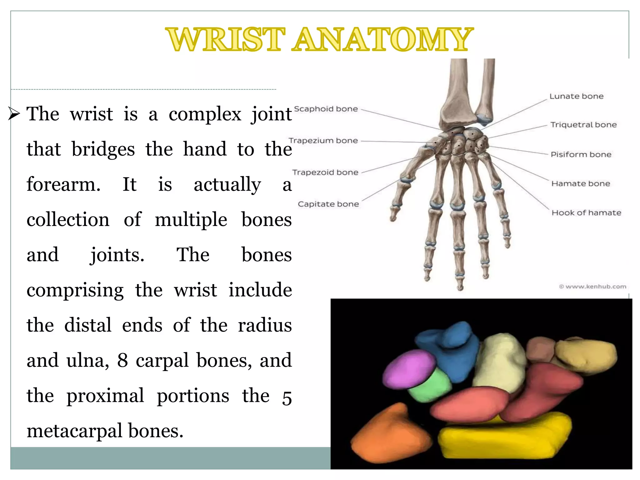
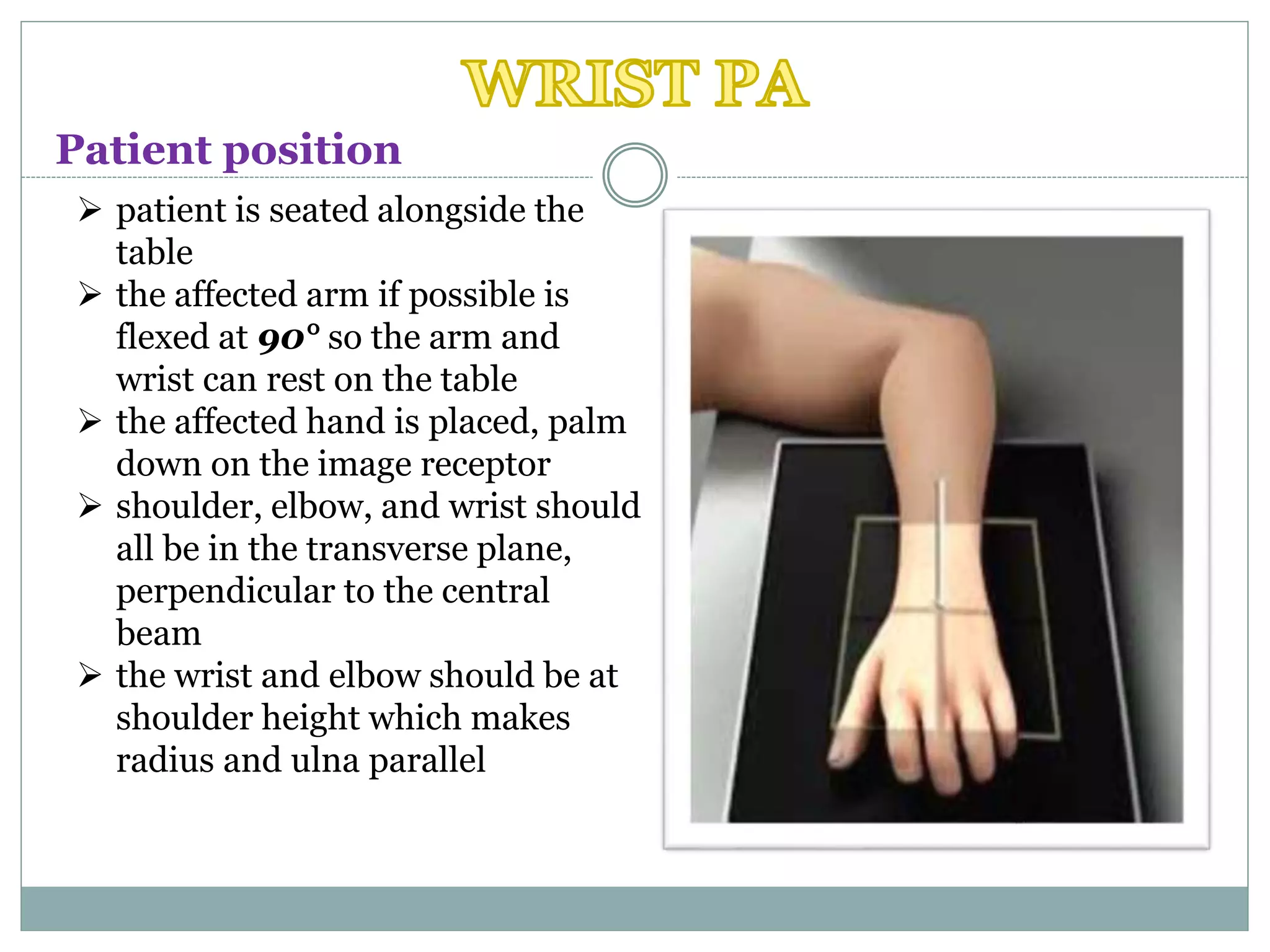
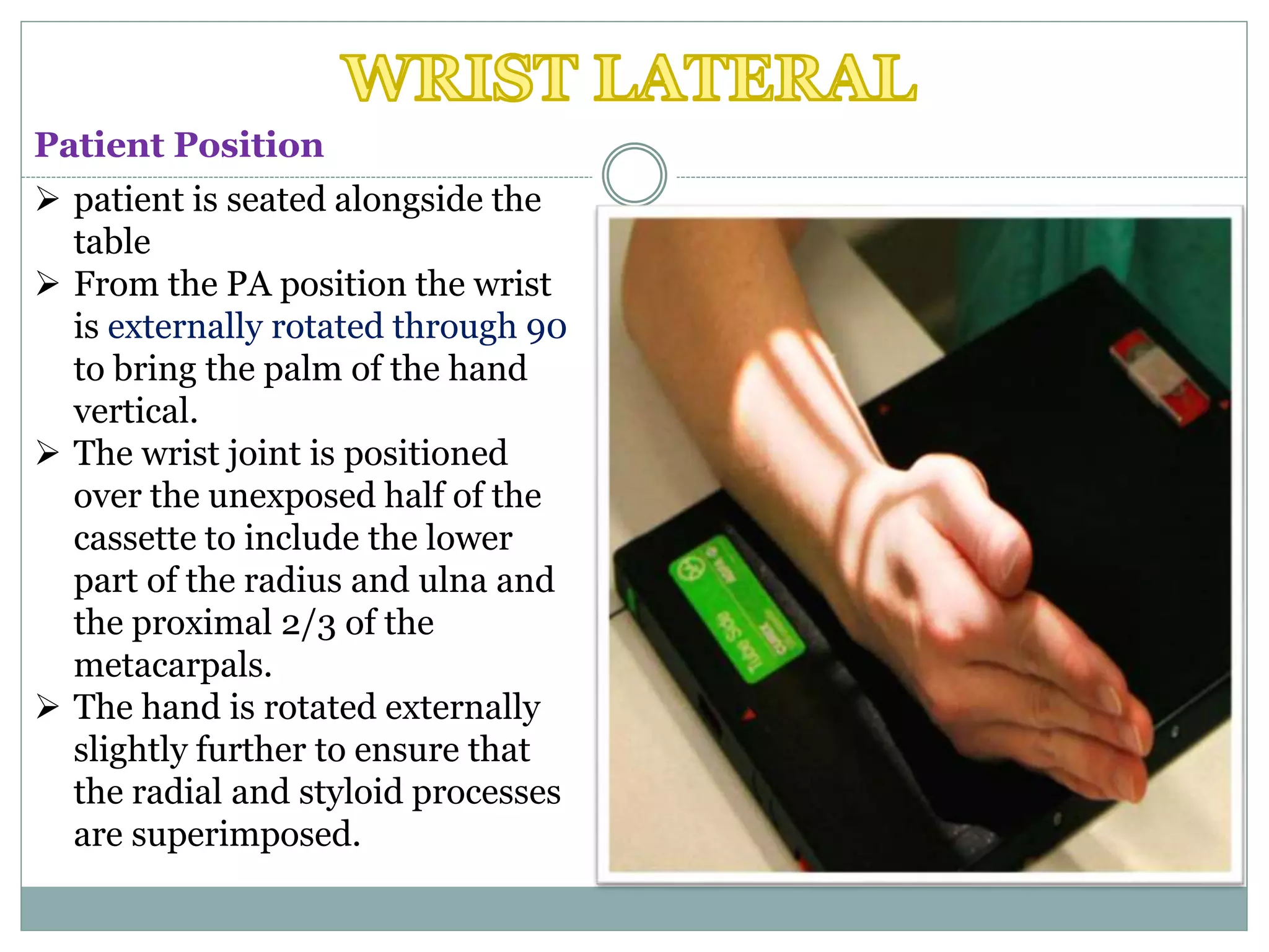
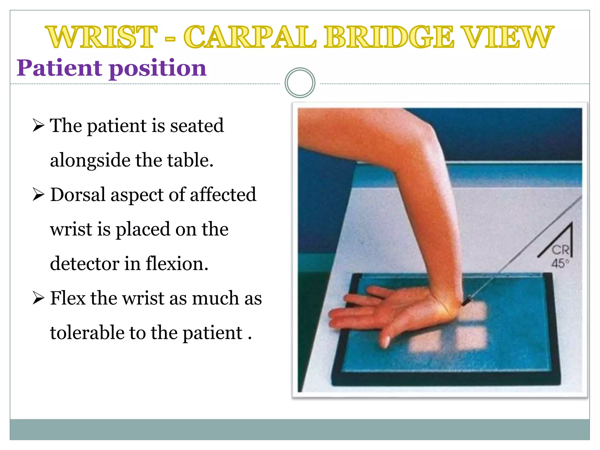
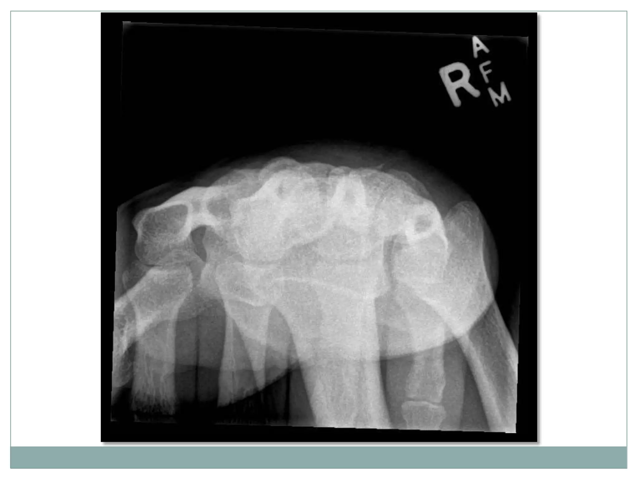
DON BENNY
BMIT- II YEAR
 The carpal tunnel refers
to the tunnel-like structure
of the wrist in which 9
tendons and the median
nerve pass through a
narrow passage under a
tight band of tissue known
as the transverse carpal
ligament.
 The boundaries of carpal
tunnel are the Carpal
bones and the flexor
retinaculum.
 Carpal tunnel syndrome is
the compression of
the median nerve as it
passes into the hand
through carpal ligaments
Carpel Tunnel
Suspected metacarpal dislocation
Foreign body detection and localisation.
Investigation of joint pain/ Hand pain.
Investigation of Carpal tunnel syndrome.
 Supero-inferior (Axial)
 Infero-superior (Axial)
The carpal tunnel view is an axial projection
to demonstrate the medial and lateral
prominences and the concavity.
 Patient is standing facing
away from the table.
 The detector is placed level
with the edge of the table
top.
 palmar surface of hand is
pressed into the detector,
with the wrist joint dorsiflexed
to approximately 135º.
 The fingers are curled
around under the table to
assist in immobilisation.
Patient Position
Carpal
Carpal TunnelCarpal Bones
Axial projection
Centring point
 Mid carpal region
 The central ray is vertical and will be
centered to the midpoint of the
dorsiflexed wrist
Collimation
 Laterally to the skin margins
 Dorsal to the skin margins
 Ventral to the carpometacarpal joint
Orientation
 portrait
Detector size
 18 cm x 24 cm
Exposure
 50-60 kVp
 3-5 mAs
The image should include the
distal end of the radius and
ulna and the proximal end of
the metacarpals.
The joint space around the
scaphoid should be
demonstrated clearly.
Patient Position
 The patient is seated
alongside the table.
 The detector is placed on top
of a plastic block approximately
8 cm high
 The lower end of the forearm
rests Against the edge of the
block, with the Wrist adducted
and dorsiflexed to 135º
 This position is assisted using
a traction bandage held by the
patients other hand
Centring point
 Mid carpal region
 The central ray is vertical and will be
centered to the midpoint of the
dorsiflexed wrist
Collimation
 Laterally to the skin margins
 Dorsal to the skin margins
 Ventral to the carpometacarpal joint
Axial projection
Orientation
 Portrait
Detector size
18 cm x 24 cm
Exposure
50-60 kVp
3-5 mAs
The image should demonstrate clearly the pisiform and the
hook of the hamate medially and the tubercle of the scaphoid
and the tubercle of the trapezium laterally.
 The wrist is a complex joint
that bridges the hand to the
forearm. It is actually a
collection of multiple bones
and joints. The bones
comprising the wrist include
the distal ends of the radius
and ulna, 8 carpal bones, and
the proximal portions the 5
metacarpal bones.
The radius articulates with
the scaphoid laterally and
the lunate medially. The lunate
is connected to the scaphoid via
the scapholunate ligament,
and provides stability to the
radiocarpal joint.
Its a complex joint that bridges the hand to the forearm
 Assessment of trauma
 To find any kind of Bone fracture
 Suspected metacarpal dislocation
 Foreign body detection and localisation
 Investigation of joint pain or deformity
BASIC PROJECTIONS
 Postero-anterior (PA)
 Oblique
 Lateral
 Horizontal Beam Lateral Wrist
 Carpal Bridge view
 Radial deviation view
SUPPLIMENTARY PROJECTIONS
 patient is seated alongside the
table
 the affected arm if possible is
flexed at 90° so the arm and
wrist can rest on the table
 the affected hand is placed, palm
down on the image receptor
 shoulder, elbow, and wrist should
all be in the transverse plane,
perpendicular to the central
beam
 the wrist and elbow should be at
shoulder height which makes
radius and ulna parallel
Patient position
Postero-anterior projection
Centring Point
 mid carpal region / Between the radial and ulnar
styloid processes.
Collimation
 laterally to the skin margins
 distal to the midway up the metacarpals
 proximal to the include one-quarter of the distal
radius and ulna
Orientation
 portrait
Detector size
 18 cm x 24 cm
Exposure
 50 - 60 kVp
 3 - 5 mAs
 The ulna head and distal radius are slight superimposed. The
proximal metacarpals 3 to 5 also being partly superimposed.
The image should demonstrate the proximal 2/3 of the
Metacarpals , the carpal bones , and the distal 1/3 of the
radius and ulna.
There should be no rotation of the wrist joint .
 patient is seated alongside the
table
 From the PA position the wrist
is externally rotated through 90
to bring the palm of the hand
vertical.
 The wrist joint is positioned
over the unexposed half of the
cassette to include the lower
part of the radius and ulna and
the proximal 2/3 of the
metacarpals.
 The hand is rotated externally
slightly further to ensure that
the radial and styloid processes
are superimposed.
Patient Position
Lateral Projection
Centring point
 mid carpal region
Collimation
 Antero-posterior to the skin
margins
 distal to the midway up the
metacarpals
 proximal to include one-
quarter of the distal radius
and ulna
Orientation
 portrait
Detector size
 18 cm x 24 cm
Exposure
 50-60 kVp
 3-5 mAs
 patient is seated alongside the
table
 the affected arm if possible is
flexed at 45° so the arm and wrist
can rest on the table
 the affected hand is placed, palm
down on the image receptor
 shoulder, elbow, and wrist should
all be in the transverse plane,
perpendicular to the central beam
 wrist and elbow should be at
shoulder height which makes
radius and ulna parallel from the
positioning of the PA projection,
the wrist is externally rotated 40°
- 45°; a sponge can be placed
under the wrist to aid stability.
Patient Position
Postero-anterior Projection
Centring point
 mid carpal region
Collimation
 laterally to the skin margins
 distal to the midway up the metacarpals
 proximal to the include one-quarter of the distal
radius and ulna.
Orientation
 portrait
Detector size
 18 cm x 24 cm
Exposure
 50-60 kVp
 3-5 mAs
 The ulna head and distal radius are slight superimposed. The
proximal metacarpals 3 to 5 also being partly superimposed.
patient is supine
affected arm is placed by the
patient's side on a pillow or
foam block
image receptor is placed
leaning against the
aforementioned foam block
or pillow with the long axis of
the image receptor in line
with the long axis of the
affected limb
Patient Position
Lateral Projection
Centring point
 The ulnar aspect of the mid
carpal region
Collimation
 Antero-posterior to the
skin margins
 distal to the mid way up the
metacarpals
 proximal to include one
quarter of the distal radius
and ulna
Orientation
 landscape
Detector size
 24 cm x 30 cm
Exposure
 50-60 kVp
 3-5 mAs
 There is superimposition of the carpal bones, including the
distal portion of the scaphoid and the pisiform. The radius
and ulna are also superimposed.
 The patient is seated
alongside the table.
 Dorsal aspect of affected
wrist is placed on the
detector in flexion.
 Flex the wrist as much as
tolerable to the patient .
Patient position
Tangential projection
Centring point
 midcarpal region
 the central ray is angled approximately 45 degrees to the
long axis of the forearm
Collimation
laterally to the skin margins
dorsal to the skin margins
ventral to the carpometacarpal joint
Orientation
 Portrait
Detector size
18 cm x 24 cm
Exposure
 50-60 kVp
 3-5 mAs
 There should be a clear outline of the dorsal aspect of the
carpal bones with no superimposition.
 patient is seated alongside the
table
 the affected arm if possible is
flexed at 90° so the arm and wrist
can rest on the table
 the affected hand is placed, palm
down on the image receptor with
hand in radial deviation
 shoulder, elbow, and wrist should
all be in the transverse plane,
perpendicular to the central beam
 the wrist and elbow should be at
shoulder height which makes
radius and ulna parallel
Patient Position
Postero-anterior Projection
Centring point
 central carpal zone
Collimation
 laterally to the skin margins
 distal to the midway up the metacarpals
 proximal to the include one-quarter of the distal radius
and ulna
Orientation
 Portrait
Detector size
 18 cm x 24 cm
Exposure
 50-60 kVp
 3-5 mAs
 hand is in radial deviation
 minor superimposition of the metacarpal base
REFERENCE
Projections of  carpal tunnel and wrist

Projections of carpal tunnel and wrist

  • 1.
  • 2.
     The carpaltunnel refers to the tunnel-like structure of the wrist in which 9 tendons and the median nerve pass through a narrow passage under a tight band of tissue known as the transverse carpal ligament.
  • 3.
     The boundariesof carpal tunnel are the Carpal bones and the flexor retinaculum.  Carpal tunnel syndrome is the compression of the median nerve as it passes into the hand through carpal ligaments
  • 4.
  • 5.
    Suspected metacarpal dislocation Foreignbody detection and localisation. Investigation of joint pain/ Hand pain. Investigation of Carpal tunnel syndrome.
  • 6.
     Supero-inferior (Axial) Infero-superior (Axial) The carpal tunnel view is an axial projection to demonstrate the medial and lateral prominences and the concavity.
  • 7.
     Patient isstanding facing away from the table.  The detector is placed level with the edge of the table top.  palmar surface of hand is pressed into the detector, with the wrist joint dorsiflexed to approximately 135º.  The fingers are curled around under the table to assist in immobilisation. Patient Position
  • 8.
  • 9.
    Axial projection Centring point Mid carpal region  The central ray is vertical and will be centered to the midpoint of the dorsiflexed wrist Collimation  Laterally to the skin margins  Dorsal to the skin margins  Ventral to the carpometacarpal joint
  • 10.
    Orientation  portrait Detector size 18 cm x 24 cm Exposure  50-60 kVp  3-5 mAs
  • 11.
    The image shouldinclude the distal end of the radius and ulna and the proximal end of the metacarpals. The joint space around the scaphoid should be demonstrated clearly.
  • 12.
    Patient Position  Thepatient is seated alongside the table.  The detector is placed on top of a plastic block approximately 8 cm high  The lower end of the forearm rests Against the edge of the block, with the Wrist adducted and dorsiflexed to 135º  This position is assisted using a traction bandage held by the patients other hand
  • 14.
    Centring point  Midcarpal region  The central ray is vertical and will be centered to the midpoint of the dorsiflexed wrist Collimation  Laterally to the skin margins  Dorsal to the skin margins  Ventral to the carpometacarpal joint Axial projection
  • 15.
    Orientation  Portrait Detector size 18cm x 24 cm Exposure 50-60 kVp 3-5 mAs
  • 16.
    The image shoulddemonstrate clearly the pisiform and the hook of the hamate medially and the tubercle of the scaphoid and the tubercle of the trapezium laterally.
  • 17.
     The wristis a complex joint that bridges the hand to the forearm. It is actually a collection of multiple bones and joints. The bones comprising the wrist include the distal ends of the radius and ulna, 8 carpal bones, and the proximal portions the 5 metacarpal bones.
  • 18.
    The radius articulateswith the scaphoid laterally and the lunate medially. The lunate is connected to the scaphoid via the scapholunate ligament, and provides stability to the radiocarpal joint.
  • 19.
    Its a complexjoint that bridges the hand to the forearm  Assessment of trauma  To find any kind of Bone fracture  Suspected metacarpal dislocation  Foreign body detection and localisation  Investigation of joint pain or deformity
  • 20.
    BASIC PROJECTIONS  Postero-anterior(PA)  Oblique  Lateral  Horizontal Beam Lateral Wrist  Carpal Bridge view  Radial deviation view SUPPLIMENTARY PROJECTIONS
  • 21.
     patient isseated alongside the table  the affected arm if possible is flexed at 90° so the arm and wrist can rest on the table  the affected hand is placed, palm down on the image receptor  shoulder, elbow, and wrist should all be in the transverse plane, perpendicular to the central beam  the wrist and elbow should be at shoulder height which makes radius and ulna parallel Patient position
  • 23.
    Postero-anterior projection Centring Point mid carpal region / Between the radial and ulnar styloid processes. Collimation  laterally to the skin margins  distal to the midway up the metacarpals  proximal to the include one-quarter of the distal radius and ulna
  • 24.
    Orientation  portrait Detector size 18 cm x 24 cm Exposure  50 - 60 kVp  3 - 5 mAs
  • 25.
     The ulnahead and distal radius are slight superimposed. The proximal metacarpals 3 to 5 also being partly superimposed. The image should demonstrate the proximal 2/3 of the Metacarpals , the carpal bones , and the distal 1/3 of the radius and ulna. There should be no rotation of the wrist joint .
  • 26.
     patient isseated alongside the table  From the PA position the wrist is externally rotated through 90 to bring the palm of the hand vertical.  The wrist joint is positioned over the unexposed half of the cassette to include the lower part of the radius and ulna and the proximal 2/3 of the metacarpals.  The hand is rotated externally slightly further to ensure that the radial and styloid processes are superimposed. Patient Position
  • 28.
    Lateral Projection Centring point mid carpal region Collimation  Antero-posterior to the skin margins  distal to the midway up the metacarpals  proximal to include one- quarter of the distal radius and ulna
  • 29.
    Orientation  portrait Detector size 18 cm x 24 cm Exposure  50-60 kVp  3-5 mAs
  • 30.
     patient isseated alongside the table  the affected arm if possible is flexed at 45° so the arm and wrist can rest on the table  the affected hand is placed, palm down on the image receptor  shoulder, elbow, and wrist should all be in the transverse plane, perpendicular to the central beam  wrist and elbow should be at shoulder height which makes radius and ulna parallel from the positioning of the PA projection, the wrist is externally rotated 40° - 45°; a sponge can be placed under the wrist to aid stability. Patient Position
  • 32.
    Postero-anterior Projection Centring point mid carpal region Collimation  laterally to the skin margins  distal to the midway up the metacarpals  proximal to the include one-quarter of the distal radius and ulna.
  • 33.
    Orientation  portrait Detector size 18 cm x 24 cm Exposure  50-60 kVp  3-5 mAs
  • 34.
     The ulnahead and distal radius are slight superimposed. The proximal metacarpals 3 to 5 also being partly superimposed.
  • 35.
    patient is supine affectedarm is placed by the patient's side on a pillow or foam block image receptor is placed leaning against the aforementioned foam block or pillow with the long axis of the image receptor in line with the long axis of the affected limb Patient Position
  • 37.
    Lateral Projection Centring point The ulnar aspect of the mid carpal region Collimation  Antero-posterior to the skin margins  distal to the mid way up the metacarpals  proximal to include one quarter of the distal radius and ulna
  • 38.
    Orientation  landscape Detector size 24 cm x 30 cm Exposure  50-60 kVp  3-5 mAs
  • 39.
     There issuperimposition of the carpal bones, including the distal portion of the scaphoid and the pisiform. The radius and ulna are also superimposed.
  • 40.
     The patientis seated alongside the table.  Dorsal aspect of affected wrist is placed on the detector in flexion.  Flex the wrist as much as tolerable to the patient . Patient position
  • 42.
    Tangential projection Centring point midcarpal region  the central ray is angled approximately 45 degrees to the long axis of the forearm Collimation laterally to the skin margins dorsal to the skin margins ventral to the carpometacarpal joint
  • 43.
    Orientation  Portrait Detector size 18cm x 24 cm Exposure  50-60 kVp  3-5 mAs
  • 44.
     There shouldbe a clear outline of the dorsal aspect of the carpal bones with no superimposition.
  • 45.
     patient isseated alongside the table  the affected arm if possible is flexed at 90° so the arm and wrist can rest on the table  the affected hand is placed, palm down on the image receptor with hand in radial deviation  shoulder, elbow, and wrist should all be in the transverse plane, perpendicular to the central beam  the wrist and elbow should be at shoulder height which makes radius and ulna parallel Patient Position
  • 47.
    Postero-anterior Projection Centring point central carpal zone Collimation  laterally to the skin margins  distal to the midway up the metacarpals  proximal to the include one-quarter of the distal radius and ulna
  • 48.
    Orientation  Portrait Detector size 18 cm x 24 cm Exposure  50-60 kVp  3-5 mAs
  • 49.
     hand isin radial deviation  minor superimposition of the metacarpal base
  • 50.

Editor's Notes

  • #3 The carpal tunnel is a fibro-osseous canal that acts as a passageway from the forearm to the anterior hand. It is found in the anterior wrist. numbness, tingling, and pain in your thumb and the first three fingers of your hand pain and burning that travels up your arm , wrist pain at night that interferes with sleep , weakness in the muscles of the hand
  • #4 Swelling inside your wrist causes the compression in carpal tunnel syndrome. It can lead to numbness, weakness, and tingling on the side of your hand near the thumb. Diabetes , thyroid dysfunction , fluid retention from pregnancy or menopause , high blood pressure ,autoimmune disorders such as rheumatoid arthritis , fractures or trauma to the wrist Causes for cts.
  • #5 Radiograph of carpal tunnel. The carpal bones forms as a tunnel shaped shape , the carpal ligament on top.
  • #6 These are shows why an carpal tunnel xray is needed.
  • #20 caused by a fall on an outstretched hand.
  • #22 (lowering the arm makes radius cross the ulna and thus relative shortening of radius)
  • #27 (lowering the arm makes radius cross the ulna and thus relative shortening of radius).
  • #31 (lowering the arm makes radius cross the ulna and thus relative shortening of radius)
  • #36 The horizontal beam lateral wrist view is a modified lateral projection when performing the three view series of the wrist and carpal bones in trauma. It is the orthogonal projection of the PA wrist without any patient movement, making it the most appropriate projection for patients in pain.
  • #46 .(lowering the arm makes radius cross the ulna and thus relative shortening of radius)