SlideShare a Scribd company logo
1 of 95
Presented by: Dr Dibya Falgoon Sarkar
 Paranasal sinuses
• Introduction
• Development
• Anatomy
• Physiology
• Examination and investigations
 Paranasal sinus pathologies
• Classification
• Description
• Complications
• Treatment
• Paranasal sinuses are the mucosa
lined air spaces present within the
bones of the face & skull.
• There are four bilaterally paired
paranasal sinuses:
- Frontal sinus
- Maxillary sinus
- Ethmoidal sinus
- Sphenoidal sinus
• Named after the bones in
which they are located
1. Frontal sinus
2. Ethmoidal sinus
3. Sphenoidal sinus
4. Maxillary sinus
• Humidifying and warming the inspired air.
• Imparts resonance to voice
• Increases surface area for absorption of noxious
gases and trapping of particles in inspired air
•Lightens the skull
•Helps in absorbing the shock of blows to the face &
thereby limiting the extent of facial injury from
trauma.
•Serves as an accessory olfactory organ by evenly
distributing the inspired air.
•Contributes to facial growth
Sinuses begin their
development at the 3rd
month of intrauterine life
as outpouchings of the
mucous membranes of
the nasal meatuses and
sphenoethmoidal recess
The early PNS expand
into the walls of the nasal
fossa by the growth of
mucous membranes into
the maxillary, sphenoid,
ethmoid and frontal
bones
Pneumatization follows.
Sinuses enlarge variably
but always retain their
original communication
with the nasal cavity
through ostia
• Largest of all the paranasal sinuses.
• Also known as the “antrum cave of
Highmore”– English physician
described an infection of sinus in
1651.
• First sinus to develop
• Pyramidal shaped - base toward the
lateral wall of nose and apex
directed laterally into the zygomatic
process
• Capacity approx. 15ml
Superior wall
• This wall forms both- roof of sinus & floor of the
orbit.
• It is thinnest wall & mostly flat slopes slightly
anteriorly and laterally.
• The Infraorbital canal runs along this wall and
crosses from back to front- accentuates the fragility
of wall
• This wall is frequently involved in orbital and
maxillary trauma.
 Anterolateral wall
• Thickness is 2-5mm.Thinnest portion over canine
fossa
• Related to facial artery and vein
• Labial levator muscles & inferior portion of
orbicularis oculi is attached to this wall above the
infra-orbital foramen.
• These attachments direct the spread of infection
from maxillary teeth.
• This wall also contains the anterior & middle
superior alveolar nerves.
• Any surgical procedure through this wall may
jeopardise the nerve supply to these teeth.
Floor of the maxillary sinus
• It is approximately 1.5 cm below
the floor of nasal cavity.
• It corresponds to the line drawn
laterally from ala of the nose.
Posterolateral wall
• Posterior superior alveolar nerves
and vessels sometimes present in
close contact with the sinus
mucosa.
• In such instances , acute sinusitis
is accompanied by pain in
multiple maxillary posterior teeth.
• Immediately posterior to this wall,
vital structures include:
-maxillary nerve & maxillary artery
- nerve of pterygoid canal
- sphenopalatine ganglion.
Ostium of maxillary sinus
• Communication between maxillary sinus
and nasal cavity.
• Average length: 5.5mm
• Located superior to the floor of the sinus.
• Opens into posterior part of hiatus
semilunaris in the middle meatus
• Arterial supply:
- Facial artery
- Infraorbital artery
- Greater palatine artery.
• Venous drainage:
- Facial vein
- Infraorbital vein
 Nerve supply:
 Infraorbital nerve
 Anterior , Middle &
Posterior superior
alveolar nerves
 Lymphatics:
 Submandibular lymph
nodes
• The frontal sinuses are rudimentary
or absent at birth.
• They are developed between 7th &
8th year of life but reach the full
size only after puberty.
• Situated between the inner and
outer tables of frontal bone
• Absent on one (15 %) or both sides
(5 %)
• Drainage pathway – naso- frontal
duct- frontal recess - situated at its
floor
• Drains into middle meatus (62%) or
ethmoid infundibulum (38%).
• The inner table is much thinner than the outer table. As a result the
chances of fracture of the inner table are more frequent ,even
without the fracture of outer table.
 Arterial supply:
- supraorbital artery
- anterior ethmoidal artery
 Venous drainage:
Anastomotic vein in the
supraorbital notch
connecting the supraorbital
& superior ophthalmic
veins.
 Lymphatic drainage:
Submandibular nodes.
 Nerve supply:
Supraorbital nerve
• Formation begins at 4th month of fetal life
• Thin-walled air cavities in the lateral masses
of ethmoid bone, between nose and the
eyes.
• Anterior ethmoidal air cells
-3- 11 drain into either the
ethmoidal infundibulum or
the frontonasal duct.
• Bullar cells (middle
ethmoidal air cells) -usually
<3 - open in ethmoidal
infundibulum.
• Posterior group :- Up to 7 -
usually drain by a single
orifice into the superior
meatus
Ethmoidal bulla:
• This is the most constant landmark
for surgery. It lies above the
infundibulum and it's lateral/inferior
surface and the superior edge of the
uncinate process forms the hiatus
semilunaris.
• The anterior ethmoid artery usually
decends across the roof of this cell.
 Arterial supply:
- Sphenopalatine artery
- Anterior and posterior
ethmoidal arteries
 Venous drainage:
Corresponding veins
 Lymphatic drainage:
• Anterior and middle groups
drain in the
submandibular nodes .
• Posterior group drains into
retropharyngeal nodes.
 Nerve supply: Anterior and
posterior ethmoidal nerves
Orbital branches of
pterygopalatine ganglion.
• They are the most anterior ethmoidal air cells.
• Its size influence the patency of the frontal recess
and the anterior middle meatus.
• Haller cells: Also called infraorbital ethmoid cells.
• Present in approx. 20 % patients.
• Clinical significance –
1. Become infected , with potential extension into
orbit.
2. Narrows the maxillary ostium.
• These are posterior ethmoidal cells extending into
the sphenoid bone, either adjacent to or impinging
upon the optic nerve.
• When these Onodi cells abut or surround the optic
nerve, the nerve is at risk when surgical excision of
these cells is performed.
• Body of sphenoid - behind the nose,
in the centre of the skull.
• Rarely symmetrical and separated by
a thin bony septum.
• Ostium of the sphenoid sinus is
situated in the upper part of its
anterior wall and drains into
sphenoethmoidal recess.
• Average size – 2 x 2 x 2 cm.
• According to Congdon sphenoid
pneumatization can be as follows:
1. Conchal – 5 %
2. Presellar – 23 %
3. Postsellar – 67%
• Arterial supply- Posterior ethmoidal
artery.
• Nerve supply- Posterior ethmoidal
nerves
• Lymphatic drainage-
Retropharyngeal nodes
• History and systemic clinical examination:
• Check general signs of health
• Systemic medical history, history of allergies, drug use and abuse
• Occupation history
• Examination of neck for lumps or swollen lymph nodes
• Palpation:
• PNS may be palpated directly to elicit any tenderness
• Tenderness over maxillary and frontal bones may indicate inflammatory
condition
• Step-off deformity indicates facial bone fracture in trauma cases
• Paresthesia is seen over midface region in injury to infraorbital nerve
1. Anterior rhinoscopy:
• Examination of nose with a nasal
speculum to check for abnormal
areas, useful in evaluation of
nasal obstruction.
• Colour of nasal mucosa and its
vascularity
• Position of septum is noted for
deviation or dislocation, as well
as septal spurs, haematoma or
perforations
• Inferior & middle turbinates seen
2. Posterior rhinoscopy:
• Assessed using a mouth mirror
and headlight in the nasopharynx
• Technique sensitive
• Low specificity and sensitivity
• Helpful in absence of endoscopy
• Decreased light transmission when
compared with normal side
suggests of significant fluid
collection or marked mucosal
edema
Transillumination
• Endoscopic examination/
Rhinoscopy:
Nasoscope/rhinoscope is a thin,
tube-like instrument with a light
and a lens for viewing.
• A special tool on the nasoscope
may be used to remove samples
of tissue. The tissues samples are
viewed under a microscope by a
pathologist.
• Helps in visualization of
inaccessible areas like middle
meatal cleft.
1. Maxillary sinus:
• Water’s view (PNS view) x-ray and CT scan are ideal for
maxillary sinus visualization.
• X-ray beam angled at 45 degree to orbitomeatal line
2. Frontal sinus:
• CT scan is helpful in trauma cases as we can appreciate both
inner and outer tables in the axial view
• PNS view x-ray is helpful to detect fluid accumulation in
sinusitis cases
3. Ethmoidal sinus: Axial views of CT scan are helpful in
detecting fractures of the ethmoid bone.
4. Sphenoid sinus: Best viewed in sagittal and axial sections of
CT scan.
“Limited CT Evaluation” – slice 3-4 mm
Maxillary sinus & Ethmoid air sinus
Axial CT scans
Frontal sinus
CT scan axial
PNS view radiograph
Sphenoid sinus
Axial CT Sagittal CT
• MRI: Excellent soft tissue definition -
evaluation of neoplastic disease.
• MRI (magnetic resonance imaging) with
gadolinium: Gadolinium is injected into
a vein. The gadolinium collects around
the cancer cells so they show up
brighter in the picture.
• Developmental variations and anomalies
• Inflammatory/infectious diseases
• Cysts
• Tumours – benign & malignant
• Other surgically relevant conditions
• Paradoxical curvature of
middle turbinate:
• Normally the convexity of the
middle turbinate is directed
medially toward the nasal
septum.
• When the convexity is directed
laterally, it is termed a
paradoxical middle turbinate .
• Most authors agree that the
paradoxical middle turbinate
can be a contributing factor to
sinusitis.
• Variation of the ethmoidal roof
anatomy
• The ethmoid roof is of critical
importance for two reasons:
1. most vulnerable to iatrogenic
cerebrospinal fluid leaks.
2. anterior ethmoid artery is vulnerable
to injury.
• The depth of the olfactory fossa is
determined by the height of the lateral
lamella of the cribriform plate.
• In 1962, Keros classified the depth of
the olfactory fossa into three types, that
is,
a) Keros type I: <3 mm
b) Keros type II: 4-7 mm
c) Keros type III: 8-16 mm - most
vulnerable to iatrogenic injury.
• Variations of uncinate process
• The uncinate process may be
medialized, lateralized, or
pneumatized/bent.
• Medialization occurs with giant
bulla ethmoidalis.
• Lateralization of the uncinate
process may obstruct the
infundibulum.
• Pneumatization (uncinate bulla)
can rarely cause obstruction of
the infundibulum.
• Sinusitis is the inflammatory condition of
the mucous membrane lining of the
sinuses. It may progress to pus formation.
• Causes :
• Rhinogenous – infection spreads from the
nasal cavity. It is the most common way
for infection and such sinusitis is the
complication of the flu.
• Odontogenic – infection spreads from
upper teeth. This way is typical only for
maxillary sinus. The pathologic process
may spreads from upper 4,5,6 teeth apex
to the inferior wall of the maxillary sinus
• Traumatic
• Hematogenic
• Allergic
1. Initial phase: reversible
2. Ostium obstruction phase
3. Bacterial phase
4. Chronic phase
Osteomeatal
complex obstruction
Decreased
ventilation of the
sinuses
Decreased drainage
of the sinuses
pO2 decrease,
pCO2 increase,
mucous stasis
Inflammation and
viscous mucous,
ciliary movement
slowing
Ciliary damage
along with increase
in number of
anaerobic organisms
• According to duration:
1. Acute : infection lasting 4 weeks, symptoms resolve completely
resolved in < 30 days.
2. Subacute :infection lasting between 4 to 12 weeks, yet resolves
completely.
3. Recurrent: ≥ 4 discrete acute episodes per year, each completely
resolved in < 30 days but recurring in cycles, with at least 10 days
between complete resolution of symptoms and initiation of a new
episode
4. Chronic: symptoms lasting more than 12 weeks.
• According to pathogen:
1. Bacterial: Hospital-acquired acute
infections are more often bacterial,
typically involving Staphylococcus aureus,
Klebsiella pneumoniae, Pseudomonas
aeruginosa
2. Viral: In immunocompetent patients - in
the community is almost always viral (eg,
rhinovirus, influenza, parainfluenza).
antibiotics given for:
• Mild to moderate sinus symptoms persisting
for ≥ 10 days
• Severe symptoms (eg, fever ≥ 39°, severe
pain) for ≥ 3 to 4 days
• Worsening sinus symptoms after initially
improving from a typical
• viral URI ("double sickening" or biphasic
illness)
3. Fungal: Usually seen in
immunocompromised patients because
of poorly controlled diabetes,
neutropenia, or HIV infection.
It is clinically of 2 types:
A)Non-invasive fungal sinusitis:
• Saprophytic fungal
infestation/colonization
• Allergic fungal rhinosinusitis (AFRS)
• Saprophytic fungus balls (mycetoma)
B)Invasive fungal sinusitis –
• Chronic invasive fungal sinusitis
• Granulomatous invasive fungal sinusitis
• Acute (fulminant) invasive fungal
sinusitis
• Risk factors:
1. Obstruction to drainage: most important
2. Defect of self-cleaning mechanism of the mucous membrane of sinus –
infections causing immobility of the cilia, Increased viscosity of secretions,
Immotile cilia syndrome, Prolonged exposure to cigarette smoke
3. Medications: First generation antihistamines (non sedating do not affect),
Anticholinergics, Aspirin, Anesthetic agents, Benzodiazepines
4. Immunodeficiency: Immunoglobulin deficiency (IgA, IgG), diabetes, HIV
infection
5. Other factors: prolonged ICU stays, severe burns, cystic fibrosis, and ciliary
dyskinesia.
According to number of
sinuses involved:
Hemisinusitis - all
sinuses on one side
Polysinusitis - several
sinuses, but not all, are
involved
Pansinusitis - all, are
involved
1.Nasal congestion and discharge
2. Sore throat and postnasal drip
3. Pain or pressure.
4. Oedema of facial tissues.
5. Bad breath or loss of smell
(hyposmia/anosmia).
6. Systemic symptoms: Malaise may be
present. Fever and chills suggest an
extension of the infection beyond the
sinuses, rise in temperature, bad appetite,
sleep disturbances, changes of the blood
(leukocytosis), Productive cough (especially
at night)
7. Complications – ocular, neurological, local
• Diagnosis is mainly clinical
• CT scan and cultures - mainly for chronic, refractory, or atypical
cases.
• Clinical diagnostic criteria include
a) 2 major factors
b) 1 major and 2 minor factors or presence of pus in nasal cavity.
Major factors Minor factors
Facial pain/pressure Headache
Facial congestion/fullness Maxillary dental pain
Nasal drainage/discharge Halitosis
Postnasal drip Cough
Fever (acute sinusitis only)
Purulence in nasal cavity on examination
Hyposmia
Treatment options measures to enhance
drainage:
1. Heat therapy: Steam inhalation and hot
beverages
2. Topical vasoconstrictors/ Nasal decongestants:
Topical nasal sprays (limit use to 3-7 days) -
Phenylephrine, Oxymetazoline.
Corticosteroids: Corticosteroid nasal sprays can
help relieve symptoms but typically take at least
10 days to be effective
3. Systemic vasoconstrictors: Systemic
vasoconstrictors, such as pseudoephedrine 30 mg
4. Nasal irrigation
5. Antihistaminics: recommended if allergy
present.
6. Hydration
7. Antibiotics: Amoxicillin 500 mg tid for 10-14
days - First line
Nasal irrigation
8. Mucoactive drugs: main purpose - increase the ability to expectorate
sputum and/or decrease mucus hypersecretion.
1. Expectorants: hypertonic saline, iodine containing compounds,
guaifenesin
2. Mucoregulators: anticholinergic drugs, glucocorticoids, macrolide
antibiotics
3. Mucolytics: classic mucolytic (N-Acetyl Cysteine), peptide mucolytic
4. Mucokinetics: bronchodilators, ambroxol
a) Nebulization: best form of physiotherapy.
b) Laser therapy: used directly over the sinuses to reduce inflammation
c) Ultrasound therapy: sound waves are conducted through a
hypoallergenic gel to reduce inflammation and loosen the accumulated
mucous.
d) Short-wave diathermy
e) Rinoflow therapy: new option, basically micronized endotracheal
wash. Used in sinusitis, rhinitis, pharyngitis, laryngitis and secretory otitis
media.
Indications:
• Sinusitis unresponsive to antibiotic therapy
• Necrotic sinusitis
• Orbital complications (abscess and phlegmon of orbit)
• Intracranial complications (meningitis, brain abscess)
• Rhinogenic sepsis
• Odontogenic sinusitis combined with maxillary osteomyelitis.
Approaches:
1. Removal of etiology (eg: removal of the infected teeth)
2. Functional endoscopic sinus surgery (FESS) is the mainstay in the surgical
treatment of sinusitis and nasal polyps, including bacterial, fungal, recurrent
acute, and chronic sinus problems.
3. Caldwell-luc procedure/ Denker’s procedure
4. Intranasal antrostomy
5. Radical surgeries
• Oro-antral communication: it is an abnormal connection between oral and
Antral cavities.
• Oro-antral fistula: long standing oro-antral communication when left open,
epithelializes to form a patent fistula between the two cavities.
• Types:
1. Alveolosinusal
2. Palato-sinusal
3. Vestibulo-sinusal
• Causes:
• Extraction – maxillary posterior teeth
• Cysts, tumors
• Osteomyelitis
• Radiation therapy
• Trauma
• Implant denture
Oro-antral Fistula
• Some patients are asymptomatic
• Unpleasant tasting discharge and odor
• Reflux of fluids and foods into the nose from mouth
• Difficulty in smoking and blowing air
• Development of chronic sinusitis in infected cases
• Valsalva manoeuvre
• Mirror fog test
• Cotton wisp test
• Nasal regurgitation of fluid
• Radiographs: Sinus floor discontinuity,
sinus opacity, focal alveolar atrophy.
 Immediate treatment: Primary purpose is
closure of defect and prevention of sinusitis
through:
• Suturing across the defect with/without
periodontal pack, warm saline rinses,
antibiotic and antihistaminic therapy with
decongestants.
• Depending on defect size:
1. Size < 5mm: non-invasive intervention
(spontaneous closure by blood clot)
2. Size 2-6mm: Collagen plug in the socket
with figure of 8 suturing
3. Size > 6mm and > 48 hours: surgical
closure : small defects – local flaps; large
defects – regional/distant flaps
Obturators: immunocompromised patients
 In long-standing OAF we must evaluate the patient for maxillary
sinusitis and treat it before closing the defect surgically
The fistulous tract must be excised prior to surgical closure
• Most common and
oldest surgical
technique
• Developed by Rehrman
• Disadvantage: Reduces
depth of buccal
vestibule
• Overall success rate
approx. 87.2%
• Full thickness flap
• Based on the greater palatine artery
• Advantages:
a) No reduction of vestibular depth
b) Lesser chance of flap breakdown
c) Anteriorly based flaps are ideal for
closure of tuberosity defects
• Disadvantage: Exposed palatal bone
takes long time to heal
• First described by Egyedi in 1977
• Helpful in small to medium sized defects
• Pedicled flap
• Complication can be avoided:
i. Graft should not be in tension
ii. Patient should be on a liquid diet
Anatomy of BFP:
It has 4 processes- buccal,
pterygoid, pterygopalatine,
superficial and deep temporal
Blood supply: Deep & superficial
temporal artery, facial artery
• Based on lingual artery and its branches
• Tongue flaps are useful :
i. When both buccal and palatal flaps have
failed
ii. Defect size is > 1.5cm
• Types of tongue flaps:
a) Anteriorly based- Defects of hard palate,
buccal mucosa
b) Posteriorly based- Defects of soft palate
• Disadvantages:
i. Second surgery is required for flap division
ii. Swallowing and speech difficulty
• Some investigators placed the patients in IMF to
reduced flap tearing
Gold foil
• By George Walter( USA, 1983) and Henri Luc
(France,1989)
• Technique to gain access to the maxillary antrum
through canine fossa of maxilla
• Indications:
Removal of tooth fragments, chronic sinusitis,
neoplasms, cysts
Controlling maxillary antrum bleed/ haematomas
Treating orbital floor fractures
Nasal Antrostomy
• Artificial ostium is made to the max. sinus with an
opening in inferior meatus
• Principle: Opening at a lower level improves
drainage
• Types : Middle meatal (child), inferior meatal,
sublabial
Presented by: Dr Dibya Falgoon Sarkar
PNS Cysts
Intrinsic cysts
Mucocele
Serous cysts
Cholesteatoma
Extrinsic cysts
Dentigerous
cysts
Radicular cyst Neurogenic cysts
Meningocele
Encephalocele
Fissural cysts
Nasopalatine
duct cyst
Globulumaxillar
cyst
Median palatal
cyst
Pierse et al Oral & Maxillofacial Surgery Clinics 2012
Mucous retention cyst Mucous extravasation cyst (Mucocele)
Most common and donot produce bone destruction Produces bone destruction
Results from obstruction of glands in the sinus lining Results from accumulation of mucous in an
obstructed sinus cavity
Site: Floor of Maxillary sinus Site predilection: Frontal & Ethmoidal sinus
Radiographically visible as a dome-shaped,
homogenous mass
Radiographically Pressure erosion seen surrounding
sinus walls. Initially sinus is opacified
• Treatment: Surgical excision along with the supplying gland
• Excellent prognosis, low recurrence
Neurogenic cysts of PNS
• Non-neoplastic lesions
• Two types : Meningocele &
encephalocele
• Both are results of congenital
herniation of intracranial meningeal
and neural tissue
• Encephalocele is more common and
contains primitive neural tissue
• Meningocele contains arachnoid and
dural tissue and CSF
• Radiographically seen as bony
dehiscences in base of skull or cranial
vault associated with a soft tissue
extension in nose and PNS
• Treatment : Surgical enucleation
Encephalocele in fronto-
ethmoidal sinus
Sinonasal
tumours
Benign tumours
Epithelial tumors
Sinonasal
papilloma
Mesenchymal
tumors
Osteoma
Angiofibroma
Haemangioma
Malignant
tumours
Epithelial
tumours
Squamous cell
carcinoma
Adenoid cystic
Carcinoma
Adenocarcinoma
Mesenchymal
tumours
Olfactory
neuroblastoama
Sarcomas
• Early : asymptomatic .
• Oral symptoms: Toothache , trismus, alveolar ridge fullness, erosion ,
malocclusion .
• Nasal findings: Obstruction, epistaxis, rhinorrhea , post nasal
discharge , anosmia, nasal mass or polyposis, mass in the medical
canthus, broadening of nasal dorsum .
• Ocular findings: Epiphora, diplopia, proptosis
• Facial signs: Paraesthesia, asymmetry
• Maxillary sinus involvement: Mass in palate or upper alveolus .
mass in upper gingivobuccal sulcus, malocclusion or loose teeth.
• Advanced :
Trismus, Orbital : Periorbital swelling , proptosis, epiphora ,
impaired ocular mobility
• Benign, localized proliferations of the
respiratory mucosa
• 10-25% of all paranasal tumors
• Cause: Some clinicians believe that these
are actually reactive hyperplasia
secondary to stimulants like allergens,
bacteria or HPV virus( type 6,11),
tobacco smoking
• Types :
1) Fungiform
2) Inverted
3) Cylindrical
Fungiform papilloma Inverted papilloma Cylindrical papilloma
Distribution Similar to oral
papillomas; 18-50% all
PNS papillomas
Most common, premalignant <7% of PNS papillomas
HPV Almost all variants are
HPV positve
HPV 6,11,16 & 18 Low frequency of HPV
association
Age/sex 20-50 years; M>F > 20 years; M>F 20-50 years; M>F
Clinical
features
Unilateral nasal
obstruction, epistaxis,
pink nodule
Unilateral nasal obstruction,
epistaxis, pink nodule, bone
erosion, locally aggressive
Unilateral nasal obstruction,
epistaxis, robust red multi
nodular
Treatment Complete surgical
excision, no malignant
transformation
Aggressive surgical excision,
Greatest potential for
malignant transformation and
destruction
Aggressive surgical excision
• Most common benign, mesenchymal tumor
of PNS
• Arises from junction of membranous &
endochondral bone in frontoethmoidal
suture region
• May have intracranial/ orbital extensions
• Histological subtypes: hard cortical & soft
cancellous
• Surgical excision
• Low recurrence
Osteoma
Hard cortical osteoma Soft cancellous
osteoma
Lobulated densities
obstructing sinus
Less dense, may be
mistaken with a soft
tissue sinus mass
• Usually an extension of juvenile
angiofibroma of nasopharynx
• Often extends into sphenoid and
ethmoidal sinuses
• Have a vertical spreading pattern
• Imaging: Appears as an enhancing
lesion in MRI
• Carotid angiography and CECT are
helpful as angiofibromas contain a
hypertrophied internal maxillary
artery
• Conservative surgical excision
• Rare mucosal neoplasm
• Neural crest origin
• Often exhibits immunological
symbiosis with the host
• Radical procedures are
contraindicated
• Treatment by endoscopic resection
• Role of radiotherapy is debatable
• Arise from the nasal cavity (septum/
alveolus)
• Multifocal, frequent recurrence
• Spreads superiorly into skull base
• Age range: both young and old
• Treatment: Craniofacial resection
• Not radiosensitive
• Classically arises from olfactory epithelium in the upper nasal vault
• The presence of a mass in the upper nasal cavity with associated skull base
erosion is typical.
• Bimodal peak in the second/third and sixth/seventh decades.
• Cervical metastases have been described in up to 23%.
• These are routinely resected in craniofacial approaches
• Endoscopic resection is being increasingly offered for this tumour particularly
when it arises from the middle and superior turbinates.
• Should always be combined with radiotherapy.
Stage I confined to primary site
Stage II presence of nodal
metastasis
Stage III presence of distant
metastasis
• 3% of all the head and neck
malignancies
• Incidence: Maxillary sinus (70%),
Ethmoid (20%), frontal (3%),
Sphenoid(1%)
• M > F ( 2 : 1 )
• Age – 6th & 7th decades
• Risk factors :
1. Wood workers
2. Leather manufacturers
3. Smoking & alcohol
• Damage from smoking and
alcohol plays a less damaging
role
Case of Small cell neuroendocrine
carcinoma of Left maxillary sinus
• About 30 per cent of patients with this
condition are woodworkers.
• These tumours usually arise in the
middle meatus and spread into the
ethmoidal sinuses
• Adenocarcinoma is generally rather
radio resistant but combined therapy is
usually offered.
• Many patients require a craniofacial but
in selected cases have been treated
successfully by an endoscopic resection
• Use of topical 5- flurouracil and surgical
debulking has been advocated
• Propensity to spread along perineural lymphatics which compromises
attempts at excision.
• Known to produce blood-borne metastases, classically to the lung while
lymphatic spread is rare.
• Treatment is generally combined surgery and radiotherapy
Clinical features:
1. Maxillary sinus ca
• Accounts for 80% of all PNS cancers
• Facial swelling, pain, paraesthesia of cheek
• Epistaxis, nasal discharge, obstruction
• Ill fitting dentures, alveolar/palatal mass
• Proptosis, diplopia, impaired vision, orbital pain
2. Ethmoid sinus ca
• Headache
• Referred pain to nasal, retrobulbar region
• Subcutaneous mass at inner canthus, diplopia & proptosis
• Nasal obstruction, discharge,
1. Tumors from maxillary sinus:
• Posterior wall > Infratemporal fossa
• Superior wall > Infraorbital canal > Orbit
• Inferiorly > Roots of upper premolars & molars > Oral cavity / Palate
2. Tumors invading ethmoid bone:
• Via lamina papyracea of ethmoid bone
• Via emissary veins present anterior to the crista galli which eventually connects
to the sagittal sinus
• Via olfactory fibres through the cribriform plate
3. Tumors invading the orbit can spread via:
• Superior orbital fissures > Cavernous sinus
• Inferior orbital fissures > Pterygopalatine fossa & infratemporal fossa
• Orbital apex > Middle cranial fossa
4. Tumors from nasal septum:
• Superiorly: Into extranasal structures
• Inferiorly: Into upper lip & gingivobuccal sulcus
Diagnostic work up for PNS carcinoma
patients:
• Routine blood examination
• Histopathology
• Chest X-ray for Adenoid cystic carcinoma
• CT/MRI
• Dental evaluation
• Baseline ophthalmologic examination
• Baseline speech & swallowing assessment
• Fibre optic endoscopic examination & biopsy
Lymphatic spread of PNS carcinoma:
• Usually sparse-10% incidence of cervical lymph
node metastasis
• First echelon: Upper jugular/ submandibular nodes
• Contralateral metastasis: extremely rare
• Tumors of nasal septum, columella and nasal
vestibule show bilateral metastases
Lymphatic drainage of sinuses
AJCC
stage
Stage
grouping
Stage description
0 Tis
N0
M0
The tumor is only in the top layer of cells lining the inside of the
maxillary sinus and has not grown any deeper (Tis).
The cancer has not spread to nearby lymph nodes (N0) or to distant
parts of the body (M0).
I T1
N0
M0
The tumor has grown deeper, but it is only in the tissue lining the sinus
(the mucosa) and does not grow into the bone. (T1).
The cancer has not spread to nearby lymph nodes (N0) or to distant
parts of the body (M0).
II T2
N0
M0
The tumor has begun to grow into some of the bones of the sinus,
other than into the bone of the back part of the sinus (T2).
The cancer has not spread to nearby lymph nodes (N0) or to distant
parts of the body (M0).
AJCC
stage
Stage
grouping
Stage description
III T3
N0
M0
The tumor has grown into the bone at the back of the sinus (called the posterior wall) or
into the ethmoid sinus, the tissues under the skin, or the side or bottom of the eye
socket (T3).
The cancer has not spread to nearby lymph nodes (N0) or to distant parts of the body
(M0).
OR
T1 to
T3
N1
M0
The tumor might or might not have grown into structures just outside the maxillary sinus
(T1 to T3).
The cancer has spread to a single lymph node on the same side of the neck as the tumor,
which is no larger than 3 centimeters (cm) across (N1).
The cancer has not spread to distant parts of the body (M0).
IVA
T4a
N0 or
N1
M0
The tumor is growing into the skin of the cheek, the front part of the eye socket, the bone at
the top of the nose (cribriform plate), the sphenoid sinus, the frontal sinus, or certain parts
of the face (the pterygoid plates or the infratemporal fossa). This is also known
as moderately advanced local disease (T4a).
The cancer has not spread to nearby lymph nodes (N0), or it has spread to a single lymph
node on the same side of the neck as the tumor, which is no larger than 3 centimeters (cm)
across (N1).
The cancer has not spread to distant parts of the body (M0).
AJCC
stage
Stage
grouping
Stage description
IVB
T4b
Any N
M0
The tumor is growing into the throat behind the nasal cavity (the nasopharynx), the
back of the eye socket, the brain, the tissue covering the brain (the dura), some parts
of the base of the skull (middle cranial fossa or clivus), or certain cranial nerves. This
is also known as very advanced local disease (T4b).
The cancer might or might not have spread to nearby lymph nodes (any N). It has not
spread to distant parts of the body (M0).
OR
Any T
N3
M0
The tumor might or might not have grown into structures outside the maxillary sinus
(any T).
The cancer has spread to at least one lymph node that is larger than 6 cm across, OR it
has spread to a lymph node and then grown outside of the lymph node (N3).
It has not spread to distant parts of the body (M0).
IVC Any T
Any N
M1
The tumor might or might not have grown into structures outside the maxillary sinus,
and it might or might not have affected a vocal cord (any T).
The cancer might or might not have spread to nearby lymph nodes (any N).
The cancer has spread to distant parts of the body (M1)
• Nodal metastases is dependent upon histological subtype of the carcinoma
 Nodal metastases has as high as 28% in SCC, 25% in adenocarcinoma, 12%
for undifferentiated carcinoma and 10% for adenoid cystic carcinoma.
( According to Kaplan et al AJCC 1999)
• Maxilary sinus carcinomas involving hard palate shows a higher incidence of
cervical node metastases
(Castelnau et al Role of Elective Neck Management in Maxillary Sinus Squamous Cell
Carcinoma. Otolaryngol (Sunnyvale). 2016)
• Most powerful prognostic factor for overall survival is T- stage followed by
nodal involvement
(Wu TH, Huang JS, Wang HM, Wang CH et al Long-term survival after surgery of stage III-IV
maxillary sinus carcinomaChang Gung Med J. 2010)
According to Jatin Shah et al 2015
(Curr Opin Otolaryngol Head Neck Surg 2015)
• Treatment failure occurs in 62% of all cases of
maxillary sinus carcinoma
• Local recurrence is the most common cause
• Thus they concluded that :
1. Elective treatment of neck is justifiable only in
advanced staged (T3-4) maxillary sinus
carcinomas
2. Elective neck dissection can be performed if
we are planning for any vascular anastomosis
in the neck during flap reconstruction
3. If neck is not entered in a case cN0 T3-4
tumor we should irradiate the neck during
post op radiotherapy of primary site.
 IMRT is the most preferable modality of
radiotherapy
4. Elective neck dissection is not recommended
in T1-2 tumors of sinus
Incisions Midfacial degloving
approach
Weber –Fergusson approach Lateral rhinotomy approach
Intra/extraoral Intraoral Extraoral Extraoral
Access Indicated in pathologies
of frontal, ethmoidal,
maxillary sinus and
infratemporal fossa
Transoral approach for
pathologies of maxilla, not
suitable for skull base /
ethmoid lesions
Indicated for approach to
maxillary, sphenoid and
ethmoidal sinuses
Advantages Excellent esthetics Easier and provides wide
area of exposure
Easier approach, scar lies
in the folds of facial skin
Disadvantages Technique sensitive Heals by scar formation, Limited access
Borle’s extension- When
temporalis flap is planned for
maxillectomy defect
reconstruction
Lynch extension- To
expose the ethmoidal
sinus
Dieffenbach extension-
When total maxillectomy is
being planned
Palatal incisaion is given 3mm lateral to
midline so that a mucoperiosteal flap is
formed which can cover the raw bony
edge
Osteotomies through zygoma, floor of
orbit, frontal process of maxilla &
pyriform aperture
• The incision is made around the base of the nose (or
entering the nostril floor for a better esthetical result)
and along the facial nasal groove (In the border of both
esthetic units).
• The dissection goes deep to the subperiosteal plane of
the frontal process of the maxilla.
• Below the nasal bone the incision is deepened through
the nasal mucosa.
• A unilateral nasomaxillary osteotomy is made
with a small osteotome, and the two osteotomy
lines are joined at the level of the naso frontal
junction.
• The septum is cut and freed from the mobilized
segment according to the resection plan
• Originally described by
Tessier
• Excellent approach for
accessing the pathologies
of frontal and ethmoidal
sinuses.
• Major risks of this approach
include injury to the
temporal and zygomatic
branches of the facial
nerve, temporal hollowing,
scalp hematoma, parietal
scalp pain, parasthesia or
anesthesia, infection, and
nasal orbital hypertrophy.
• First reported by Smith in 1954
• Has become a ‘gold standard’ for tumors affecting the anterior skull
base ( According to Stell & Maran)
• Principle: En bloc resection of the tumor including the ethmoidal
sinuses, superior nasal septum and floor of anterior cranial fossa.
• Laterally part of the bony orbit may also be included in anterolateral
resection.
• Indications: Malignant tumors requiring surgical resection and
involving the anterior skull base
• Contraindications:
1. Extensive frontal lobe involvement
2. Bilateral orbital invasion/ optic chiasm
3. Distant metastasis cases
4. Tumors with poor prognosis like malignant melanoma
• Incision: An extended lateral
rhinotomy incision is made on
the side of maximal tumor
involvement.
• IMMEDIATE:
– convulsions
– haemorrhage
– air embolism
• INTERMEDIATE:
– cerebrovascular accident
– confusion
– pulmonary embolism
– meningitis
• LONG TERM:
– haemorrhage
– frontal abscess/encephalitis
– bone necrosis/fistula
– cerebrospinal fluid leak
– epilepsy
– epiphora
Brown’s classification of Maxillectomy, 2010 (Lancet Oncol 2010; 11: 1001–08)
Vertical classification: I—maxillectomy not causing an oronasal fistula; II—not involving the orbit; III—involving the
orbital adnexae with orbital retention; IV—with orbital enucleation or exenteration; V—orbitomaxillary defect; VI—
nasomaxillary defect.
Horizontal classification: a—palatal defect only, not involving the dental alveolus; b—less than or equal to 1/2
unilateral; c—less than or equal to 1/2 bilateral or transverse anterior; d—greater than 1/2 maxillectomy. Letters refer to
the increasing complexity of the dentoalveolar and palatal defect, and qualify the vertical dimension.
Algorithm for Maxillary Reconstruction
(Brown JS, Richard Shaw; Lancet Oncol 2010; 11: 1001–08)
• Class I defects: Most reconstructive techniques reported for class I
defects are the fasciocutaneous radial forearm flap or obturators
• Class II defects:
1) If the defect is less than half of the lateral alveolus and palate
(class IIb), very good results can be achieved with either
obturation or reconstruction.
2) Zygomatic implants are also efficient in Class II defects
• Class III defects:
a) With class III defects, orbital support is lost as well as anterior
support of the cheek and dental arch. Very little role for a
prosthetics.
b) Vascularized bone grafts are ideal especially DCIA with internal
oblique flap.
• Class IV: Involve patients with stage IV disease and poor prognosis
• Class V: Reconstruction with temporalis flap is adequate
• Class VI: Nasomaxillary defects that involve the nasal bone with or
without skin require reconstruction. Osteocut. RFFP is ideal.
Summary of published methods of
reconstruction from 1998-2009, in number of
reported cases
Recommended methods by Brown et al.
Management algorithm of the PNS tumors
invading the orbit (Stell & Maran)
• Can be used in resection of PNS tumours
• May need combination with craniofacial
resection if:
1. Dural invasion
2. Infiltration of superior sagittal sinus
• Principles: Complete resection of the
tumour including a wide field of clearance of
adjacent mucosa, bone and cartilage
• Can be used to repair skull base bone and
dural defects
• Extent of surgery: A complete fronto-ethmo-
sphenoidectomy is undertaken with lateral
nasal wall removal
• Advantage: low morbidity and short hospital
stay
• Disadvantage: technique sensitive
• Extension through anterior cranial fossa
• Involvement of both optic nerve
• Posterior extension into sphenoid sinus
• Invasion of middle cranial fossa
• Inoperable neck node & distant metastasis
• Well differentiated adenocarcinoma
• Early maxillary tumors
• Olfactory neuroblastoma
• SCC arising from inverted papiloma
Paranasal sinus diseases and their management

More Related Content

What's hot

surgical management of sinusitis
surgical management of sinusitissurgical management of sinusitis
surgical management of sinusitishitesh verma
 
Csf rhinorrhea ppt
Csf rhinorrhea ppt Csf rhinorrhea ppt
Csf rhinorrhea ppt TONY SCARIA
 
surgical approaches to frontal sinus ppt
surgical approaches to frontal sinus pptsurgical approaches to frontal sinus ppt
surgical approaches to frontal sinus pptVaibhav Lahane
 
Pterygopalatine fossa and approaches by Dr.Ashwin Menon
Pterygopalatine fossa and approaches by Dr.Ashwin MenonPterygopalatine fossa and approaches by Dr.Ashwin Menon
Pterygopalatine fossa and approaches by Dr.Ashwin MenonDr.Ashwin Menon
 
Hypopharyngeal pouch and stylalgia
Hypopharyngeal pouch and stylalgiaHypopharyngeal pouch and stylalgia
Hypopharyngeal pouch and stylalgiaDr Krishna Koirala
 
Measurements of nasal airway(dr ravindra daggupati),rhinomanometry,acoustic r...
Measurements of nasal airway(dr ravindra daggupati),rhinomanometry,acoustic r...Measurements of nasal airway(dr ravindra daggupati),rhinomanometry,acoustic r...
Measurements of nasal airway(dr ravindra daggupati),rhinomanometry,acoustic r...Ravindra Daggupati
 
Sphenopalatine Neuralgia
Sphenopalatine NeuralgiaSphenopalatine Neuralgia
Sphenopalatine NeuralgiaShazeena Qaiser
 
Surgical treatment of paraphyrngeal and retropharyngeal abscesses
Surgical treatment of paraphyrngeal and retropharyngeal abscessesSurgical treatment of paraphyrngeal and retropharyngeal abscesses
Surgical treatment of paraphyrngeal and retropharyngeal abscessesMamoon Ameen
 
Submandibular salivary gland dr chithra
Submandibular salivary gland dr chithraSubmandibular salivary gland dr chithra
Submandibular salivary gland dr chithraDr. Chithra P
 
Nose and paranasal sinuses
Nose and paranasal sinusesNose and paranasal sinuses
Nose and paranasal sinusesAnil Narayanam
 
Septal perforation
Septal perforationSeptal perforation
Septal perforationJunaid Ahmad
 

What's hot (20)

surgical management of sinusitis
surgical management of sinusitissurgical management of sinusitis
surgical management of sinusitis
 
Csf rhinorrhea ppt
Csf rhinorrhea ppt Csf rhinorrhea ppt
Csf rhinorrhea ppt
 
surgical approaches to frontal sinus ppt
surgical approaches to frontal sinus pptsurgical approaches to frontal sinus ppt
surgical approaches to frontal sinus ppt
 
Embryology nose and paranasal sinuses
Embryology nose and paranasal sinusesEmbryology nose and paranasal sinuses
Embryology nose and paranasal sinuses
 
Cavenous sinus thrombosis
Cavenous sinus thrombosis Cavenous sinus thrombosis
Cavenous sinus thrombosis
 
Nasolabial cyst
Nasolabial cystNasolabial cyst
Nasolabial cyst
 
Diseases of external ear
Diseases of external earDiseases of external ear
Diseases of external ear
 
Pterygopalatine fossa and approaches by Dr.Ashwin Menon
Pterygopalatine fossa and approaches by Dr.Ashwin MenonPterygopalatine fossa and approaches by Dr.Ashwin Menon
Pterygopalatine fossa and approaches by Dr.Ashwin Menon
 
Hypopharyngeal pouch and stylalgia
Hypopharyngeal pouch and stylalgiaHypopharyngeal pouch and stylalgia
Hypopharyngeal pouch and stylalgia
 
Measurements of nasal airway(dr ravindra daggupati),rhinomanometry,acoustic r...
Measurements of nasal airway(dr ravindra daggupati),rhinomanometry,acoustic r...Measurements of nasal airway(dr ravindra daggupati),rhinomanometry,acoustic r...
Measurements of nasal airway(dr ravindra daggupati),rhinomanometry,acoustic r...
 
Sphenopalatine Neuralgia
Sphenopalatine NeuralgiaSphenopalatine Neuralgia
Sphenopalatine Neuralgia
 
Ca maxilla
Ca maxillaCa maxilla
Ca maxilla
 
Surgical treatment of paraphyrngeal and retropharyngeal abscesses
Surgical treatment of paraphyrngeal and retropharyngeal abscessesSurgical treatment of paraphyrngeal and retropharyngeal abscesses
Surgical treatment of paraphyrngeal and retropharyngeal abscesses
 
Glomus tumors
Glomus tumorsGlomus tumors
Glomus tumors
 
Radical neck dissection
Radical neck dissectionRadical neck dissection
Radical neck dissection
 
Submandibular salivary gland dr chithra
Submandibular salivary gland dr chithraSubmandibular salivary gland dr chithra
Submandibular salivary gland dr chithra
 
Adenoids
AdenoidsAdenoids
Adenoids
 
Nose and paranasal sinuses
Nose and paranasal sinusesNose and paranasal sinuses
Nose and paranasal sinuses
 
MAXILLECTOMY
MAXILLECTOMYMAXILLECTOMY
MAXILLECTOMY
 
Septal perforation
Septal perforationSeptal perforation
Septal perforation
 

Similar to Paranasal sinus diseases and their management

PARANASAL AIR SINUS.pptx
PARANASAL AIR SINUS.pptxPARANASAL AIR SINUS.pptx
PARANASAL AIR SINUS.pptxSanaKhwaja2
 
Imaging of paranasal sinuses (including anatomy and varaints)pk1 pdf ppt
Imaging of paranasal sinuses (including anatomy and varaints)pk1 pdf  pptImaging of paranasal sinuses (including anatomy and varaints)pk1 pdf  ppt
Imaging of paranasal sinuses (including anatomy and varaints)pk1 pdf pptDr pradeep Kumar
 
Nose and Paranasal Sinus.pptx
Nose and Paranasal Sinus.pptxNose and Paranasal Sinus.pptx
Nose and Paranasal Sinus.pptxMathew Joseph
 
Anatomy of the nose ppt-1.pptx
Anatomy of the nose ppt-1.pptxAnatomy of the nose ppt-1.pptx
Anatomy of the nose ppt-1.pptxBaksantino123
 
Nasal septum &amp; paranasal sinuses
Nasal septum &amp; paranasal sinusesNasal septum &amp; paranasal sinuses
Nasal septum &amp; paranasal sinusesMathew Joseph
 
Anatomy of external and middle ear by dr. faisal rahman
Anatomy of external and middle ear by dr. faisal rahmanAnatomy of external and middle ear by dr. faisal rahman
Anatomy of external and middle ear by dr. faisal rahmanFaisalRahman153
 
Endoscopic nasal anatomy
Endoscopic nasal anatomyEndoscopic nasal anatomy
Endoscopic nasal anatomyKode Sashanka
 
PARANASAL AIR SINUSES.pptx
PARANASAL AIR SINUSES.pptxPARANASAL AIR SINUSES.pptx
PARANASAL AIR SINUSES.pptxKAZIMAHBUBURRASHID
 
EAR-ANATOMY.pdf
EAR-ANATOMY.pdfEAR-ANATOMY.pdf
EAR-ANATOMY.pdfsdgaf
 
EAR-ANATOMY.pdf
EAR-ANATOMY.pdfEAR-ANATOMY.pdf
EAR-ANATOMY.pdfKBashaIrfan
 
Anatomy of the Internal Ear -Medical Presentation
Anatomy of the Internal Ear -Medical PresentationAnatomy of the Internal Ear -Medical Presentation
Anatomy of the Internal Ear -Medical Presentationshubhammishra10627
 
Dentist in pune.(BDS. MDS) - Dr. Amit T. Suryawanshi. Nose & Paranasal sinuses.
Dentist in pune.(BDS. MDS) - Dr. Amit T. Suryawanshi. Nose & Paranasal sinuses.Dentist in pune.(BDS. MDS) - Dr. Amit T. Suryawanshi. Nose & Paranasal sinuses.
Dentist in pune.(BDS. MDS) - Dr. Amit T. Suryawanshi. Nose & Paranasal sinuses.All Good Things
 
12130764 nose
12130764 nose12130764 nose
12130764 noseabctutor
 
12130764 nose
12130764 nose12130764 nose
12130764 noseYoAmoNYC
 
Anatomy of nose and paranasal sinus
Anatomy of nose and paranasal sinus Anatomy of nose and paranasal sinus
Anatomy of nose and paranasal sinus KevinMungasia
 

Similar to Paranasal sinus diseases and their management (20)

paranasal air sinuses
paranasal air sinusesparanasal air sinuses
paranasal air sinuses
 
PARANASAL AIR SINUS.pptx
PARANASAL AIR SINUS.pptxPARANASAL AIR SINUS.pptx
PARANASAL AIR SINUS.pptx
 
Imaging of paranasal sinuses (including anatomy and varaints)pk1 pdf ppt
Imaging of paranasal sinuses (including anatomy and varaints)pk1 pdf  pptImaging of paranasal sinuses (including anatomy and varaints)pk1 pdf  ppt
Imaging of paranasal sinuses (including anatomy and varaints)pk1 pdf ppt
 
Functional endoscopic sinus surgery
Functional endoscopic sinus surgeryFunctional endoscopic sinus surgery
Functional endoscopic sinus surgery
 
Nose and Paranasal Sinus.pptx
Nose and Paranasal Sinus.pptxNose and Paranasal Sinus.pptx
Nose and Paranasal Sinus.pptx
 
Anatomy of the nose ppt-1.pptx
Anatomy of the nose ppt-1.pptxAnatomy of the nose ppt-1.pptx
Anatomy of the nose ppt-1.pptx
 
Nasal septum &amp; paranasal sinuses
Nasal septum &amp; paranasal sinusesNasal septum &amp; paranasal sinuses
Nasal septum &amp; paranasal sinuses
 
Anatomy of external and middle ear by dr. faisal rahman
Anatomy of external and middle ear by dr. faisal rahmanAnatomy of external and middle ear by dr. faisal rahman
Anatomy of external and middle ear by dr. faisal rahman
 
Endoscopic nasal anatomy
Endoscopic nasal anatomyEndoscopic nasal anatomy
Endoscopic nasal anatomy
 
PARANASAL AIR SINUSES.pptx
PARANASAL AIR SINUSES.pptxPARANASAL AIR SINUSES.pptx
PARANASAL AIR SINUSES.pptx
 
Maxillary sinus sinus
Maxillary sinus sinus Maxillary sinus sinus
Maxillary sinus sinus
 
EAR-ANATOMY.pdf
EAR-ANATOMY.pdfEAR-ANATOMY.pdf
EAR-ANATOMY.pdf
 
EAR-ANATOMY.pdf
EAR-ANATOMY.pdfEAR-ANATOMY.pdf
EAR-ANATOMY.pdf
 
Anatomy of the Internal Ear -Medical Presentation
Anatomy of the Internal Ear -Medical PresentationAnatomy of the Internal Ear -Medical Presentation
Anatomy of the Internal Ear -Medical Presentation
 
Paranasal sinuses
Paranasal sinuses Paranasal sinuses
Paranasal sinuses
 
Dentist in pune.(BDS. MDS) - Dr. Amit T. Suryawanshi. Nose & Paranasal sinuses.
Dentist in pune.(BDS. MDS) - Dr. Amit T. Suryawanshi. Nose & Paranasal sinuses.Dentist in pune.(BDS. MDS) - Dr. Amit T. Suryawanshi. Nose & Paranasal sinuses.
Dentist in pune.(BDS. MDS) - Dr. Amit T. Suryawanshi. Nose & Paranasal sinuses.
 
12130764 nose
12130764 nose12130764 nose
12130764 nose
 
12130764 nose
12130764 nose12130764 nose
12130764 nose
 
Cavernous sinus
Cavernous sinusCavernous sinus
Cavernous sinus
 
Anatomy of nose and paranasal sinus
Anatomy of nose and paranasal sinus Anatomy of nose and paranasal sinus
Anatomy of nose and paranasal sinus
 

More from Dibya Falgoon Sarkar

Surgical approaches for condylar fractures related to facial nerve injury: de...
Surgical approaches for condylar fractures related to facial nerve injury: de...Surgical approaches for condylar fractures related to facial nerve injury: de...
Surgical approaches for condylar fractures related to facial nerve injury: de...Dibya Falgoon Sarkar
 
Genioglossus muscle advancement and simultaneous sliding genioplasty in the m...
Genioglossus muscle advancement and simultaneous sliding genioplasty in the m...Genioglossus muscle advancement and simultaneous sliding genioplasty in the m...
Genioglossus muscle advancement and simultaneous sliding genioplasty in the m...Dibya Falgoon Sarkar
 
Corseting: A new technique for the management of diffuse venous malformations...
Corseting: A new technique for the management of diffuse venous malformations...Corseting: A new technique for the management of diffuse venous malformations...
Corseting: A new technique for the management of diffuse venous malformations...Dibya Falgoon Sarkar
 
More harm than benefit of perioperative dexamethasone on recovery following ...
More harm than benefit of perioperative  dexamethasone on recovery following ...More harm than benefit of perioperative  dexamethasone on recovery following ...
More harm than benefit of perioperative dexamethasone on recovery following ...Dibya Falgoon Sarkar
 
Validity of sentinel node biopsy in early oral and oropharyngeal carcinoma
Validity of sentinel node biopsy in early oral and oropharyngeal carcinomaValidity of sentinel node biopsy in early oral and oropharyngeal carcinoma
Validity of sentinel node biopsy in early oral and oropharyngeal carcinomaDibya Falgoon Sarkar
 
Naso-orbital-ethmoid (NOE) fractures: Management principles, options and rec...
Naso-orbital-ethmoid (NOE) fractures: Management principles, options  and rec...Naso-orbital-ethmoid (NOE) fractures: Management principles, options  and rec...
Naso-orbital-ethmoid (NOE) fractures: Management principles, options and rec...Dibya Falgoon Sarkar
 
Temporomandibular joint ankylosis and its management
Temporomandibular joint ankylosis and its managementTemporomandibular joint ankylosis and its management
Temporomandibular joint ankylosis and its managementDibya Falgoon Sarkar
 
Maxillofacial space infections
Maxillofacial space infectionsMaxillofacial space infections
Maxillofacial space infectionsDibya Falgoon Sarkar
 
Management of Impacted Third Molars
Management of Impacted Third MolarsManagement of Impacted Third Molars
Management of Impacted Third MolarsDibya Falgoon Sarkar
 
Surgical Anatomy of Temporomandibular Joint
Surgical Anatomy of Temporomandibular JointSurgical Anatomy of Temporomandibular Joint
Surgical Anatomy of Temporomandibular JointDibya Falgoon Sarkar
 
Surgical Anatomy of Salivary Glands and its Applied aspects
Surgical Anatomy of Salivary Glands and its Applied aspectsSurgical Anatomy of Salivary Glands and its Applied aspects
Surgical Anatomy of Salivary Glands and its Applied aspectsDibya Falgoon Sarkar
 
Evolution of oral and maxillofacial surgery
Evolution of oral and maxillofacial surgeryEvolution of oral and maxillofacial surgery
Evolution of oral and maxillofacial surgeryDibya Falgoon Sarkar
 
Diagnosis for edentulous patients
Diagnosis  for edentulous patientsDiagnosis  for edentulous patients
Diagnosis for edentulous patientsDibya Falgoon Sarkar
 
Implants : An Overview, Biomechanics & Treatment Planning
Implants : An Overview, Biomechanics & Treatment PlanningImplants : An Overview, Biomechanics & Treatment Planning
Implants : An Overview, Biomechanics & Treatment PlanningDibya Falgoon Sarkar
 
Orthodontic Study Model Analysis
Orthodontic Study Model Analysis Orthodontic Study Model Analysis
Orthodontic Study Model Analysis Dibya Falgoon Sarkar
 

More from Dibya Falgoon Sarkar (18)

Surgical approaches for condylar fractures related to facial nerve injury: de...
Surgical approaches for condylar fractures related to facial nerve injury: de...Surgical approaches for condylar fractures related to facial nerve injury: de...
Surgical approaches for condylar fractures related to facial nerve injury: de...
 
Genioglossus muscle advancement and simultaneous sliding genioplasty in the m...
Genioglossus muscle advancement and simultaneous sliding genioplasty in the m...Genioglossus muscle advancement and simultaneous sliding genioplasty in the m...
Genioglossus muscle advancement and simultaneous sliding genioplasty in the m...
 
Corseting: A new technique for the management of diffuse venous malformations...
Corseting: A new technique for the management of diffuse venous malformations...Corseting: A new technique for the management of diffuse venous malformations...
Corseting: A new technique for the management of diffuse venous malformations...
 
More harm than benefit of perioperative dexamethasone on recovery following ...
More harm than benefit of perioperative  dexamethasone on recovery following ...More harm than benefit of perioperative  dexamethasone on recovery following ...
More harm than benefit of perioperative dexamethasone on recovery following ...
 
Validity of sentinel node biopsy in early oral and oropharyngeal carcinoma
Validity of sentinel node biopsy in early oral and oropharyngeal carcinomaValidity of sentinel node biopsy in early oral and oropharyngeal carcinoma
Validity of sentinel node biopsy in early oral and oropharyngeal carcinoma
 
Naso-orbital-ethmoid (NOE) fractures: Management principles, options and rec...
Naso-orbital-ethmoid (NOE) fractures: Management principles, options  and rec...Naso-orbital-ethmoid (NOE) fractures: Management principles, options  and rec...
Naso-orbital-ethmoid (NOE) fractures: Management principles, options and rec...
 
Temporomandibular joint ankylosis and its management
Temporomandibular joint ankylosis and its managementTemporomandibular joint ankylosis and its management
Temporomandibular joint ankylosis and its management
 
Maxillofacial space infections
Maxillofacial space infectionsMaxillofacial space infections
Maxillofacial space infections
 
Management of Impacted Third Molars
Management of Impacted Third MolarsManagement of Impacted Third Molars
Management of Impacted Third Molars
 
Oral mucosal lesions
Oral mucosal lesionsOral mucosal lesions
Oral mucosal lesions
 
Surgical Anatomy of Orbit
Surgical Anatomy of OrbitSurgical Anatomy of Orbit
Surgical Anatomy of Orbit
 
Surgical Anatomy of Temporomandibular Joint
Surgical Anatomy of Temporomandibular JointSurgical Anatomy of Temporomandibular Joint
Surgical Anatomy of Temporomandibular Joint
 
Surgical Anatomy of Salivary Glands and its Applied aspects
Surgical Anatomy of Salivary Glands and its Applied aspectsSurgical Anatomy of Salivary Glands and its Applied aspects
Surgical Anatomy of Salivary Glands and its Applied aspects
 
Evolution of oral and maxillofacial surgery
Evolution of oral and maxillofacial surgeryEvolution of oral and maxillofacial surgery
Evolution of oral and maxillofacial surgery
 
Diagnosis for edentulous patients
Diagnosis  for edentulous patientsDiagnosis  for edentulous patients
Diagnosis for edentulous patients
 
Implants : An Overview, Biomechanics & Treatment Planning
Implants : An Overview, Biomechanics & Treatment PlanningImplants : An Overview, Biomechanics & Treatment Planning
Implants : An Overview, Biomechanics & Treatment Planning
 
Oro-antral Fistula Poster
Oro-antral Fistula PosterOro-antral Fistula Poster
Oro-antral Fistula Poster
 
Orthodontic Study Model Analysis
Orthodontic Study Model Analysis Orthodontic Study Model Analysis
Orthodontic Study Model Analysis
 

Recently uploaded

Vip Call Girls Anna Salai Chennai 👉 8250192130 ❣️💯 Top Class Girls Available
Vip Call Girls Anna Salai Chennai 👉 8250192130 ❣️💯 Top Class Girls AvailableVip Call Girls Anna Salai Chennai 👉 8250192130 ❣️💯 Top Class Girls Available
Vip Call Girls Anna Salai Chennai 👉 8250192130 ❣️💯 Top Class Girls AvailableNehru place Escorts
 
Call Girl Number in Panvel Mumbai📲 9833363713 💞 Full Night Enjoy
Call Girl Number in Panvel Mumbai📲 9833363713 💞 Full Night EnjoyCall Girl Number in Panvel Mumbai📲 9833363713 💞 Full Night Enjoy
Call Girl Number in Panvel Mumbai📲 9833363713 💞 Full Night Enjoybabeytanya
 
Call Girl Coimbatore Prisha☎️ 8250192130 Independent Escort Service Coimbatore
Call Girl Coimbatore Prisha☎️  8250192130 Independent Escort Service CoimbatoreCall Girl Coimbatore Prisha☎️  8250192130 Independent Escort Service Coimbatore
Call Girl Coimbatore Prisha☎️ 8250192130 Independent Escort Service Coimbatorenarwatsonia7
 
Call Girls Colaba Mumbai ❤️ 9920874524 👈 Cash on Delivery
Call Girls Colaba Mumbai ❤️ 9920874524 👈 Cash on DeliveryCall Girls Colaba Mumbai ❤️ 9920874524 👈 Cash on Delivery
Call Girls Colaba Mumbai ❤️ 9920874524 👈 Cash on Deliverynehamumbai
 
Bangalore Call Girl Whatsapp Number 100% Complete Your Sexual Needs
Bangalore Call Girl Whatsapp Number 100% Complete Your Sexual NeedsBangalore Call Girl Whatsapp Number 100% Complete Your Sexual Needs
Bangalore Call Girl Whatsapp Number 100% Complete Your Sexual NeedsGfnyt
 
Call Girls Ooty Just Call 9907093804 Top Class Call Girl Service Available
Call Girls Ooty Just Call 9907093804 Top Class Call Girl Service AvailableCall Girls Ooty Just Call 9907093804 Top Class Call Girl Service Available
Call Girls Ooty Just Call 9907093804 Top Class Call Girl Service AvailableDipal Arora
 
Premium Call Girls Cottonpet Whatsapp 7001035870 Independent Escort Service
Premium Call Girls Cottonpet Whatsapp 7001035870 Independent Escort ServicePremium Call Girls Cottonpet Whatsapp 7001035870 Independent Escort Service
Premium Call Girls Cottonpet Whatsapp 7001035870 Independent Escort Servicevidya singh
 
Bangalore Call Girls Hebbal Kempapura Number 7001035870 Meetin With Bangalor...
Bangalore Call Girls Hebbal Kempapura Number 7001035870  Meetin With Bangalor...Bangalore Call Girls Hebbal Kempapura Number 7001035870  Meetin With Bangalor...
Bangalore Call Girls Hebbal Kempapura Number 7001035870 Meetin With Bangalor...narwatsonia7
 
VIP Call Girls Indore Kirti 💚😋 9256729539 🚀 Indore Escorts
VIP Call Girls Indore Kirti 💚😋  9256729539 🚀 Indore EscortsVIP Call Girls Indore Kirti 💚😋  9256729539 🚀 Indore Escorts
VIP Call Girls Indore Kirti 💚😋 9256729539 🚀 Indore Escortsaditipandeya
 
Book Paid Powai Call Girls Mumbai 𖠋 9930245274 𖠋Low Budget Full Independent H...
Book Paid Powai Call Girls Mumbai 𖠋 9930245274 𖠋Low Budget Full Independent H...Book Paid Powai Call Girls Mumbai 𖠋 9930245274 𖠋Low Budget Full Independent H...
Book Paid Powai Call Girls Mumbai 𖠋 9930245274 𖠋Low Budget Full Independent H...Call Girls in Nagpur High Profile
 
♛VVIP Hyderabad Call Girls Chintalkunta🖕7001035870🖕Riya Kappor Top Call Girl ...
♛VVIP Hyderabad Call Girls Chintalkunta🖕7001035870🖕Riya Kappor Top Call Girl ...♛VVIP Hyderabad Call Girls Chintalkunta🖕7001035870🖕Riya Kappor Top Call Girl ...
♛VVIP Hyderabad Call Girls Chintalkunta🖕7001035870🖕Riya Kappor Top Call Girl ...astropune
 
All Time Service Available Call Girls Marine Drive 📳 9820252231 For 18+ VIP C...
All Time Service Available Call Girls Marine Drive 📳 9820252231 For 18+ VIP C...All Time Service Available Call Girls Marine Drive 📳 9820252231 For 18+ VIP C...
All Time Service Available Call Girls Marine Drive 📳 9820252231 For 18+ VIP C...Arohi Goyal
 
Call Girls Nagpur Just Call 9907093804 Top Class Call Girl Service Available
Call Girls Nagpur Just Call 9907093804 Top Class Call Girl Service AvailableCall Girls Nagpur Just Call 9907093804 Top Class Call Girl Service Available
Call Girls Nagpur Just Call 9907093804 Top Class Call Girl Service AvailableDipal Arora
 
Chandrapur Call girls 8617370543 Provides all area service COD available
Chandrapur Call girls 8617370543 Provides all area service COD availableChandrapur Call girls 8617370543 Provides all area service COD available
Chandrapur Call girls 8617370543 Provides all area service COD availableDipal Arora
 
Top Rated Bangalore Call Girls Mg Road ⟟ 8250192130 ⟟ Call Me For Genuine Sex...
Top Rated Bangalore Call Girls Mg Road ⟟ 8250192130 ⟟ Call Me For Genuine Sex...Top Rated Bangalore Call Girls Mg Road ⟟ 8250192130 ⟟ Call Me For Genuine Sex...
Top Rated Bangalore Call Girls Mg Road ⟟ 8250192130 ⟟ Call Me For Genuine Sex...narwatsonia7
 
Night 7k to 12k Chennai City Center Call Girls 👉👉 7427069034⭐⭐ 100% Genuine E...
Night 7k to 12k Chennai City Center Call Girls 👉👉 7427069034⭐⭐ 100% Genuine E...Night 7k to 12k Chennai City Center Call Girls 👉👉 7427069034⭐⭐ 100% Genuine E...
Night 7k to 12k Chennai City Center Call Girls 👉👉 7427069034⭐⭐ 100% Genuine E...hotbabesbook
 
Call Girls Horamavu WhatsApp Number 7001035870 Meeting With Bangalore Escorts
Call Girls Horamavu WhatsApp Number 7001035870 Meeting With Bangalore EscortsCall Girls Horamavu WhatsApp Number 7001035870 Meeting With Bangalore Escorts
Call Girls Horamavu WhatsApp Number 7001035870 Meeting With Bangalore Escortsvidya singh
 
VIP Russian Call Girls in Varanasi Samaira 8250192130 Independent Escort Serv...
VIP Russian Call Girls in Varanasi Samaira 8250192130 Independent Escort Serv...VIP Russian Call Girls in Varanasi Samaira 8250192130 Independent Escort Serv...
VIP Russian Call Girls in Varanasi Samaira 8250192130 Independent Escort Serv...Neha Kaur
 
VIP Service Call Girls Sindhi Colony 📳 7877925207 For 18+ VIP Call Girl At Th...
VIP Service Call Girls Sindhi Colony 📳 7877925207 For 18+ VIP Call Girl At Th...VIP Service Call Girls Sindhi Colony 📳 7877925207 For 18+ VIP Call Girl At Th...
VIP Service Call Girls Sindhi Colony 📳 7877925207 For 18+ VIP Call Girl At Th...jageshsingh5554
 

Recently uploaded (20)

Vip Call Girls Anna Salai Chennai 👉 8250192130 ❣️💯 Top Class Girls Available
Vip Call Girls Anna Salai Chennai 👉 8250192130 ❣️💯 Top Class Girls AvailableVip Call Girls Anna Salai Chennai 👉 8250192130 ❣️💯 Top Class Girls Available
Vip Call Girls Anna Salai Chennai 👉 8250192130 ❣️💯 Top Class Girls Available
 
Call Girl Number in Panvel Mumbai📲 9833363713 💞 Full Night Enjoy
Call Girl Number in Panvel Mumbai📲 9833363713 💞 Full Night EnjoyCall Girl Number in Panvel Mumbai📲 9833363713 💞 Full Night Enjoy
Call Girl Number in Panvel Mumbai📲 9833363713 💞 Full Night Enjoy
 
Call Girl Coimbatore Prisha☎️ 8250192130 Independent Escort Service Coimbatore
Call Girl Coimbatore Prisha☎️  8250192130 Independent Escort Service CoimbatoreCall Girl Coimbatore Prisha☎️  8250192130 Independent Escort Service Coimbatore
Call Girl Coimbatore Prisha☎️ 8250192130 Independent Escort Service Coimbatore
 
Call Girls Colaba Mumbai ❤️ 9920874524 👈 Cash on Delivery
Call Girls Colaba Mumbai ❤️ 9920874524 👈 Cash on DeliveryCall Girls Colaba Mumbai ❤️ 9920874524 👈 Cash on Delivery
Call Girls Colaba Mumbai ❤️ 9920874524 👈 Cash on Delivery
 
Bangalore Call Girl Whatsapp Number 100% Complete Your Sexual Needs
Bangalore Call Girl Whatsapp Number 100% Complete Your Sexual NeedsBangalore Call Girl Whatsapp Number 100% Complete Your Sexual Needs
Bangalore Call Girl Whatsapp Number 100% Complete Your Sexual Needs
 
Call Girls Ooty Just Call 9907093804 Top Class Call Girl Service Available
Call Girls Ooty Just Call 9907093804 Top Class Call Girl Service AvailableCall Girls Ooty Just Call 9907093804 Top Class Call Girl Service Available
Call Girls Ooty Just Call 9907093804 Top Class Call Girl Service Available
 
Premium Call Girls Cottonpet Whatsapp 7001035870 Independent Escort Service
Premium Call Girls Cottonpet Whatsapp 7001035870 Independent Escort ServicePremium Call Girls Cottonpet Whatsapp 7001035870 Independent Escort Service
Premium Call Girls Cottonpet Whatsapp 7001035870 Independent Escort Service
 
Bangalore Call Girls Hebbal Kempapura Number 7001035870 Meetin With Bangalor...
Bangalore Call Girls Hebbal Kempapura Number 7001035870  Meetin With Bangalor...Bangalore Call Girls Hebbal Kempapura Number 7001035870  Meetin With Bangalor...
Bangalore Call Girls Hebbal Kempapura Number 7001035870 Meetin With Bangalor...
 
VIP Call Girls Indore Kirti 💚😋 9256729539 🚀 Indore Escorts
VIP Call Girls Indore Kirti 💚😋  9256729539 🚀 Indore EscortsVIP Call Girls Indore Kirti 💚😋  9256729539 🚀 Indore Escorts
VIP Call Girls Indore Kirti 💚😋 9256729539 🚀 Indore Escorts
 
Russian Call Girls in Delhi Tanvi ➡️ 9711199012 💋📞 Independent Escort Service...
Russian Call Girls in Delhi Tanvi ➡️ 9711199012 💋📞 Independent Escort Service...Russian Call Girls in Delhi Tanvi ➡️ 9711199012 💋📞 Independent Escort Service...
Russian Call Girls in Delhi Tanvi ➡️ 9711199012 💋📞 Independent Escort Service...
 
Book Paid Powai Call Girls Mumbai 𖠋 9930245274 𖠋Low Budget Full Independent H...
Book Paid Powai Call Girls Mumbai 𖠋 9930245274 𖠋Low Budget Full Independent H...Book Paid Powai Call Girls Mumbai 𖠋 9930245274 𖠋Low Budget Full Independent H...
Book Paid Powai Call Girls Mumbai 𖠋 9930245274 𖠋Low Budget Full Independent H...
 
♛VVIP Hyderabad Call Girls Chintalkunta🖕7001035870🖕Riya Kappor Top Call Girl ...
♛VVIP Hyderabad Call Girls Chintalkunta🖕7001035870🖕Riya Kappor Top Call Girl ...♛VVIP Hyderabad Call Girls Chintalkunta🖕7001035870🖕Riya Kappor Top Call Girl ...
♛VVIP Hyderabad Call Girls Chintalkunta🖕7001035870🖕Riya Kappor Top Call Girl ...
 
All Time Service Available Call Girls Marine Drive 📳 9820252231 For 18+ VIP C...
All Time Service Available Call Girls Marine Drive 📳 9820252231 For 18+ VIP C...All Time Service Available Call Girls Marine Drive 📳 9820252231 For 18+ VIP C...
All Time Service Available Call Girls Marine Drive 📳 9820252231 For 18+ VIP C...
 
Call Girls Nagpur Just Call 9907093804 Top Class Call Girl Service Available
Call Girls Nagpur Just Call 9907093804 Top Class Call Girl Service AvailableCall Girls Nagpur Just Call 9907093804 Top Class Call Girl Service Available
Call Girls Nagpur Just Call 9907093804 Top Class Call Girl Service Available
 
Chandrapur Call girls 8617370543 Provides all area service COD available
Chandrapur Call girls 8617370543 Provides all area service COD availableChandrapur Call girls 8617370543 Provides all area service COD available
Chandrapur Call girls 8617370543 Provides all area service COD available
 
Top Rated Bangalore Call Girls Mg Road ⟟ 8250192130 ⟟ Call Me For Genuine Sex...
Top Rated Bangalore Call Girls Mg Road ⟟ 8250192130 ⟟ Call Me For Genuine Sex...Top Rated Bangalore Call Girls Mg Road ⟟ 8250192130 ⟟ Call Me For Genuine Sex...
Top Rated Bangalore Call Girls Mg Road ⟟ 8250192130 ⟟ Call Me For Genuine Sex...
 
Night 7k to 12k Chennai City Center Call Girls 👉👉 7427069034⭐⭐ 100% Genuine E...
Night 7k to 12k Chennai City Center Call Girls 👉👉 7427069034⭐⭐ 100% Genuine E...Night 7k to 12k Chennai City Center Call Girls 👉👉 7427069034⭐⭐ 100% Genuine E...
Night 7k to 12k Chennai City Center Call Girls 👉👉 7427069034⭐⭐ 100% Genuine E...
 
Call Girls Horamavu WhatsApp Number 7001035870 Meeting With Bangalore Escorts
Call Girls Horamavu WhatsApp Number 7001035870 Meeting With Bangalore EscortsCall Girls Horamavu WhatsApp Number 7001035870 Meeting With Bangalore Escorts
Call Girls Horamavu WhatsApp Number 7001035870 Meeting With Bangalore Escorts
 
VIP Russian Call Girls in Varanasi Samaira 8250192130 Independent Escort Serv...
VIP Russian Call Girls in Varanasi Samaira 8250192130 Independent Escort Serv...VIP Russian Call Girls in Varanasi Samaira 8250192130 Independent Escort Serv...
VIP Russian Call Girls in Varanasi Samaira 8250192130 Independent Escort Serv...
 
VIP Service Call Girls Sindhi Colony 📳 7877925207 For 18+ VIP Call Girl At Th...
VIP Service Call Girls Sindhi Colony 📳 7877925207 For 18+ VIP Call Girl At Th...VIP Service Call Girls Sindhi Colony 📳 7877925207 For 18+ VIP Call Girl At Th...
VIP Service Call Girls Sindhi Colony 📳 7877925207 For 18+ VIP Call Girl At Th...
 

Paranasal sinus diseases and their management

  • 1. Presented by: Dr Dibya Falgoon Sarkar
  • 2.  Paranasal sinuses • Introduction • Development • Anatomy • Physiology • Examination and investigations  Paranasal sinus pathologies • Classification • Description • Complications • Treatment
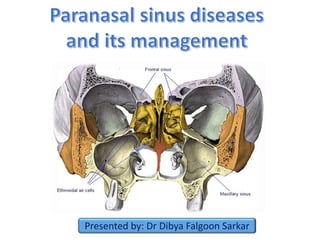
  • 3. • Paranasal sinuses are the mucosa lined air spaces present within the bones of the face & skull. • There are four bilaterally paired paranasal sinuses: - Frontal sinus - Maxillary sinus - Ethmoidal sinus - Sphenoidal sinus • Named after the bones in which they are located
  • 4. 1. Frontal sinus 2. Ethmoidal sinus 3. Sphenoidal sinus 4. Maxillary sinus
  • 5. • Humidifying and warming the inspired air. • Imparts resonance to voice • Increases surface area for absorption of noxious gases and trapping of particles in inspired air •Lightens the skull •Helps in absorbing the shock of blows to the face & thereby limiting the extent of facial injury from trauma. •Serves as an accessory olfactory organ by evenly distributing the inspired air. •Contributes to facial growth
  • 6. Sinuses begin their development at the 3rd month of intrauterine life as outpouchings of the mucous membranes of the nasal meatuses and sphenoethmoidal recess The early PNS expand into the walls of the nasal fossa by the growth of mucous membranes into the maxillary, sphenoid, ethmoid and frontal bones Pneumatization follows. Sinuses enlarge variably but always retain their original communication with the nasal cavity through ostia
  • 7.
  • 8. • Largest of all the paranasal sinuses. • Also known as the “antrum cave of Highmore”– English physician described an infection of sinus in 1651. • First sinus to develop • Pyramidal shaped - base toward the lateral wall of nose and apex directed laterally into the zygomatic process • Capacity approx. 15ml
  • 9. Superior wall • This wall forms both- roof of sinus & floor of the orbit. • It is thinnest wall & mostly flat slopes slightly anteriorly and laterally. • The Infraorbital canal runs along this wall and crosses from back to front- accentuates the fragility of wall • This wall is frequently involved in orbital and maxillary trauma.  Anterolateral wall • Thickness is 2-5mm.Thinnest portion over canine fossa • Related to facial artery and vein • Labial levator muscles & inferior portion of orbicularis oculi is attached to this wall above the infra-orbital foramen. • These attachments direct the spread of infection from maxillary teeth. • This wall also contains the anterior & middle superior alveolar nerves. • Any surgical procedure through this wall may jeopardise the nerve supply to these teeth.
  • 10. Floor of the maxillary sinus • It is approximately 1.5 cm below the floor of nasal cavity. • It corresponds to the line drawn laterally from ala of the nose. Posterolateral wall • Posterior superior alveolar nerves and vessels sometimes present in close contact with the sinus mucosa. • In such instances , acute sinusitis is accompanied by pain in multiple maxillary posterior teeth. • Immediately posterior to this wall, vital structures include: -maxillary nerve & maxillary artery - nerve of pterygoid canal - sphenopalatine ganglion.
  • 11. Ostium of maxillary sinus • Communication between maxillary sinus and nasal cavity. • Average length: 5.5mm • Located superior to the floor of the sinus. • Opens into posterior part of hiatus semilunaris in the middle meatus • Arterial supply: - Facial artery - Infraorbital artery - Greater palatine artery. • Venous drainage: - Facial vein - Infraorbital vein  Nerve supply:  Infraorbital nerve  Anterior , Middle & Posterior superior alveolar nerves  Lymphatics:  Submandibular lymph nodes
  • 12. • The frontal sinuses are rudimentary or absent at birth. • They are developed between 7th & 8th year of life but reach the full size only after puberty. • Situated between the inner and outer tables of frontal bone • Absent on one (15 %) or both sides (5 %) • Drainage pathway – naso- frontal duct- frontal recess - situated at its floor • Drains into middle meatus (62%) or ethmoid infundibulum (38%).
  • 13. • The inner table is much thinner than the outer table. As a result the chances of fracture of the inner table are more frequent ,even without the fracture of outer table.  Arterial supply: - supraorbital artery - anterior ethmoidal artery  Venous drainage: Anastomotic vein in the supraorbital notch connecting the supraorbital & superior ophthalmic veins.  Lymphatic drainage: Submandibular nodes.  Nerve supply: Supraorbital nerve
  • 14. • Formation begins at 4th month of fetal life • Thin-walled air cavities in the lateral masses of ethmoid bone, between nose and the eyes. • Anterior ethmoidal air cells -3- 11 drain into either the ethmoidal infundibulum or the frontonasal duct. • Bullar cells (middle ethmoidal air cells) -usually <3 - open in ethmoidal infundibulum. • Posterior group :- Up to 7 - usually drain by a single orifice into the superior meatus
  • 15. Ethmoidal bulla: • This is the most constant landmark for surgery. It lies above the infundibulum and it's lateral/inferior surface and the superior edge of the uncinate process forms the hiatus semilunaris. • The anterior ethmoid artery usually decends across the roof of this cell.  Arterial supply: - Sphenopalatine artery - Anterior and posterior ethmoidal arteries  Venous drainage: Corresponding veins  Lymphatic drainage: • Anterior and middle groups drain in the submandibular nodes . • Posterior group drains into retropharyngeal nodes.  Nerve supply: Anterior and posterior ethmoidal nerves Orbital branches of pterygopalatine ganglion.
  • 16. • They are the most anterior ethmoidal air cells. • Its size influence the patency of the frontal recess and the anterior middle meatus. • Haller cells: Also called infraorbital ethmoid cells. • Present in approx. 20 % patients. • Clinical significance – 1. Become infected , with potential extension into orbit. 2. Narrows the maxillary ostium. • These are posterior ethmoidal cells extending into the sphenoid bone, either adjacent to or impinging upon the optic nerve. • When these Onodi cells abut or surround the optic nerve, the nerve is at risk when surgical excision of these cells is performed.
  • 17. • Body of sphenoid - behind the nose, in the centre of the skull. • Rarely symmetrical and separated by a thin bony septum. • Ostium of the sphenoid sinus is situated in the upper part of its anterior wall and drains into sphenoethmoidal recess. • Average size – 2 x 2 x 2 cm. • According to Congdon sphenoid pneumatization can be as follows: 1. Conchal – 5 % 2. Presellar – 23 % 3. Postsellar – 67% • Arterial supply- Posterior ethmoidal artery. • Nerve supply- Posterior ethmoidal nerves • Lymphatic drainage- Retropharyngeal nodes
  • 18. • History and systemic clinical examination: • Check general signs of health • Systemic medical history, history of allergies, drug use and abuse • Occupation history • Examination of neck for lumps or swollen lymph nodes • Palpation: • PNS may be palpated directly to elicit any tenderness • Tenderness over maxillary and frontal bones may indicate inflammatory condition • Step-off deformity indicates facial bone fracture in trauma cases • Paresthesia is seen over midface region in injury to infraorbital nerve
  • 19. 1. Anterior rhinoscopy: • Examination of nose with a nasal speculum to check for abnormal areas, useful in evaluation of nasal obstruction. • Colour of nasal mucosa and its vascularity • Position of septum is noted for deviation or dislocation, as well as septal spurs, haematoma or perforations • Inferior & middle turbinates seen 2. Posterior rhinoscopy: • Assessed using a mouth mirror and headlight in the nasopharynx • Technique sensitive
  • 20. • Low specificity and sensitivity • Helpful in absence of endoscopy • Decreased light transmission when compared with normal side suggests of significant fluid collection or marked mucosal edema Transillumination
  • 21. • Endoscopic examination/ Rhinoscopy: Nasoscope/rhinoscope is a thin, tube-like instrument with a light and a lens for viewing. • A special tool on the nasoscope may be used to remove samples of tissue. The tissues samples are viewed under a microscope by a pathologist. • Helps in visualization of inaccessible areas like middle meatal cleft.
  • 22. 1. Maxillary sinus: • Water’s view (PNS view) x-ray and CT scan are ideal for maxillary sinus visualization. • X-ray beam angled at 45 degree to orbitomeatal line 2. Frontal sinus: • CT scan is helpful in trauma cases as we can appreciate both inner and outer tables in the axial view • PNS view x-ray is helpful to detect fluid accumulation in sinusitis cases 3. Ethmoidal sinus: Axial views of CT scan are helpful in detecting fractures of the ethmoid bone. 4. Sphenoid sinus: Best viewed in sagittal and axial sections of CT scan. “Limited CT Evaluation” – slice 3-4 mm
  • 23. Maxillary sinus & Ethmoid air sinus Axial CT scans
  • 24. Frontal sinus CT scan axial PNS view radiograph
  • 25. Sphenoid sinus Axial CT Sagittal CT
  • 26. • MRI: Excellent soft tissue definition - evaluation of neoplastic disease. • MRI (magnetic resonance imaging) with gadolinium: Gadolinium is injected into a vein. The gadolinium collects around the cancer cells so they show up brighter in the picture.
  • 27. • Developmental variations and anomalies • Inflammatory/infectious diseases • Cysts • Tumours – benign & malignant • Other surgically relevant conditions
  • 28. • Paradoxical curvature of middle turbinate: • Normally the convexity of the middle turbinate is directed medially toward the nasal septum. • When the convexity is directed laterally, it is termed a paradoxical middle turbinate . • Most authors agree that the paradoxical middle turbinate can be a contributing factor to sinusitis.
  • 29. • Variation of the ethmoidal roof anatomy • The ethmoid roof is of critical importance for two reasons: 1. most vulnerable to iatrogenic cerebrospinal fluid leaks. 2. anterior ethmoid artery is vulnerable to injury. • The depth of the olfactory fossa is determined by the height of the lateral lamella of the cribriform plate. • In 1962, Keros classified the depth of the olfactory fossa into three types, that is, a) Keros type I: <3 mm b) Keros type II: 4-7 mm c) Keros type III: 8-16 mm - most vulnerable to iatrogenic injury.
  • 30. • Variations of uncinate process • The uncinate process may be medialized, lateralized, or pneumatized/bent. • Medialization occurs with giant bulla ethmoidalis. • Lateralization of the uncinate process may obstruct the infundibulum. • Pneumatization (uncinate bulla) can rarely cause obstruction of the infundibulum.
  • 31. • Sinusitis is the inflammatory condition of the mucous membrane lining of the sinuses. It may progress to pus formation. • Causes : • Rhinogenous – infection spreads from the nasal cavity. It is the most common way for infection and such sinusitis is the complication of the flu. • Odontogenic – infection spreads from upper teeth. This way is typical only for maxillary sinus. The pathologic process may spreads from upper 4,5,6 teeth apex to the inferior wall of the maxillary sinus • Traumatic • Hematogenic • Allergic
  • 32. 1. Initial phase: reversible 2. Ostium obstruction phase 3. Bacterial phase 4. Chronic phase Osteomeatal complex obstruction Decreased ventilation of the sinuses Decreased drainage of the sinuses pO2 decrease, pCO2 increase, mucous stasis Inflammation and viscous mucous, ciliary movement slowing Ciliary damage along with increase in number of anaerobic organisms
  • 33. • According to duration: 1. Acute : infection lasting 4 weeks, symptoms resolve completely resolved in < 30 days. 2. Subacute :infection lasting between 4 to 12 weeks, yet resolves completely. 3. Recurrent: ≥ 4 discrete acute episodes per year, each completely resolved in < 30 days but recurring in cycles, with at least 10 days between complete resolution of symptoms and initiation of a new episode 4. Chronic: symptoms lasting more than 12 weeks.
  • 34. • According to pathogen: 1. Bacterial: Hospital-acquired acute infections are more often bacterial, typically involving Staphylococcus aureus, Klebsiella pneumoniae, Pseudomonas aeruginosa 2. Viral: In immunocompetent patients - in the community is almost always viral (eg, rhinovirus, influenza, parainfluenza). antibiotics given for: • Mild to moderate sinus symptoms persisting for ≥ 10 days • Severe symptoms (eg, fever ≥ 39°, severe pain) for ≥ 3 to 4 days • Worsening sinus symptoms after initially improving from a typical • viral URI ("double sickening" or biphasic illness)
  • 35. 3. Fungal: Usually seen in immunocompromised patients because of poorly controlled diabetes, neutropenia, or HIV infection. It is clinically of 2 types: A)Non-invasive fungal sinusitis: • Saprophytic fungal infestation/colonization • Allergic fungal rhinosinusitis (AFRS) • Saprophytic fungus balls (mycetoma) B)Invasive fungal sinusitis – • Chronic invasive fungal sinusitis • Granulomatous invasive fungal sinusitis • Acute (fulminant) invasive fungal sinusitis
  • 36. • Risk factors: 1. Obstruction to drainage: most important 2. Defect of self-cleaning mechanism of the mucous membrane of sinus – infections causing immobility of the cilia, Increased viscosity of secretions, Immotile cilia syndrome, Prolonged exposure to cigarette smoke 3. Medications: First generation antihistamines (non sedating do not affect), Anticholinergics, Aspirin, Anesthetic agents, Benzodiazepines 4. Immunodeficiency: Immunoglobulin deficiency (IgA, IgG), diabetes, HIV infection 5. Other factors: prolonged ICU stays, severe burns, cystic fibrosis, and ciliary dyskinesia. According to number of sinuses involved: Hemisinusitis - all sinuses on one side Polysinusitis - several sinuses, but not all, are involved Pansinusitis - all, are involved
  • 37. 1.Nasal congestion and discharge 2. Sore throat and postnasal drip 3. Pain or pressure. 4. Oedema of facial tissues. 5. Bad breath or loss of smell (hyposmia/anosmia). 6. Systemic symptoms: Malaise may be present. Fever and chills suggest an extension of the infection beyond the sinuses, rise in temperature, bad appetite, sleep disturbances, changes of the blood (leukocytosis), Productive cough (especially at night) 7. Complications – ocular, neurological, local
  • 38. • Diagnosis is mainly clinical • CT scan and cultures - mainly for chronic, refractory, or atypical cases. • Clinical diagnostic criteria include a) 2 major factors b) 1 major and 2 minor factors or presence of pus in nasal cavity. Major factors Minor factors Facial pain/pressure Headache Facial congestion/fullness Maxillary dental pain Nasal drainage/discharge Halitosis Postnasal drip Cough Fever (acute sinusitis only) Purulence in nasal cavity on examination Hyposmia
  • 39. Treatment options measures to enhance drainage: 1. Heat therapy: Steam inhalation and hot beverages 2. Topical vasoconstrictors/ Nasal decongestants: Topical nasal sprays (limit use to 3-7 days) - Phenylephrine, Oxymetazoline. Corticosteroids: Corticosteroid nasal sprays can help relieve symptoms but typically take at least 10 days to be effective 3. Systemic vasoconstrictors: Systemic vasoconstrictors, such as pseudoephedrine 30 mg 4. Nasal irrigation 5. Antihistaminics: recommended if allergy present. 6. Hydration 7. Antibiotics: Amoxicillin 500 mg tid for 10-14 days - First line Nasal irrigation
  • 40. 8. Mucoactive drugs: main purpose - increase the ability to expectorate sputum and/or decrease mucus hypersecretion. 1. Expectorants: hypertonic saline, iodine containing compounds, guaifenesin 2. Mucoregulators: anticholinergic drugs, glucocorticoids, macrolide antibiotics 3. Mucolytics: classic mucolytic (N-Acetyl Cysteine), peptide mucolytic 4. Mucokinetics: bronchodilators, ambroxol a) Nebulization: best form of physiotherapy. b) Laser therapy: used directly over the sinuses to reduce inflammation c) Ultrasound therapy: sound waves are conducted through a hypoallergenic gel to reduce inflammation and loosen the accumulated mucous. d) Short-wave diathermy e) Rinoflow therapy: new option, basically micronized endotracheal wash. Used in sinusitis, rhinitis, pharyngitis, laryngitis and secretory otitis media.
  • 41. Indications: • Sinusitis unresponsive to antibiotic therapy • Necrotic sinusitis • Orbital complications (abscess and phlegmon of orbit) • Intracranial complications (meningitis, brain abscess) • Rhinogenic sepsis • Odontogenic sinusitis combined with maxillary osteomyelitis. Approaches: 1. Removal of etiology (eg: removal of the infected teeth) 2. Functional endoscopic sinus surgery (FESS) is the mainstay in the surgical treatment of sinusitis and nasal polyps, including bacterial, fungal, recurrent acute, and chronic sinus problems. 3. Caldwell-luc procedure/ Denker’s procedure 4. Intranasal antrostomy 5. Radical surgeries
  • 42. • Oro-antral communication: it is an abnormal connection between oral and Antral cavities. • Oro-antral fistula: long standing oro-antral communication when left open, epithelializes to form a patent fistula between the two cavities. • Types: 1. Alveolosinusal 2. Palato-sinusal 3. Vestibulo-sinusal • Causes: • Extraction – maxillary posterior teeth • Cysts, tumors • Osteomyelitis • Radiation therapy • Trauma • Implant denture Oro-antral Fistula
  • 43. • Some patients are asymptomatic • Unpleasant tasting discharge and odor • Reflux of fluids and foods into the nose from mouth • Difficulty in smoking and blowing air • Development of chronic sinusitis in infected cases • Valsalva manoeuvre • Mirror fog test • Cotton wisp test • Nasal regurgitation of fluid • Radiographs: Sinus floor discontinuity, sinus opacity, focal alveolar atrophy.
  • 44.  Immediate treatment: Primary purpose is closure of defect and prevention of sinusitis through: • Suturing across the defect with/without periodontal pack, warm saline rinses, antibiotic and antihistaminic therapy with decongestants. • Depending on defect size: 1. Size < 5mm: non-invasive intervention (spontaneous closure by blood clot) 2. Size 2-6mm: Collagen plug in the socket with figure of 8 suturing 3. Size > 6mm and > 48 hours: surgical closure : small defects – local flaps; large defects – regional/distant flaps Obturators: immunocompromised patients
  • 45.  In long-standing OAF we must evaluate the patient for maxillary sinusitis and treat it before closing the defect surgically The fistulous tract must be excised prior to surgical closure
  • 46. • Most common and oldest surgical technique • Developed by Rehrman • Disadvantage: Reduces depth of buccal vestibule • Overall success rate approx. 87.2%
  • 47. • Full thickness flap • Based on the greater palatine artery • Advantages: a) No reduction of vestibular depth b) Lesser chance of flap breakdown c) Anteriorly based flaps are ideal for closure of tuberosity defects • Disadvantage: Exposed palatal bone takes long time to heal
  • 48. • First described by Egyedi in 1977 • Helpful in small to medium sized defects • Pedicled flap • Complication can be avoided: i. Graft should not be in tension ii. Patient should be on a liquid diet Anatomy of BFP: It has 4 processes- buccal, pterygoid, pterygopalatine, superficial and deep temporal Blood supply: Deep & superficial temporal artery, facial artery
  • 49. • Based on lingual artery and its branches • Tongue flaps are useful : i. When both buccal and palatal flaps have failed ii. Defect size is > 1.5cm • Types of tongue flaps: a) Anteriorly based- Defects of hard palate, buccal mucosa b) Posteriorly based- Defects of soft palate • Disadvantages: i. Second surgery is required for flap division ii. Swallowing and speech difficulty • Some investigators placed the patients in IMF to reduced flap tearing
  • 51. • By George Walter( USA, 1983) and Henri Luc (France,1989) • Technique to gain access to the maxillary antrum through canine fossa of maxilla • Indications: Removal of tooth fragments, chronic sinusitis, neoplasms, cysts Controlling maxillary antrum bleed/ haematomas Treating orbital floor fractures Nasal Antrostomy • Artificial ostium is made to the max. sinus with an opening in inferior meatus • Principle: Opening at a lower level improves drainage • Types : Middle meatal (child), inferior meatal, sublabial
  • 52. Presented by: Dr Dibya Falgoon Sarkar
  • 53. PNS Cysts Intrinsic cysts Mucocele Serous cysts Cholesteatoma Extrinsic cysts Dentigerous cysts Radicular cyst Neurogenic cysts Meningocele Encephalocele Fissural cysts Nasopalatine duct cyst Globulumaxillar cyst Median palatal cyst Pierse et al Oral & Maxillofacial Surgery Clinics 2012
  • 54. Mucous retention cyst Mucous extravasation cyst (Mucocele) Most common and donot produce bone destruction Produces bone destruction Results from obstruction of glands in the sinus lining Results from accumulation of mucous in an obstructed sinus cavity Site: Floor of Maxillary sinus Site predilection: Frontal & Ethmoidal sinus Radiographically visible as a dome-shaped, homogenous mass Radiographically Pressure erosion seen surrounding sinus walls. Initially sinus is opacified • Treatment: Surgical excision along with the supplying gland • Excellent prognosis, low recurrence
  • 55. Neurogenic cysts of PNS • Non-neoplastic lesions • Two types : Meningocele & encephalocele • Both are results of congenital herniation of intracranial meningeal and neural tissue • Encephalocele is more common and contains primitive neural tissue • Meningocele contains arachnoid and dural tissue and CSF • Radiographically seen as bony dehiscences in base of skull or cranial vault associated with a soft tissue extension in nose and PNS • Treatment : Surgical enucleation Encephalocele in fronto- ethmoidal sinus
  • 56.
  • 57. Sinonasal tumours Benign tumours Epithelial tumors Sinonasal papilloma Mesenchymal tumors Osteoma Angiofibroma Haemangioma Malignant tumours Epithelial tumours Squamous cell carcinoma Adenoid cystic Carcinoma Adenocarcinoma Mesenchymal tumours Olfactory neuroblastoama Sarcomas
  • 58. • Early : asymptomatic . • Oral symptoms: Toothache , trismus, alveolar ridge fullness, erosion , malocclusion . • Nasal findings: Obstruction, epistaxis, rhinorrhea , post nasal discharge , anosmia, nasal mass or polyposis, mass in the medical canthus, broadening of nasal dorsum . • Ocular findings: Epiphora, diplopia, proptosis • Facial signs: Paraesthesia, asymmetry • Maxillary sinus involvement: Mass in palate or upper alveolus . mass in upper gingivobuccal sulcus, malocclusion or loose teeth. • Advanced : Trismus, Orbital : Periorbital swelling , proptosis, epiphora , impaired ocular mobility
  • 59. • Benign, localized proliferations of the respiratory mucosa • 10-25% of all paranasal tumors • Cause: Some clinicians believe that these are actually reactive hyperplasia secondary to stimulants like allergens, bacteria or HPV virus( type 6,11), tobacco smoking • Types : 1) Fungiform 2) Inverted 3) Cylindrical
  • 60. Fungiform papilloma Inverted papilloma Cylindrical papilloma Distribution Similar to oral papillomas; 18-50% all PNS papillomas Most common, premalignant <7% of PNS papillomas HPV Almost all variants are HPV positve HPV 6,11,16 & 18 Low frequency of HPV association Age/sex 20-50 years; M>F > 20 years; M>F 20-50 years; M>F Clinical features Unilateral nasal obstruction, epistaxis, pink nodule Unilateral nasal obstruction, epistaxis, pink nodule, bone erosion, locally aggressive Unilateral nasal obstruction, epistaxis, robust red multi nodular Treatment Complete surgical excision, no malignant transformation Aggressive surgical excision, Greatest potential for malignant transformation and destruction Aggressive surgical excision
  • 61. • Most common benign, mesenchymal tumor of PNS • Arises from junction of membranous & endochondral bone in frontoethmoidal suture region • May have intracranial/ orbital extensions • Histological subtypes: hard cortical & soft cancellous • Surgical excision • Low recurrence Osteoma Hard cortical osteoma Soft cancellous osteoma Lobulated densities obstructing sinus Less dense, may be mistaken with a soft tissue sinus mass
  • 62. • Usually an extension of juvenile angiofibroma of nasopharynx • Often extends into sphenoid and ethmoidal sinuses • Have a vertical spreading pattern • Imaging: Appears as an enhancing lesion in MRI • Carotid angiography and CECT are helpful as angiofibromas contain a hypertrophied internal maxillary artery • Conservative surgical excision
  • 63. • Rare mucosal neoplasm • Neural crest origin • Often exhibits immunological symbiosis with the host • Radical procedures are contraindicated • Treatment by endoscopic resection • Role of radiotherapy is debatable
  • 64. • Arise from the nasal cavity (septum/ alveolus) • Multifocal, frequent recurrence • Spreads superiorly into skull base • Age range: both young and old • Treatment: Craniofacial resection • Not radiosensitive
  • 65. • Classically arises from olfactory epithelium in the upper nasal vault • The presence of a mass in the upper nasal cavity with associated skull base erosion is typical. • Bimodal peak in the second/third and sixth/seventh decades. • Cervical metastases have been described in up to 23%. • These are routinely resected in craniofacial approaches • Endoscopic resection is being increasingly offered for this tumour particularly when it arises from the middle and superior turbinates. • Should always be combined with radiotherapy. Stage I confined to primary site Stage II presence of nodal metastasis Stage III presence of distant metastasis
  • 66. • 3% of all the head and neck malignancies • Incidence: Maxillary sinus (70%), Ethmoid (20%), frontal (3%), Sphenoid(1%) • M > F ( 2 : 1 ) • Age – 6th & 7th decades • Risk factors : 1. Wood workers 2. Leather manufacturers 3. Smoking & alcohol • Damage from smoking and alcohol plays a less damaging role Case of Small cell neuroendocrine carcinoma of Left maxillary sinus
  • 67. • About 30 per cent of patients with this condition are woodworkers. • These tumours usually arise in the middle meatus and spread into the ethmoidal sinuses • Adenocarcinoma is generally rather radio resistant but combined therapy is usually offered. • Many patients require a craniofacial but in selected cases have been treated successfully by an endoscopic resection • Use of topical 5- flurouracil and surgical debulking has been advocated
  • 68. • Propensity to spread along perineural lymphatics which compromises attempts at excision. • Known to produce blood-borne metastases, classically to the lung while lymphatic spread is rare. • Treatment is generally combined surgery and radiotherapy
  • 69. Clinical features: 1. Maxillary sinus ca • Accounts for 80% of all PNS cancers • Facial swelling, pain, paraesthesia of cheek • Epistaxis, nasal discharge, obstruction • Ill fitting dentures, alveolar/palatal mass • Proptosis, diplopia, impaired vision, orbital pain 2. Ethmoid sinus ca • Headache • Referred pain to nasal, retrobulbar region • Subcutaneous mass at inner canthus, diplopia & proptosis • Nasal obstruction, discharge,
  • 70. 1. Tumors from maxillary sinus: • Posterior wall > Infratemporal fossa • Superior wall > Infraorbital canal > Orbit • Inferiorly > Roots of upper premolars & molars > Oral cavity / Palate 2. Tumors invading ethmoid bone: • Via lamina papyracea of ethmoid bone • Via emissary veins present anterior to the crista galli which eventually connects to the sagittal sinus • Via olfactory fibres through the cribriform plate 3. Tumors invading the orbit can spread via: • Superior orbital fissures > Cavernous sinus • Inferior orbital fissures > Pterygopalatine fossa & infratemporal fossa • Orbital apex > Middle cranial fossa 4. Tumors from nasal septum: • Superiorly: Into extranasal structures • Inferiorly: Into upper lip & gingivobuccal sulcus
  • 71. Diagnostic work up for PNS carcinoma patients: • Routine blood examination • Histopathology • Chest X-ray for Adenoid cystic carcinoma • CT/MRI • Dental evaluation • Baseline ophthalmologic examination • Baseline speech & swallowing assessment • Fibre optic endoscopic examination & biopsy Lymphatic spread of PNS carcinoma: • Usually sparse-10% incidence of cervical lymph node metastasis • First echelon: Upper jugular/ submandibular nodes • Contralateral metastasis: extremely rare • Tumors of nasal septum, columella and nasal vestibule show bilateral metastases Lymphatic drainage of sinuses
  • 72.
  • 73. AJCC stage Stage grouping Stage description 0 Tis N0 M0 The tumor is only in the top layer of cells lining the inside of the maxillary sinus and has not grown any deeper (Tis). The cancer has not spread to nearby lymph nodes (N0) or to distant parts of the body (M0). I T1 N0 M0 The tumor has grown deeper, but it is only in the tissue lining the sinus (the mucosa) and does not grow into the bone. (T1). The cancer has not spread to nearby lymph nodes (N0) or to distant parts of the body (M0). II T2 N0 M0 The tumor has begun to grow into some of the bones of the sinus, other than into the bone of the back part of the sinus (T2). The cancer has not spread to nearby lymph nodes (N0) or to distant parts of the body (M0).
  • 74. AJCC stage Stage grouping Stage description III T3 N0 M0 The tumor has grown into the bone at the back of the sinus (called the posterior wall) or into the ethmoid sinus, the tissues under the skin, or the side or bottom of the eye socket (T3). The cancer has not spread to nearby lymph nodes (N0) or to distant parts of the body (M0). OR T1 to T3 N1 M0 The tumor might or might not have grown into structures just outside the maxillary sinus (T1 to T3). The cancer has spread to a single lymph node on the same side of the neck as the tumor, which is no larger than 3 centimeters (cm) across (N1). The cancer has not spread to distant parts of the body (M0). IVA T4a N0 or N1 M0 The tumor is growing into the skin of the cheek, the front part of the eye socket, the bone at the top of the nose (cribriform plate), the sphenoid sinus, the frontal sinus, or certain parts of the face (the pterygoid plates or the infratemporal fossa). This is also known as moderately advanced local disease (T4a). The cancer has not spread to nearby lymph nodes (N0), or it has spread to a single lymph node on the same side of the neck as the tumor, which is no larger than 3 centimeters (cm) across (N1). The cancer has not spread to distant parts of the body (M0).
  • 75. AJCC stage Stage grouping Stage description IVB T4b Any N M0 The tumor is growing into the throat behind the nasal cavity (the nasopharynx), the back of the eye socket, the brain, the tissue covering the brain (the dura), some parts of the base of the skull (middle cranial fossa or clivus), or certain cranial nerves. This is also known as very advanced local disease (T4b). The cancer might or might not have spread to nearby lymph nodes (any N). It has not spread to distant parts of the body (M0). OR Any T N3 M0 The tumor might or might not have grown into structures outside the maxillary sinus (any T). The cancer has spread to at least one lymph node that is larger than 6 cm across, OR it has spread to a lymph node and then grown outside of the lymph node (N3). It has not spread to distant parts of the body (M0). IVC Any T Any N M1 The tumor might or might not have grown into structures outside the maxillary sinus, and it might or might not have affected a vocal cord (any T). The cancer might or might not have spread to nearby lymph nodes (any N). The cancer has spread to distant parts of the body (M1)
  • 76.
  • 77. • Nodal metastases is dependent upon histological subtype of the carcinoma  Nodal metastases has as high as 28% in SCC, 25% in adenocarcinoma, 12% for undifferentiated carcinoma and 10% for adenoid cystic carcinoma. ( According to Kaplan et al AJCC 1999) • Maxilary sinus carcinomas involving hard palate shows a higher incidence of cervical node metastases (Castelnau et al Role of Elective Neck Management in Maxillary Sinus Squamous Cell Carcinoma. Otolaryngol (Sunnyvale). 2016) • Most powerful prognostic factor for overall survival is T- stage followed by nodal involvement (Wu TH, Huang JS, Wang HM, Wang CH et al Long-term survival after surgery of stage III-IV maxillary sinus carcinomaChang Gung Med J. 2010)
  • 78. According to Jatin Shah et al 2015 (Curr Opin Otolaryngol Head Neck Surg 2015) • Treatment failure occurs in 62% of all cases of maxillary sinus carcinoma • Local recurrence is the most common cause • Thus they concluded that : 1. Elective treatment of neck is justifiable only in advanced staged (T3-4) maxillary sinus carcinomas 2. Elective neck dissection can be performed if we are planning for any vascular anastomosis in the neck during flap reconstruction 3. If neck is not entered in a case cN0 T3-4 tumor we should irradiate the neck during post op radiotherapy of primary site.  IMRT is the most preferable modality of radiotherapy 4. Elective neck dissection is not recommended in T1-2 tumors of sinus
  • 79. Incisions Midfacial degloving approach Weber –Fergusson approach Lateral rhinotomy approach Intra/extraoral Intraoral Extraoral Extraoral Access Indicated in pathologies of frontal, ethmoidal, maxillary sinus and infratemporal fossa Transoral approach for pathologies of maxilla, not suitable for skull base / ethmoid lesions Indicated for approach to maxillary, sphenoid and ethmoidal sinuses Advantages Excellent esthetics Easier and provides wide area of exposure Easier approach, scar lies in the folds of facial skin Disadvantages Technique sensitive Heals by scar formation, Limited access
  • 80.
  • 81.
  • 82. Borle’s extension- When temporalis flap is planned for maxillectomy defect reconstruction Lynch extension- To expose the ethmoidal sinus Dieffenbach extension- When total maxillectomy is being planned
  • 83. Palatal incisaion is given 3mm lateral to midline so that a mucoperiosteal flap is formed which can cover the raw bony edge Osteotomies through zygoma, floor of orbit, frontal process of maxilla & pyriform aperture
  • 84. • The incision is made around the base of the nose (or entering the nostril floor for a better esthetical result) and along the facial nasal groove (In the border of both esthetic units). • The dissection goes deep to the subperiosteal plane of the frontal process of the maxilla. • Below the nasal bone the incision is deepened through the nasal mucosa. • A unilateral nasomaxillary osteotomy is made with a small osteotome, and the two osteotomy lines are joined at the level of the naso frontal junction. • The septum is cut and freed from the mobilized segment according to the resection plan
  • 85. • Originally described by Tessier • Excellent approach for accessing the pathologies of frontal and ethmoidal sinuses. • Major risks of this approach include injury to the temporal and zygomatic branches of the facial nerve, temporal hollowing, scalp hematoma, parietal scalp pain, parasthesia or anesthesia, infection, and nasal orbital hypertrophy.
  • 86. • First reported by Smith in 1954 • Has become a ‘gold standard’ for tumors affecting the anterior skull base ( According to Stell & Maran) • Principle: En bloc resection of the tumor including the ethmoidal sinuses, superior nasal septum and floor of anterior cranial fossa. • Laterally part of the bony orbit may also be included in anterolateral resection. • Indications: Malignant tumors requiring surgical resection and involving the anterior skull base • Contraindications: 1. Extensive frontal lobe involvement 2. Bilateral orbital invasion/ optic chiasm 3. Distant metastasis cases 4. Tumors with poor prognosis like malignant melanoma
  • 87. • Incision: An extended lateral rhinotomy incision is made on the side of maximal tumor involvement.
  • 88. • IMMEDIATE: – convulsions – haemorrhage – air embolism • INTERMEDIATE: – cerebrovascular accident – confusion – pulmonary embolism – meningitis • LONG TERM: – haemorrhage – frontal abscess/encephalitis – bone necrosis/fistula – cerebrospinal fluid leak – epilepsy – epiphora
  • 89. Brown’s classification of Maxillectomy, 2010 (Lancet Oncol 2010; 11: 1001–08) Vertical classification: I—maxillectomy not causing an oronasal fistula; II—not involving the orbit; III—involving the orbital adnexae with orbital retention; IV—with orbital enucleation or exenteration; V—orbitomaxillary defect; VI— nasomaxillary defect. Horizontal classification: a—palatal defect only, not involving the dental alveolus; b—less than or equal to 1/2 unilateral; c—less than or equal to 1/2 bilateral or transverse anterior; d—greater than 1/2 maxillectomy. Letters refer to the increasing complexity of the dentoalveolar and palatal defect, and qualify the vertical dimension.
  • 90. Algorithm for Maxillary Reconstruction (Brown JS, Richard Shaw; Lancet Oncol 2010; 11: 1001–08) • Class I defects: Most reconstructive techniques reported for class I defects are the fasciocutaneous radial forearm flap or obturators • Class II defects: 1) If the defect is less than half of the lateral alveolus and palate (class IIb), very good results can be achieved with either obturation or reconstruction. 2) Zygomatic implants are also efficient in Class II defects • Class III defects: a) With class III defects, orbital support is lost as well as anterior support of the cheek and dental arch. Very little role for a prosthetics. b) Vascularized bone grafts are ideal especially DCIA with internal oblique flap. • Class IV: Involve patients with stage IV disease and poor prognosis • Class V: Reconstruction with temporalis flap is adequate • Class VI: Nasomaxillary defects that involve the nasal bone with or without skin require reconstruction. Osteocut. RFFP is ideal.
  • 91. Summary of published methods of reconstruction from 1998-2009, in number of reported cases Recommended methods by Brown et al.
  • 92. Management algorithm of the PNS tumors invading the orbit (Stell & Maran)
  • 93. • Can be used in resection of PNS tumours • May need combination with craniofacial resection if: 1. Dural invasion 2. Infiltration of superior sagittal sinus • Principles: Complete resection of the tumour including a wide field of clearance of adjacent mucosa, bone and cartilage • Can be used to repair skull base bone and dural defects • Extent of surgery: A complete fronto-ethmo- sphenoidectomy is undertaken with lateral nasal wall removal • Advantage: low morbidity and short hospital stay • Disadvantage: technique sensitive
  • 94. • Extension through anterior cranial fossa • Involvement of both optic nerve • Posterior extension into sphenoid sinus • Invasion of middle cranial fossa • Inoperable neck node & distant metastasis • Well differentiated adenocarcinoma • Early maxillary tumors • Olfactory neuroblastoma • SCC arising from inverted papiloma

Editor's Notes

  1. Walls- a) Superior wall b) Anterolateralwall c) Posterolateralwall d) Inferior wall.
  2. Inferomedially- anterior lacrimal and post lacrimal crests
  3. Relations : i)Superiorly– optic chiasma & pituitary gland. ii)Inferiorly– roof of pharynx iii)On either side – cavernous sinus & internal carotid artery iv) Anteriorly – sphenoethmoidal recess v) Posteriorly – Pons & medulla
  4. Maxillary sinus- Light placed in sublabial region Frontal sinus- Light placed over supraorbital rim
  5. Affected sinus must be irrigated copiously with physiologic saline 3 times/day until inflammation subsides Antibiotics like Augmentin must be used Caldwell luc procedure must be done to removed infected sinus membrane prior surgical closure of OAC
  6. Cholesteatoma : Hyperkeratinised epithelium which completely replaces normal mucous membrane
  7. Neuroendocrine tumor, metabolites like VMA may be detected in urine Treatment: Craniofacial resection
  8. Radiotherapy delays recurrence
  9. 3 parts: intracartilaginous, transfixion incision, vestibular incision Limits of resecetion are posteriorly wall of sphenoid, pterygoid plates, superiorly skull base and laterally coronoid process of mandible Can be combined with coronal incision for craniofacial resection
  10. Extends 1cm lateral to lateral canthus & 3mm below lower eyelash Too close to lashes: ectropion; Too far: lower lid edema Orbicularis oculi is left intact but dissection is made at bony infraorbital rim Infraorbital neurovascular bundle is cut
  11. Described by Moure in 1902 Excellent access to nasal cavity through which a medial maxillectomy can be done Ant skull base, ethmoids, sphenoid & maxillary sinuses
  12. The soft tissues of the face are mobilized by subperiostial elevation to expose the nasal bones, frontal processes of the maxilla and frontal bone up to the hairline via an extended lateral rhinotomy. • Through the lateral rhinotomy, the upper lateral cartilage is separated from the nasal bone to allow complete retraction of the nasal ala. • The orbital periosteum is elevated to expose the lacrimal fossa and the medial orbital wall. The nasolacrimal duct is often transacted obliquely at this point • anterior and posterior ethmoidal arteries are divided after bipolar coagulation. • If the lamina has been eroded by tumour, the adjacent periorbita should be resected for frozen section assessment A shield-shaped craniotomy is performed above the level of the supraorbital rim to include the frontal sinus. usually approximately 3x3x3.5 cm size. • The frontal sinus which has been opened by this manoeuvre is cleared of its mucosa and the posterior wall removed combined with a wide dissection of the dura. The anterior and posterior ethmoidal arteries are coagulated with the bipolar diathermy although care must be exercised as the optic nerve is approached. Osteotomies are performed around the cribriform plate through the ethmoidal and sphenoid roofs. • The posterior osteotomy crosses the planum sphenoidale to include the anterior face of the sphenoid and the nasal septum is separated by quadrilateral cuts. • The specimen is mobilized this can be removed, haemostasis achieved and the cavity inspected for further resection. • fashion a large middle meatal antrostomy to prevent subsequent infection. • dura has small defects which can be repaired primarily but more with fascia lata held in place with fibrin glue to which a split-skin graft taken from the thigh is applied inferiorly.
  13. The most complex and controversial area in head and neck reconstructive surgery is the management of maxillary, midface, and skull-base tumours
  14. Should be regarded as major surgery equivalent to CRF resection via an endonasal route