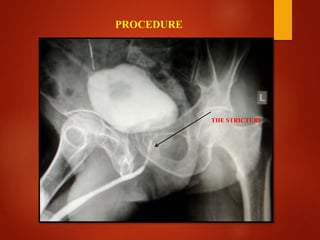
GOOD MORNING TO ALL….
OPPOSING URETHROGRAM (OUG)
MISS . SOWMIYA G,
DRDT 2ND YEAR,
GOVT.KMCH
CHENNAI-10.
 The opposing urethrogram is the radiographic study to
evaluate and document the stricture and define the
stricture recurrence.
 Combination of ascending urethrogram (AUG) and
micturating cystourethrography (MCU)
 This procedure helps to access length of the stricture.
INTRODUCTION
 The adult male urethra is approximately 18 cm long, with
the posterior urethra comprising the proximal 3 cm.
 The anterior urethra comprising the remaining 15 cm, with
the division point between the two located at the perineal
membrane.
ANATOMY OF MALE URETHRA
Posterior urethra:
1) Prostatic urethra
2) Membranous urethra
Anterior urethra:
1) Bulbous urethra
2) Pendulous urethra
3) Fossa naviculari
THE URETHRA SUBDIVIDED INTO FIVE SEGMENTS
RADIOGRAPHIC ANATOMY
1) Stricture
2) Urethral trauma
3) Fistulae or false passage
4) Vesico ureteric reflux
5) Study of the urethra during micturition
6) Bladder leak post surgery or trauma
7) Urodynamic studies,e.g.for incontinence.
8) Congenital abnormalities
INDICATIONS
 Acute urinary tract Infection
 Recent instrumentation
CONTRAINDICATIONS
 High osmolar contrast material or LOCM 300 mg/I
200-300 ml.
 LOCM 200–300mg/I 10-20ml.
 Pre-warming the contrast medium will help reduce
the incidence of spasm of the external sphincter.
CONTRAST MEDIUM
CONTRAST MEDIUM
 Fluoroscopy unit with spot Film
device and tilting table
 Video recorder (for urodynamics)
 Suprapubic catheter
 Foleys catheter 8-F
EQUIPMENT
ALLENGERS 600mA FLUROSCOPY MACHINE
Suprapubic catheter Foleys catheter
 Shave and clean lower abdomen and pelvis area.
 Take informed consent from patient or stander.
 The patient empties their bladder prior to the examination.
 Preliminary image Coned view of the bladder and Urethra
PATIENT PREPARATION
 Patient supine.
 It may be possible to fill the bladder via the
suprapubic catheter. Using aseptic technique the bladder is
catheterized. Residual urine is drained.
 To demonstrate Vesico-ureteric reflux (this
indication is almost exclusively confined to children)
TECHNIQUE
 Contrast medium is slowly injected or dripped in with the
patient supine and Bladder filling is observed by intermittent
fluoroscopy.
 It is important that early filling is monitored by fluoroscopy.
 Intermittent monitoring is also necessary to identify transient
reflux. Any reflux should be recorded.
 Following of this procedure, then
 Using aseptic technique the tip of the catheter is inserted so
that the balloon lies in the fossa navicularis.
 The catheter is connected to a 20 ml syringe containing
contrast medium and flushed to eliminate air bubbles.
 And its balloon is inflated with 2–3 ml of water to anchor the
catheter and occlude the meatus.
 Gentle traction on the catheter is used to straighten the penis
over the ipsilateral leg and prevent urethral overlap.
 Contrast medium is injected under fluoroscopic control
and steep (30–45°) oblique films taken.
 And the patient is instructed to strain
 Depending on the clinical indication, ascending
urethrography (AUG) may be followed by descending
micturating cystourethrography (MCU) to demonstrate the
proximal urethra and bladder, assuming there is no
contraindication to bladder catheterization.
Examples :
1. False passage
2. Urethral stricture
PROCEDURE
THE STRICTURE
Contrast reaction ( due to absorption through bladder mucosa)
Urethral trauma or rupture.
Extravasation of contrast – due to use of excessive pressure
in stricture.
COMPLICATIONS
AFTERCARE
Warned - of rare dysuria , retention.
Reflux - Antibiotics.
CONCLUSIONS
 Opposing Urethrogram (OUG) is the only procedure in
x-ray to accesses the length of the Urethral stricture.
 And we can calculate and measures the abscesses in male
urethra.
THANKING YOU……

OPPOSING URETHROGRAM (OUG)

  • 1.
  • 2.
    OPPOSING URETHROGRAM (OUG) MISS. SOWMIYA G, DRDT 2ND YEAR, GOVT.KMCH CHENNAI-10.
  • 3.
     The opposingurethrogram is the radiographic study to evaluate and document the stricture and define the stricture recurrence.  Combination of ascending urethrogram (AUG) and micturating cystourethrography (MCU)  This procedure helps to access length of the stricture. INTRODUCTION
  • 4.
     The adultmale urethra is approximately 18 cm long, with the posterior urethra comprising the proximal 3 cm.  The anterior urethra comprising the remaining 15 cm, with the division point between the two located at the perineal membrane. ANATOMY OF MALE URETHRA
  • 5.
    Posterior urethra: 1) Prostaticurethra 2) Membranous urethra Anterior urethra: 1) Bulbous urethra 2) Pendulous urethra 3) Fossa naviculari THE URETHRA SUBDIVIDED INTO FIVE SEGMENTS
  • 6.
  • 7.
    1) Stricture 2) Urethraltrauma 3) Fistulae or false passage 4) Vesico ureteric reflux 5) Study of the urethra during micturition 6) Bladder leak post surgery or trauma 7) Urodynamic studies,e.g.for incontinence. 8) Congenital abnormalities INDICATIONS
  • 8.
     Acute urinarytract Infection  Recent instrumentation CONTRAINDICATIONS
  • 9.
     High osmolarcontrast material or LOCM 300 mg/I 200-300 ml.  LOCM 200–300mg/I 10-20ml.  Pre-warming the contrast medium will help reduce the incidence of spasm of the external sphincter. CONTRAST MEDIUM
  • 10.
  • 11.
     Fluoroscopy unitwith spot Film device and tilting table  Video recorder (for urodynamics)  Suprapubic catheter  Foleys catheter 8-F EQUIPMENT
  • 12.
  • 13.
  • 14.
     Shave andclean lower abdomen and pelvis area.  Take informed consent from patient or stander.  The patient empties their bladder prior to the examination.  Preliminary image Coned view of the bladder and Urethra PATIENT PREPARATION
  • 15.
     Patient supine. It may be possible to fill the bladder via the suprapubic catheter. Using aseptic technique the bladder is catheterized. Residual urine is drained.  To demonstrate Vesico-ureteric reflux (this indication is almost exclusively confined to children) TECHNIQUE
  • 16.
     Contrast mediumis slowly injected or dripped in with the patient supine and Bladder filling is observed by intermittent fluoroscopy.  It is important that early filling is monitored by fluoroscopy.  Intermittent monitoring is also necessary to identify transient reflux. Any reflux should be recorded.
  • 17.
     Following ofthis procedure, then  Using aseptic technique the tip of the catheter is inserted so that the balloon lies in the fossa navicularis.  The catheter is connected to a 20 ml syringe containing contrast medium and flushed to eliminate air bubbles.  And its balloon is inflated with 2–3 ml of water to anchor the catheter and occlude the meatus.
  • 18.
     Gentle tractionon the catheter is used to straighten the penis over the ipsilateral leg and prevent urethral overlap.  Contrast medium is injected under fluoroscopic control and steep (30–45°) oblique films taken.  And the patient is instructed to strain
  • 19.
     Depending onthe clinical indication, ascending urethrography (AUG) may be followed by descending micturating cystourethrography (MCU) to demonstrate the proximal urethra and bladder, assuming there is no contraindication to bladder catheterization. Examples : 1. False passage 2. Urethral stricture
  • 20.
  • 21.
    Contrast reaction (due to absorption through bladder mucosa) Urethral trauma or rupture. Extravasation of contrast – due to use of excessive pressure in stricture. COMPLICATIONS
  • 22.
    AFTERCARE Warned - ofrare dysuria , retention. Reflux - Antibiotics.
  • 23.
    CONCLUSIONS  Opposing Urethrogram(OUG) is the only procedure in x-ray to accesses the length of the Urethral stricture.  And we can calculate and measures the abscesses in male urethra.
  • 24.