Downloaded 352 times
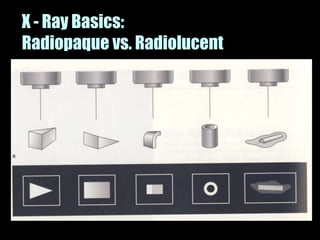
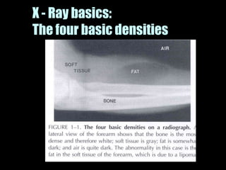
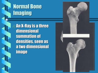
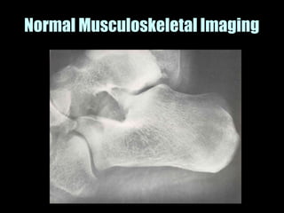
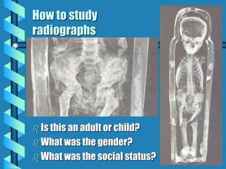
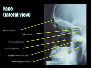
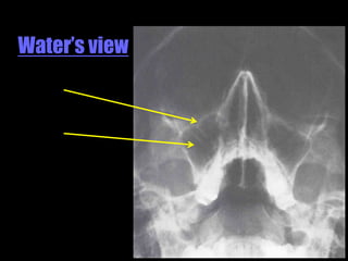
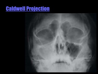
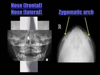
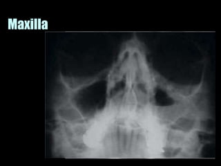
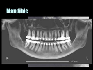
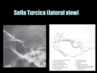
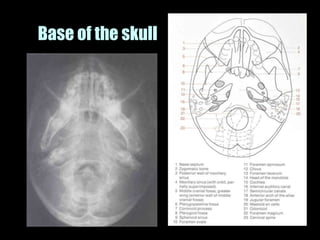
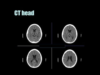
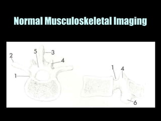
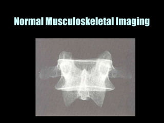
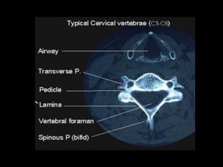
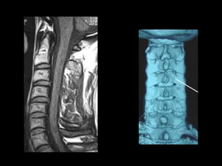
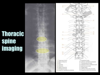
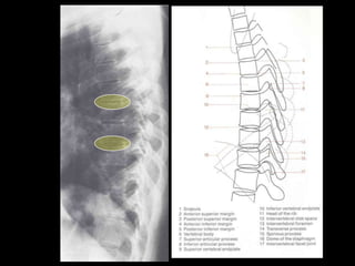
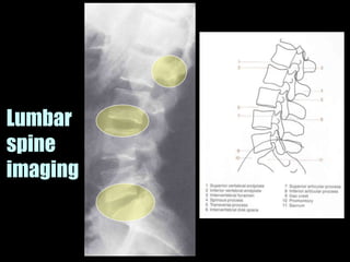
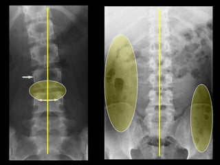
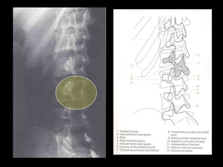
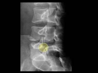
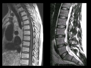
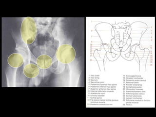
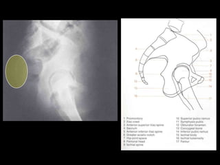
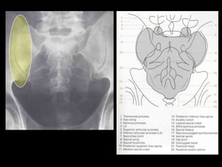
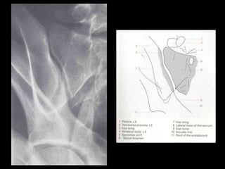
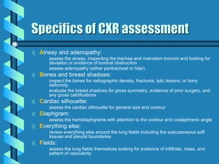
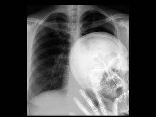
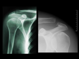
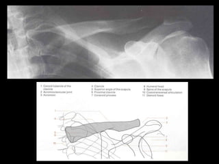
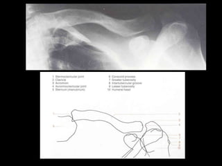
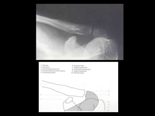
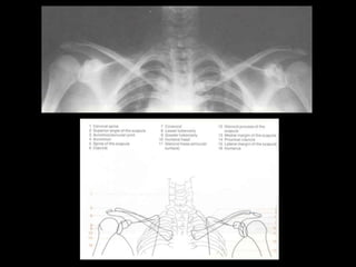
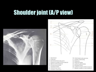
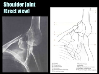
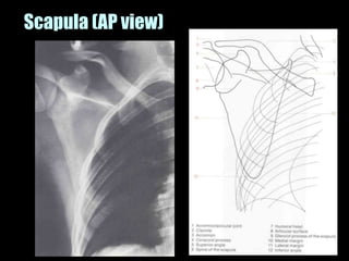
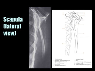
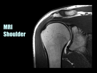
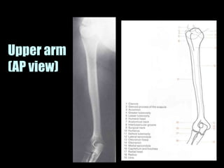
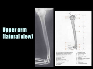
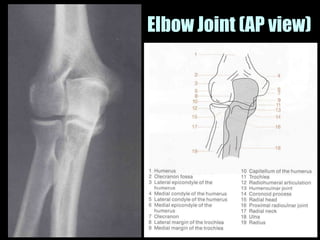
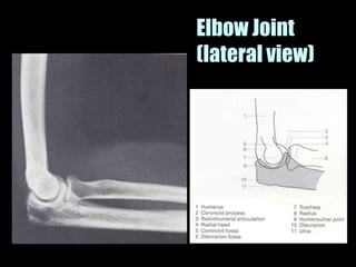
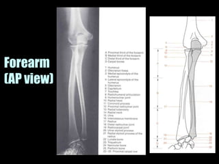
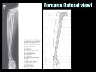
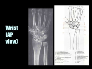
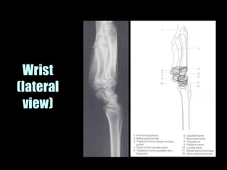
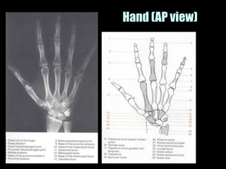

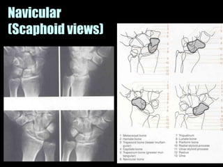
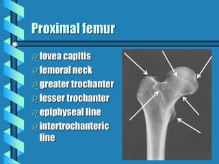
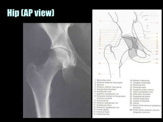
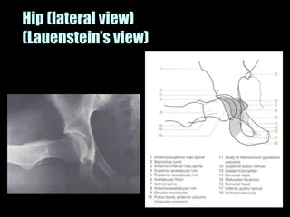
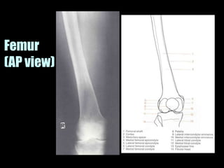
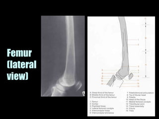
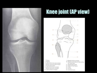
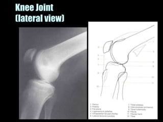
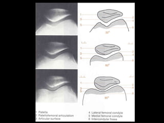
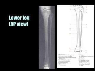
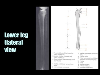
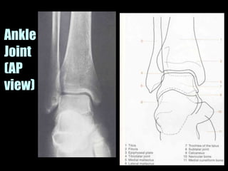
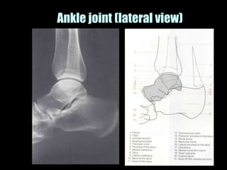
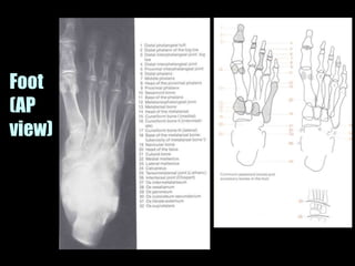
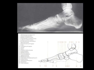
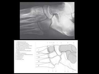
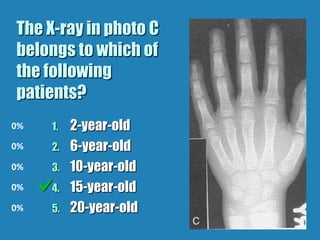
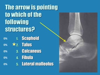


This document provides an overview of normal musculoskeletal imaging. It discusses basic x-ray concepts and densities. It then reviews normal anatomy as seen on x-rays of the skull, spine, pelvis, chest, and extremities. Key anatomical structures are labeled on example x-rays for the shoulder, hip, knee, and foot. Quizzes are included to test recognition of anatomical structures and patient age based on x-rays.


