SlideShare a Scribd company logo
PARANASAL SINUS PATHOLOGIES
Presented by
Dr. Cathrine Diana PG-III
contents
 Paranasal sinuses
 Introduction
 Development
 Anatomy
 Physiology
 Examination and investigations
 Paranasal sinus pathologies
 Classification
 Description
 Complications
 Treatment
INTRODUCTION
 Sinus (Latin) - fold or pocket.
 Paranasal sinuses - four paired,
hollow air filled spaces in various
cranio-facial bones
 Named after the bones in which
they are located
INTRODUCTION Clinically, Divided into two groups:
1. Anterior group: maxillary, frontal, anterior and middle ethmoidal air
cells. They all open in the middle meatus.
2. Posterior group: Posterior ethmoidal and the sphenoid sinus
DEVELOPMENT
 Excavation of bone by air-filled sacs (pneumatic diverticula) from
the nasal cavity.
 Begins prenatally - continues through lifetime.
ANATOMY– MAXILLARYSINUS
 MAXILLARY ANTRECHEA / ANTRUM
OF HIGHMORE
 Largest
 Maxilla - under the eyes, on either
side of the nose.
 Pyramidal - base toward the lateral
wall of nose and apex directed
laterally into the zygomatic process.
 Capacity of 15 ml (average).
FRONTAL SINUS
 Situated deep to the inner and outer table of
frontal bone
 Drain- frontal recess to the middle meatus
 Absent on one (15 %) or both sides (5 %)
 Drainage pathway – naso- frontal duct-
frontal recess - situated at its floor – drains
into middle meatus (62%) or ethmoid
infundibulum (38%).
Ethmoid sinus
Thin-walled air cavities in the lateral masses
of ethmoid bone, between nose and the
eyes.
1. Anterior ethmoidal air cells –3- 11 drain
into either the ethmoidal infundibulum
or the frontonasal duct.
2. Bullar cells (middle ethmoidal air cells)
- usually <3 - open in ethmoidal
infundibulum.
3. Posterior group :- Up to 7 - usually drain
by a single orifice into the superior
meatus.
Agar nasi cells
 They are the most anterior ethmoidal air
cells. .
 Its size influence the patency of the frontal
recess and the anterior middle meatus.
 Haller cells:
 Also called infraorbital ethmoid cells.
 Present in approx. 20 % pateints.
Clinical significance –
 Become infected , with potential extension
into orbit.
 Narrows the maxillary ostium.
Onodi cells
 These are posterior ethmoidal cells
extending into the sphenoid
bone ,either adjacent to or impinging
upon the optic nerve.
 When these Onodi cells abut or
surround the optic nerve, the nerve is
at risk when surgical excision of these
cells is performed.
 It is also a potential cause of
incomplete sphenoidectomy.
SPHENOID SINUS
 Body of sphenoid - behind the nose, in the
center of the skull.
 Rarely symmetrical and separated by a
thin bony septum.
 Ostium of the sphenoid sinus is situated
in the upper part of its anterior wall and
drains into sphenoethmoidal recess.
 Average size – 2 x 2 x 2 cm.
 According to Congdon sphenoid
pneumatization can be as follows
 Conchal – 5 %
 Presellar – 23 %
 Post-sellar – 67%
ANATOMY- LATERAL NASALWALL
 3 projections - superior, middle
and inferior concha.
 Meatus - space below each
concha.
 Inferior meatus: nasolacrimal
duct
 Middle meatus:
 Maxillary sinus
 Frontal sinus
 Anterior ethmoid sinuses
 Superior meatus: posterior
ethmoid sinuses
 Sphenoethmoidal recess:
sphenoid sinus
OSTEOMEATAL COMPLEX
 It is a common channel that links
the frontal sinus, anterior and
middle ethmoid sinuses and the maxillary
sinus to the middle meatus. It is composed
of five structures:
 Maxillary ostium
 Infundibilum
 Ethmoidal bulla
 Uncinate process
 Hiatus semilunaris
PHYSIOLOGY– SINUS EPITHELIUM
 Respiratory epithelium - ciliated pseudostratified columnar epithelium,
goblet cells, and submucosal glands
 Produce a protective mucous blanket - traps bacteria and noxious
materials, which are carried by ciliary motion to the ostium and into the
nose for elimination
 Ciliary movements: 50- 300 cilia/ cell; 8-20
beat/ second.
 For maximum ciliary activity:
Humidity: >85%, Temperature: 18- 40 degree
C, pH: 7- 8.
 The orientation of the cilia within a given
sinus is specific as secretions are propelled
towards the natural sinus ostia and from
there to the nasopharynx and oropharynx
where they are subsequently cleared by
swallowing.
SINUS HEALTH
 Composition of gas content in the maxillary sinus is similar to venous
blood, with high CO2 and lower O2 level compared to breathing air.
 Sinus health depends on:
1. Mucous secretion of normal viscosity, volume, and composition
2. Normal muco-ciliary flow to prevent mucous stasis and subsequent
infection
3. Open sinus ostia to allow adequate drainage and aeration.
 Negative factors:
 Dryness of air, Cigarette, Temperature variations, hypoxia, hypercapnia,
Hypertonic/ hypotonic fluids, Dehydration, pH changes, diseases (like
Cystic fibrosis and Primary ciliary dyskinesia), Drugs (phenylephrine,
adrenaline, lidocaine, atropine, antihistaminic), Infections, Anatomic
obstruction (septal deviation, enlarged or irregular turbinate), Foreign
bodies and Nasal polyps.
PATHOPHYSIOLOGICALSTAGES OF
SINUS DISEASES
Initial phase: - reversible
Ostium obstruction phase
Bacterial phase
Chronic phase
Osteomeatal complex obstruction
↓
Decreased ventilation of the sinuses
↓
Decreased drainage of the sinuses
↓
pO2 decrease, pCO2 increase, mucous stasis
↓
Inflammation and viscous mucous, ciliary movement slowing
↓
Stasis and proteolytic enzymes
↓
Ciliary damage
↓
Anaerobic microorganisms
↓
More damage
FUNCTIONS OF SINUS
1. Reduction of weight of skull
2. Increasing resonance of the voice
3. Providing a buffer against blows to the face.
4. Insulating sensitive structures like dental roots and eyes from rapid
temperature fluctuations in the nasal cavity.
5. Humidifying and heating of inhaled air because of slow air turnover in this
region.
6. Regulation of intranasal and serum gas pressures
7. Increasing surface area for olfaction
8. Contribute to facial growth
EXAMINATION
 History and systemic clinical examination:
 Check general signs of health
 Systemic medical history, history of
allergies, drug use and abuse
 Occupation history
 Examination of and neck for lumps or
swollen lymph nodes
Examination
 Local examination of the nose,
face, and neck:
1. Anterior Rhinoscopy:
Examination of nose with a
nasal speculum to check
for abnormal areas, useful in
evaluation of nasal
obstruction.
2. Posterior rhinoscopy: With a
mouth mirror in the
nasopharynx
Examination - transillumination
 Normal transillumination decreases chance of pus in the sinus.
 No light reflex suggests mucopurulent material or thickening of
nasal mucosa.
 Inexpensive screening tool
Transillumination of Frontal Sinus
Transillumination of Maxillary Sinus
24
Examination - endoscopy
 Endoscopic examination/ Rhinoscopy:
 nasoscope/rhinoscope is a thin, tube-like instrument with a light and
a lens for viewing. A special tool on the nasoscope may be used to
remove samples of tissue. The tissues samples are viewed under a
microscope by a pathologist.
EXAMINATION – PLAIN RADIOGRAPHS
 Plain radiographs:
 to check for Sinus opacifications, Air-fluid level, Mass, Fractures
 Caldwell view: PA view/ “forehead-nose” view to evaluate maxilla, maxillary
and frontal sinus, ethmoid air cells, lamina papyracea
 Water’s view: chin-nose” or “occipito-
mental” view for evaluation of the
paranasal sinuses.
submento-vertical” view to evaluate the
sphenoid, the posterior ethmoids, the
maxillary and frontal sinuses
CT SCANS
 CT scans: Excellent views of the sinuses, best for
osteomeatal complex and ethmoidal disease
 “Limited CT Evaluation” – slice 3-4 mm
 CT navigation:
 A computer is used to identify the 3-
dimensional location of a probe tip placed
within the patient's nose or sinuses..
 Improves anatomical identification and avoid
damage to vital neighbouring structures such
as the brain and eyes.
AXIAL CT
CORONAL CT
SAGITTAL CT
EXAMINATION
 MRI:
 Excellent soft tissue definition
- evaluation of neoplastic
disease.
 MRI (magnetic resonance
imaging) with gadolinium:
Gadolinium is injected into a
vein. The gadolinium collects
around the cancer cells so
they show up brighter in the
picture.
 PET scan (positron emission tomography scan): A small amount
of radioactive glucose is injected into a vein. The PET scanner rotates
around the body and makes a picture of where glucose is being used
in the body. Malignant tumor cells show up brighter in the picture
because they are more active and take up more glucose than normal
cells do.
 Stuckensen and colleagues found a sensitivity of 70%, 84%, and 66%
and a specificity of 82%, 68%, and 74% for PET, ultrasound, and CT
scan in terms of nodal metastasis
9. Histological examination:
 Fine-needle aspiration (FNA) biopsy,
Incisional biopsy and excisional biopsy are
done from pathologic tissues.
10. Culture examination: Correlation of
routine nasal culture and sinus culture is
poor. Endoscopically guided aspiration of
cultures from medial meatus do correlate
with sinus culture.
Silver stained section showing invasive
fungal sinusitis (aspergillus)
Allergic mucin of allergic fungal
sinusitis
Classification
 DEVELOPMENTAL VARIATIONS AND ANOMALIES
 INFLAMMATORY/INFECTIOUS DISEASES
 CYSTS
 TUMORS
 OTHER SURGICALLY RELEVANT CONDITIONS
Developmental variations and anomalies
 Paradoxical curvature of middle turbinate
 Concha bullosa in middle turbinate
 Lateralization and pneumatisation of uncinate process
 Variations of ethmoidal roof anatomy
 Bulla ethmoidalis – torus ethmoidalis and giant bulla
 Others: Agenesis of sphenoid sinus, pneumatisation of greater wings
of sphenoid and crista galli.
DEVELOPMENTALVARIATIONSAND
ANOMALIES
Paradoxical curvature:
Normally the convexity of the middle
turbinate is directed medially toward
the nasal septum.
When the convexity is directed
laterally, it is termed a paradoxical
middle turbinate .
Most authors agree that the
paradoxical middle turbinate can be a
contributing factor to sinusitis.
DEVELOPMENTALVARIATIONS
ANDANOMALIES
Concha bullosa:
When pneumatization involves the
bulbous portion of the middle turbinate
it is termed concha bullosa.
If only the attachment portion of the
middle turbinate is pneumatized, it is
termed lamellar concha .
A concha bullosa may obstruct the
ethmoid infundibulum.
Variations of uncinate process
The uncinate process may be medialized,
lateralized, or pneumatized/bent.
Medialization occurs with giant bulla
ethmoidalis.
Lateralization of the uncinate process
may obstruct the infundibulum.
Pneumatization (uncinate bulla) can
rarely cause obstruction of the
infundibulum.
Variation of the ethmoidal roof anatomy
The ethmoid roof is of critical importance for two
reasons.
 most vulnerable to iatrogenic cerebrospinal
fluid leaks.
 anterior ethmoid artery is vulnerable to injury.
The depth of the olfactory fossa is determined by
the height of the lateral lamella of the
cribriform plate.
In 1962, Keros classified the depth of the olfactory
fossa into three types, that is,
Keros type I: <3 mm
Keros type II: 4-7 mm
Keros type III: 8-16 mm - most vulnerable to
iatrogenic injury.
Variations of sphenoid sinus … Agenesis of sphenoid sinus
 Pneumatisation of other bones
 The crista galli is normally bony.
When aerated, it may communicate with
the frontal recess, causing obstruction of
the ostium and thus lead to chronic
sinusitis and mucocele formation
Bullae ethmoidalis
 The bulla ethmoidalis is a
prominent anterior ethmoid air
cell.
 Failure to pneumatise - torus
ethmoidalis.
 A 'giant bulla' may fill the entire
middle meatus and force its way
between the uncinate process and
the middle turbinate.
INFLAMMATORY/ INFECTIOUS
CONDITIONS
 POLYPS
 SINUSITIS
 GRANULOMATOUS DISEASES
POLYPS
 They are fleshy outgrowths of the nasal
mucosa that form at the site of dependent
edema in the lamina propria of the mucous
membrane, usually around the ostia of the
maxillary sinuses.
 usually start near the ethmoid sinuses and
grow into the open areas.
 Large polyps can block the sinuses or nasal
airway.
 Risk factors: Aspirin sensitivity (wheezing),
Asthma, Acute and Chronic sinus infections,
Cystic fibrosis, Hay fever (allergic rhinitis).
 Clinical features:
 Nasal obstruction and mouth breathing
 Nasal congestion and postnasal drainage
 Anosmia, hyposmia
 Sneezing, rhinorrhea
 Facial pain
 Ocular itching
 Bleeding polyps occur in rhinosporidiosis
 Unilateral polyps occasionally occur in association with or represent
benign or malignant tumors of the nose or paranasal sinuses, or in
response to a foreign body.
 Diagnosis:
 physical examination - A developing polyp is teardrop-shaped; when
mature, it resembles a peeled seedless grape.
 CT scans
 Treatment
1. Steroids –may shrink or eliminate polyps
 Topical corticosteroid spray - mometasone [30
mcg/spray], beclomethasone [42 mcg/spray], flunisolide -
given as 1 or 2 sprays bid in each nasal cavity
 1-wk tapered course of oral corticosteroids.
2. Surgery :
 FESS
 Steroid therapy after surgery - to retard recurrence.
 In severe recurrent cases- maxillary sinusotomy or
ethmoidectomy, usually done endoscopically.
3. Removal of etiology – control of underlying allergy or infection.
SINUSITIS
 Definition: Sinusitis is the
inflammatory condition of the
mucous membrane lining of the
sinuses
 RHINOSINUSITIS is a better term
because:
 Allergic or non-allergic rhinitis
nearly always precedes sinusitis
 Sinusitis without rhinitis is rare
 Nasal discharge and congestion
are prominent symptoms of
sinusitis
 Nasal mucosa and sinus mucosa
are similar and are contiguous
Classifications
ACCORDING TO DURATION:
1. Acute :infection lasting 4 weeks, symptoms resolve completely resolved
in < 30 days.
2. Subacute :infection lasting between 4 to 12 weeks, yet resolves
completely.
3. Recurrent: ≥ 4 discrete acute episodes per year, each completely resolved
in < 30 days but recurring in cycles, with at least 10 days between complete
resolution of symptoms and initiation of a new episode
4. Chronic: symptoms lasting more than 12 weeks.
 ACCORDING TO PATHOGEN:
1. Bacterial: Hospital-acquired acute infections are more often
bacterial, typically involving Staphylococcus aureus, Klebsiella
pneumoniae, Pseudomonas aeruginosa
2. Viral: In immunocompetent patients - in the community is almost
always viral (eg, rhinovirus, influenza, parainfluenza).
antibiotics given for:
 Mild to moderate sinus symptoms persisting for ≥ 10 days
 Severe symptoms (eg, fever ≥ 39°, severe pain) for ≥ 3 to 4 days
 Worsening sinus symptoms after initially improving from a typical
viral URI ("double sickening" or biphasic illness)
3. Fungal: Usually seen in immunocompromised patients because
of poorly controlled diabetes, neutropenia, or HIV infection. It is
clinically of 2 types:
 Non-invasive fungal sinusitis:
 Saprophytic fungal infestation/colonization
 Allergic fungal rhinosinusitis (AFRS)
 Saprophytic fungus balls (mycetoma)
 Invasive fungal sinusitis –
 Chronic invasive fungal sinusitis
 Granulomatous invasive fungal sinusitis
 Acute (fulminant) invasive fungal sinusitis
sinusitis
 According to source:
 Primary
 Secondary
 According to number of sinuses
involved:
 Hemisinusitis –all sinuses on one side
 Polisinusitis – several sinuses, but
not all, are involved
 Pansinusitis
 According to source of infection:
 Rhinogenous
 Odontogenic
 Traumatic
 Hematogenic
 Allergic
Risk factors:
1. Obstruction to drainage: most important
2. Defect of self-cleaning mechanism of the mucous membrane of sinus –
infections causing immobility of the cilia, Increased viscosity of
secretions, Immotile cilia syndrome, Prolonged exposure to cigarette
smoke
3. Medications
 First generation antihistamines (non sedating do not affect)
 Anticholinergics, Aspirin, Anesthetic agents, Benzodiazepines
4. Immunodeficiency: Immunoglobulin deficiency (IgA, IgG), diabetes, HIV
infection
5. Other factors: prolonged ICU stays, severe burns, cystic fibrosis, and
ciliary dyskinesia.
Clinical Features:
1. Nasal congestion and discharge
2. Sore throat and postnasal drip
3. Pain or pressure.
4. Oedema of facial tissues.
5. Bad breath or loss of smell (hyposmia/anosmia).
6. Systemic symptoms: Malaise may be present. Fever and chills suggest an
extension of the infection beyond the sinuses, rise in temperature, bad
appetite, sleep disturbances, changes of the blood (leukocytosis),
Productive cough (especially at night)
7. Complications – ocular, neurological, local
Diagnosis
1. Diagnosis is clinical;
2. CT and cultures - mainly for chronic, refractory, or atypical cases.
3. X-rays of the apices of the teeth
Clinical diagnostic criteria include 2major factors, I major and 2 minor
factors or presence of pus in nasal cavity.
Major Factors Minor Factors
Facial pain/pressure Headache
Facial congestion/fullness Maxillary dental pain
Nasal drainage/discharge Cough
Postnasal drip Halitosis
Nasal obstruction/blockage Fatigue
Hyposmia/anosmia Ear pain/ pressure/ fullness
Fever (acute sinusitis only) Fever
Purulence in nasal cavity on examination
(diagnostic by itself)
TREATMENT OPTIONS
MEASURES TO ENHANCE DRAINAGE:
1. Heat therapy: Steam inhalation; hot, wet towels over the affected
sinuses; and hot beverages
2. Topical vasoconstrictors/ Nasal decongestants:
 Topical nasal sprays (limit use to 3-7 days) - Phenylephrine,
Oxymetazoline, Naphthazoline, Tetrahydrozoline, Zylometazoline.
Phenylephrine(0.25%) spray q 3 h or oxymetazoline q 8 to 12 h, are
effective but should be used for a maximum of 5 days or for a
repeating cycle of 3 days on and 3 days off until the sinusitis is
resolved
 CORTICOSTEROIDS: Corticosteroid nasal sprays can help relieve
symptoms but typically take at least 10 days to be effective
3. Systemic vasoconstrictors: Systemic vasoconstrictors, such
as pseudoephedrine 30 mg po (for adults) q 4 to 6 h, are less effective.
4. Nasal irrigation:
 Commercial buffered sprays, Bulb syringe, waterpik and ceramic
irrigators with lavage tip or disposable enema bucket
 Washes away irritants and moistens the dry nose.
 cumbersome and uncomfortable - better for patients with recurrent
sinusitis.
 ANTIHISTAMINICS: recommended if allergy present. They can be oral or
topical
 HYDRATION
5. Mucoactive drugs:
 main purpose - increase the ability to expectorate sputum and/or
decrease mucus hypersecretion.
1. Expectorants: hypertonic saline, iodine containing compounds,
guaifenesin (glyceryl guaiacolate), ion channel modifiers (tricyclic
nucleotides)
2. Mucoregulators: carbocysteine, anticholinergic drugs,
glucocorticoids, macrolide antibiotics
3. Mucolytics: classic mucolytic (N-Acetyl Cysteine), peptide mucolytic,
non destructive mucolytics
4. Mucokinetics: bronchodialators, ambroxol
a) Nebulization: best form of physiotherapy. Can be done using
compressors or ultrasonic nebulisers. Normal saline solutions
are nebulized which has a hydrating effect on the mucous
lining. Ultrasonic nebulisers can set the rate according to need.
b) Laser therapy: used directly over the sinuses to reduce
inflammation
c) Ultrasound therapy: sound waves are conducted through a
hypoallergenic gel to reduce inflammation and loosen the
accumulated mucous.
d) Short-wave diathermy
e) Rinoflow therapy: new option, basically micronized
endotracheal wash. Used in sinusitis, rhinitis, pharyngitis,
laryngitis and secretory otitis media.
Antibiotics and antifungal drugs:
 Amoxicillin 500 mg tid for 10-14 days - First line
 Beta-lactanase resistance - Amoxicillin/clavulanate, Cefuroxime,
Cefpodoxime, Cefprozil
Surgery
 Indications:
 Sinusitis unresponsive to antibiotic therapy
 Necrotic sinusitis
 Orbital complications (abscess and phlegmon of orbit)
 Intracranial complications (meningitis, brain abscess)
 Rhinogenic sepsis
 Odontogenic sinusitis combined with maxillary osteomyelitis.
 Approaches:
1. Removal of etiology
2. Fess
3. Caldwell-luc procedure
4. Intranasal antrostomy
5. Radical surgeries
Removal of etiology:
 Treatment of affected tooth.
 Caldwell luc approach may be used.
 Intra-nasal antrostomy may be needed.
Caldwell - luc
 Caldwell-Luc is the fenestration of the anterior wall
of the maxillary sinus and the surgical drainage of
this sinus into the nose via an antrostomy.
 a middle meatus antrostomy is being utilized as a
more physiologic antrostomy..
Denkers procedure
FESS:
 Functional endoscopic sinus surgery (FESS) is the mainstay in the surgical
treatment of sinusitis and nasal polyps, including bacterial, fungal,
recurrent acute, and chronic sinus problems.
 Nasal endoscopes through the nostrils to avoid cutting the skin.
 Telescope diameters - 4mm (adult use) and 2.7mm (pediatric use)
 Viewing angles - 0 degrees to 30, 45, 70, 90, and 120 degrees
 Carry: High definition cameras attached to monitors, tiny articulating
instruments - cutting, suction, biopsy, curettage
 All the sinuses can be accessed at least to some degree by means of FESS.
 Extended approaches: Paranasal sinuses are found to a relatively low-
morbidity approach to selected tumors even inside the skull or brain. This
can be divided into approaches to: anterior cranial fossa, mid cranial fossa,
posterior cranial fossa, infratemporal fossa (incl. pterygopalatine fissure),
sella turcica, orbital access, and optic nerve access.
 Complications:
 Proximity of the sinuses to the eyes, optic nerves, brain and internal
carotid arteries
 Serious risks are rare occurrences
Before FESS 2 months after FESS
 Principal element - granuloma formation - a conglomerate of
macrophages, epithelioid cells, and multinucleated giant cells.
1. Infectious:
 spirochetes (syphilis, yaws)
 mycobacteria [tuberculosis, leprosy]
 bacterial [rhinoscleroma]
 fungus [aspergillus]
2. Inflammatory
 Wegener granulomatosis
 sarcoidosis,
 Churg-Strauss syndrome
 cocaine induced midline destructive lesions
Granulomatous diseases
Cysts
 Mucocele and Pyocele
 Retention cyst
 Pseudocyst
 Post-operative maxillary cyst
Mucocele and pyocele
 Mucocele:
 formed when drainage of mucus from one of the paranasal sinuses
becomes blocked by obstruction of its ostium.
 Contents: clear serous fluid, thick mucoid material, or, if hemorrhage has
occurred, thick brown material
 Pyocele/mucopyocele:
 Infected mucocele
 Contents: mixture of mucus and pus from which a causative organism
may or may not be seen on smear or subsequently cultured.
Pathogenesis:
 Blockage of the ostium pressure develops within sinus
 expansion of the sinus space erosion, and displacement of bone.
 Encroachment of the mucocele upon contiguous structure
 Sinus lining – remains normal or becomes attenuated to undergo
metaplasia to low cuboidal or squamous cells.
 Most commonly: frontal and anterior ethmoid sinuses
 Etiology: chronic inflammation, osteomas, fractures, tumors, polyps,
scarring, and congenital abnormalities.
Management
 Decompression by complete removal and curettage
 Marsupialisation via endoscopic approach through the middle meatus
 Prophylactic Nasofrontal duct obstruction
 Retention cyst and Pseudocyst: Together called antral cyst or mucosal
cyst
 Indistinguishable on radiological or clinical examination.
 Quite common.
 Mostly - single cysts, but in a few instances - they may be multiple
and bilateral.
 Clinical features:
 Usually asymptomatic – discovered during radiographic
examination
 Symptoms – similar to chronic sinusitis
 Sometimes an antral cyst may produce a swelling
 Diagnosis :CT based
 Spherical, ovoid or dome-shaped radiopacities that have a
smooth and uniform outline
 narrow or broad base.
 Size from minute to very large
 Usually remain static
 Many regress spontaneously
POST-OPERATIVE MAXILLARYCYST
(SURGICALCILIATEDCYSTOFMAXILLA)
 Delayed complication arising years after surgery of maxillary sinus.
 Causes:
 Caldwell–Luc procedure including a nasal antrostomy
 Gun- shot injuries
 Fractures of the malar–maxillary complex
 Mid-face osteotomies.
 Clinical features:
 Pain, discomfort or swelling in the cheek or face, or intra-orally in the
palate or alveolus.
 Pus discharge.
 Radiographs - well-defined radiolucent area closely related to the
maxillary sinus.
 Treatment: enucleation.
TUMORS
 0.2% of all malignancies
 80% - maxillary sinus.
 Men>> women.
 40 and 70 years.
 Carcinomas >> sarcomas
 Metastases are relatively rare.
 Tumours of the sphenoid and frontal sinuses are extremely rare - no
standard staging system
 Most common - squamous cell carcinoma
WHO HISTOLOGICAL CLASSIFICATION OF NASAL
AND PNS TUMORS
TNM CLASSIFICATION OF CARCINOMAS OF
NOSE AND PARANASAL SINUSES
.
Risk factors:
1. Woodworking (carpentry), Shoemaking, Metal-plating, Flour mill or bakery
work.
2. Human papillomavirus (HPV) infection
3. Male
4. Older than 40 years.
5. Smoking.
Clinical features
 No signs or symptoms in the early stages.
 later:
 Blocked sinuses that do not clear, or sinus pressure.
 Headaches or pain in the sinus areas.
 Rhinitis and epistaxis
 A lump or sore inside the nose that does not heal.
 A lump on the face or roof of the mouth.
 Numbness or tingling in the face.
 Swelling or other trouble with the eyes, such as double vision or the
eyes pointing in different directions.
 Pain in the upper teeth, loose teeth, or dentures that no longer fit
well.
 Pain or pressure in the ear.
Treatment
1. Surgery: For all stages of paranasal sinus and
nasal cavity cancer.
 Transfacial approaches
 1. lateral rhinotomy/ weberfergussen incision
 2. diffenbech extension
 3. Lynch extension with the modification
 External ethmoidectomy/frontoehtmoidectomy
 Bicoronal with Mid face degloving
 Subcranial approach
 Acess osteotomies
 Trans oral approach:
 Caldwell lue
 Denker
 Janson – horgan approach
 Trans nasal:
 Transseptal approach
 Endoscopic approaches
Draw backs of endoscopic
approach
 -Not indicated in case of extensive involvement
 Trans orbital extension
 Scar tissue due to previous surgery
 in case to reduce the bony fracture
 TRANSORAL ROBOTIC SURGERY
 New advances in technology facilitate minimal access
 To avoid large transcervical or face-splitting incisions.
 Transoral robotic surgery allows access to tumors within the posterior oral
cavity and oropharynx via multiple robotic arms and a high-definition/
magnification camera.
 The operator sits at a separate console and via remote control can operate
the various instruments.
 Electrocautery, laser, and standard dissection instruments can be used with
the robot.
 Advantages include surgical access via minimal approaches, resulting in
definitive pathologic assessment while minimizing transection and resection
of critical swallowing musculature
Oral Maxillofacial Surg Clin N Am 24 (2012) 307–316
Trans oral Robotic surgery
 more-precise movements in narrow spaces and the capability to work
around corners.
 This result in preservation of maximum amount of healthy muscular
and neurovascular tissue which helps the patient swallow on their
own sooner and discharged home earlier.
1. Radiation therapy: External radiation therapy, Internal radiation
therapy - depending on the type and stage of the cancer being treated.
 IMRT:
 Intensity-modulated radiation therapy (IMRT) can deliver high doses of
radiation with precision while minimizing damage to surrounding
tissues.
 IMRT can conform to the irregular shape of a tumor, delivering higher
doses directly to the tumor cells and potentially destroying more tumor
cells.
 The technique requires more precise planning due to the sharp dose
falloff gradient between the gross tumor and the surrounding normal
tissue.
 IMRT provides locoregional control (90%) and is well tolerated by
patients.
Oral Maxillofacial Surg Clin N Am 24 (2012) 307–316
Chemotherapy:
 Systemic chemotherapy: When chemotherapy drug is given PO, IV or
IM the drugs enter the bloodstream and can reach cancer cells
throughout the body
 Regional chemotherapy: When chemotherapy is placed directly into
the cerebrospinal fluid, an organ, or a body cavity, the drugs mainly
affect cancer cells in those areas.
 Combination chemotherapy is treatment using more than one
anticancer drug.
 The way the chemotherapy is given depends on the type and stage of
the cancer being treated.
 Intra arterial cisplastin therapy: As a primary therapy, the complete
response rate was 83.3%, The 2-year local control rate was 63.0%, and
the 2-year overall survival rate was 75.5%. The 2-year preservation
rate of the hard palate was 97.1%, that of the eyeball was 97.2%, and
that of visual function was 94.4%. This treatment regimen can
contribute to improving the quality of life of patients without reducing
the curability of the therapy.
Int. J. Oral Maxillofac. Surg. 2015; 44: 697–704.
Other surgically relevant conditions
 Oro-antral communication/fistula
 Antral foreign bodies
 Hypertrophied maxillary sinus
Oro-Antral Fistula (oaf)
 Oro-antral communication: it is an abnormal connection between oral
and Antral cavities.
 Oro-antral fistula: long standing oro-antral communication when left
open, epithelializes to form a patent fistula between the two cavities.
 Types:
 ALVEOLOSINUSAL
 PALATO-SINUSAL
 VESTIBULO-SINUSAL
Oro-Antral Communications
CAUSES:
 Extraction – maxillary posterior teeth
 Cysts, tumors
 Osteomyelitis
 Radiation therapy
 Trauma
 Implant denture
CLINICAL FEATURES:
 Some patients are asymptomatic
 Unpleasant tasting discharge and odor
 Reflux of fluids and foods into the nose from mouth
 Leakage of air
 Difficulty in smoking and blowing air
 Development of chronic sinusitis in infected cases
Diagnosis:
 Valsalva maneauver
 Mirror fog test
 Cotton wisp test
 Nasal regurgitation of fluid
 Radiographs: Sinus floor discontinuity, Sinus opacity, Focal alveolar
atrophy, Associated periodontal disease
Oro-Antral Communications (UL7)
Treatment:
 Immediate treatment: Primary purpose is closure of defect and
prevention of sinusitis through:
 Suturing across the defect with/without periodontal pack, warm
saline rinses, antibiotic and antihistaminic therapy with
decongestants.
 Size < 5mm: non-invasive intervention (spontaneous closure by
blood clot)
 Size > 5mm, and > 48 hours - : surgical closure : small defects –
local flaps; large defects – regional/distant flaps.
 Obturators
 Buccal Advancement Flap most common.
 Described by Rehrmann & made popular by Berger.
 Trapezoidal sliding flap – Moczair
 buccal osteoperiosteal flapOro-Antral Communications
 Palatal flaps
1. Palatal rotational advancement flap most common
2. V-shaped palatal flap (kruger) &
3. Split-thickness palatal flap (ito & hara).Oro-Antral Communications
 Combination flaps
 Inverted periosteal flap
 Tongue flap, temporalis flap
 BFP closure
 PRF membrane coverage
 Autologous bone grafts: press-fit
technique
 Autologous cartilage grafts: auricular
cartilage, auricular cartilage
 Alloplastic materials
 Transplantation of a mature wisdom
tooth (followed by root canal treatment of
the tooth 5 - 6 weeks later)
 Laser bio-stimulation (over 5 days)
Oro-Antral Communication
ANTRAL FOREIGN BODIES:
 Gutta-percha points, tooth roots,
impression materials, dental
burs, bone pieces , implants etc..
Treatment has been direct
explored by Caldwell-Luc
approach, with or without nasal
antrostomy. FESS may be done
for sinusitis.
HYPERTROPHIED MAXILLARY SINUS:
 Not pathology but causes
difficulty in implant supported
rehabilitation of posterior
maxilla, with the risk of
subsequent development of sinus
pathologies. The management
includes direct and indirect sinus
lifting.
Oro-Antral Communications (UL8)
Complications
 Because of the proximity of the paranasal sinuses to the eyes and
brain, complications of sinusitis are divided into
 orbital,
 neurological and
 local complications.
Orbital complications
 highest frequency - in children under 6 years of age.
 Infection usually originates from the ethmoids and occurs through:
(1) direct extension through the orbital wall
(2) retrograde spread through veins between the sinuses and the orbit.
 Lymphatic spread – not significant
Orbital complications
1. Preseptal cellulitis, or periorbital cellulitis
2. Orbital cellulitis and edema
3. Subperiosteal abscess
4. orbital abscess
5. Cavernous sinus thrombosis: direct extension or retrograde
thrombophlebitis (via the ophthalmic vein) of ethmoid or sphenoid
infections.
 restriction of extra ocular mobility, proptosis, chemosis, and visual loss
 cranial neuropathies and signs of meningitis
Neurological complications
 Less frequently than orbital
 Most commonly related to the
frontal or sphenoid sinuses.
 Via - direct spread or retrograde
thrombophlebitis.
A – osteomyelitis
B - periorbital abscess
C – epidural abscess
D – subdural abscess
E – brain abscess
F – meningitis
G - septic thrombosis of superior sagittal
sinus
Local complications
 Osteomyelitis - complication of frontal sinusitis.
 Tender, doughy, erythematous swelling over the forehead.
 Treatment of choice - surgical eradication of the affected bone under
antibiotic coverage.
REFERENCES
1. References:
2. Peterson’s principles of oral and maxillofacial surgery
 DISEASES of the SINUSES Diagnosis and Management- DAVIDW. KENNEDY, MD, FACS
1. WHO classification of head and neck tumors
2. PL Dhingra - Disease of ear, nose & throat 4th edition
3. Rosai and Ackerman’s Surgical Pathology (9th edition)
4. David L. Daniels et.al. The Frontal Sinus Drainage Pathway and Related Structures, AJNR: 24,
August 2003.
5. Interactive Atlas. http://uwmsk.org/sinusanatomy2/axial/axial.html
6. (Adapted from Chow AW, Benninger MS, Brook I, et al: IDSA clinical practice guideline for acute
bacterial rhinosinusitis in children and adults. Clinical Infectious Diseases 54 (8):1041–5 (2012).)
7. Thompson and Patterson: Fungal disease of the nose and paranasal sinuses:J Allergy Clin
Immunol 2012;129:321-6.
8. Multiplanar Sinus CT: A Systematic Approach to Imaging Before Functional Endoscopic Sinus
Surgery Jenny K. Hoang, James D. Eastwood, Christopher L. Tebbit, and Christine M. Glastonbury
American Journal of Roentgenology 2010 194:6, W527-W536
 Raef S. Ahmed, Roger Ove, Jun Duan, Richard Popple, Glenn B. Cobb Intensity-modulated
radiotherapy (IMRT) for carcinoma of the maxillary sinus: A comparison of IMRT planning systems
Medical Dosimetry, Volume 31, Issue 3, Autumn 2006, Pages 224-232
 Management of rhinomaxillary mucormycosis with Posaconazole in immunocompetent patients
Sachin Rai *, Shikha Yadav, Dinesh

 Kumar, Vijay Kumar, Vidya Rattan Journal of Oral Biology and Craniofacial Research xxx (2016) xxx
 Modified transnanal endoscopic maxillectomy: a novel surgery style of maxillary malignant tumor
Yonghua Bi1,2, Shuangba He1, Tao Guo1, Jingwu Sun Int J Clin Exp Med 2016;9(6):11361-11366
 Modified double-layered flap technique for closure of anoroantral fistula: Surgical procedure and
case reportAlberto Merlinia, Joseph Garibaldia, Matteo Piazzaia, Luca Giorgisb, British Journal of
Oral and Maxillofacial Surgery 54 (2016) 959–961
 Repair of Oroantral Communication by Use of a Combined Surgical Approach Functional
Endoscopic Surgery and Buccal Advancement Flap/Buccal Fat Pad Graft Timothy Adams, DDS,*
Daniel Taub, DDS, MD,y and Marc Rosen, MD. J Oral Maxillofac Surg 73:1452-1456, 2015


 An Update on Squamous Carcinoma of the Oral Cavity,Oropharynx, and Maxillary SinusJoshua E.
Lubek, DDS, MDa,*, Lewis Clayman, DMD, MD Oral Maxillofacial Surg Clin N Am 24 (2012) 307–316
Thank you

More Related Content

What's hot

Smr and septoplasty
Smr and septoplastySmr and septoplasty
Smr and septoplasty
humra shamim
 
Fess
FessFess
Cortical mastoidectomy
Cortical mastoidectomy Cortical mastoidectomy
Cortical mastoidectomy
Mamoon Ameen
 
Diseases of external ear
Diseases of external earDiseases of external ear
Diseases of external ear
Balasubramanian Thiagarajan
 
Rhinomanometry
RhinomanometryRhinomanometry
RhinomanometrySupreet Sn
 
Mucosal folds of the middle ear
Mucosal folds of the middle earMucosal folds of the middle ear
Mucosal folds of the middle ear
Dʀ Smruti Ranjan Samal
 
Functional endoscopic sinus surgery
Functional endoscopic sinus surgeryFunctional endoscopic sinus surgery
Functional endoscopic sinus surgery
Dʀ Smruti Ranjan Samal
 
External ear,tympanic membrane and auditory tube Dr.N.Mugunthan.M.S.,
External ear,tympanic membrane and auditory tube  Dr.N.Mugunthan.M.S.,External ear,tympanic membrane and auditory tube  Dr.N.Mugunthan.M.S.,
External ear,tympanic membrane and auditory tube Dr.N.Mugunthan.M.S.,
mgmcri1234
 
Hypopharynx anatomy
Hypopharynx anatomyHypopharynx anatomy
Hypopharynx anatomy
Mohammed Nishad N
 
Canal wall up Mastoidectomy ( Intact Bridge Mastoidectomy) by Dr.Aditya Tiwari
Canal wall up Mastoidectomy ( Intact Bridge Mastoidectomy) by Dr.Aditya TiwariCanal wall up Mastoidectomy ( Intact Bridge Mastoidectomy) by Dr.Aditya Tiwari
Canal wall up Mastoidectomy ( Intact Bridge Mastoidectomy) by Dr.Aditya Tiwari
Aditya Tiwari
 
Mastoidectomy (by drdhiru456)
Mastoidectomy (by drdhiru456)Mastoidectomy (by drdhiru456)
Mastoidectomy (by drdhiru456)
Dr Dhirendra Patil
 
Nasal septum & septoplasty
Nasal  septum & septoplastyNasal  septum & septoplasty
Nasal septum & septoplasty
Dr Soumya Singh
 
Tympanoplasty; Indications, types, anesthesia, surgical procedure.
Tympanoplasty; Indications, types, anesthesia, surgical procedure.Tympanoplasty; Indications, types, anesthesia, surgical procedure.
Tympanoplasty; Indications, types, anesthesia, surgical procedure.
Prasanna Datta
 
Eustachian tube, anatomy, test and disorders, dr.vijaya sundarm, 20.03.17
Eustachian tube, anatomy, test and disorders, dr.vijaya sundarm,   20.03.17Eustachian tube, anatomy, test and disorders, dr.vijaya sundarm,   20.03.17
Eustachian tube, anatomy, test and disorders, dr.vijaya sundarm, 20.03.17
ophthalmgmcri
 
Complications of csom
Complications of csomComplications of csom
Complications of csom
Ajay Manickam
 
Radiology in ENT
Radiology in ENTRadiology in ENT
Radiology in ENT
Anwaaar
 
surgical approaches to frontal sinus ppt
surgical approaches to frontal sinus pptsurgical approaches to frontal sinus ppt
surgical approaches to frontal sinus ppt
Vaibhav Lahane
 
Examination of nose and pns
Examination of nose and pnsExamination of nose and pns
Examination of nose and pns
Manpreet Nanda
 
Cholesteatoma
CholesteatomaCholesteatoma
Embryology & anatomy of external ear
Embryology &  anatomy of external earEmbryology &  anatomy of external ear
Embryology & anatomy of external ear
Dr. Pruthvi Raj S
 

What's hot (20)

Smr and septoplasty
Smr and septoplastySmr and septoplasty
Smr and septoplasty
 
Fess
FessFess
Fess
 
Cortical mastoidectomy
Cortical mastoidectomy Cortical mastoidectomy
Cortical mastoidectomy
 
Diseases of external ear
Diseases of external earDiseases of external ear
Diseases of external ear
 
Rhinomanometry
RhinomanometryRhinomanometry
Rhinomanometry
 
Mucosal folds of the middle ear
Mucosal folds of the middle earMucosal folds of the middle ear
Mucosal folds of the middle ear
 
Functional endoscopic sinus surgery
Functional endoscopic sinus surgeryFunctional endoscopic sinus surgery
Functional endoscopic sinus surgery
 
External ear,tympanic membrane and auditory tube Dr.N.Mugunthan.M.S.,
External ear,tympanic membrane and auditory tube  Dr.N.Mugunthan.M.S.,External ear,tympanic membrane and auditory tube  Dr.N.Mugunthan.M.S.,
External ear,tympanic membrane and auditory tube Dr.N.Mugunthan.M.S.,
 
Hypopharynx anatomy
Hypopharynx anatomyHypopharynx anatomy
Hypopharynx anatomy
 
Canal wall up Mastoidectomy ( Intact Bridge Mastoidectomy) by Dr.Aditya Tiwari
Canal wall up Mastoidectomy ( Intact Bridge Mastoidectomy) by Dr.Aditya TiwariCanal wall up Mastoidectomy ( Intact Bridge Mastoidectomy) by Dr.Aditya Tiwari
Canal wall up Mastoidectomy ( Intact Bridge Mastoidectomy) by Dr.Aditya Tiwari
 
Mastoidectomy (by drdhiru456)
Mastoidectomy (by drdhiru456)Mastoidectomy (by drdhiru456)
Mastoidectomy (by drdhiru456)
 
Nasal septum & septoplasty
Nasal  septum & septoplastyNasal  septum & septoplasty
Nasal septum & septoplasty
 
Tympanoplasty; Indications, types, anesthesia, surgical procedure.
Tympanoplasty; Indications, types, anesthesia, surgical procedure.Tympanoplasty; Indications, types, anesthesia, surgical procedure.
Tympanoplasty; Indications, types, anesthesia, surgical procedure.
 
Eustachian tube, anatomy, test and disorders, dr.vijaya sundarm, 20.03.17
Eustachian tube, anatomy, test and disorders, dr.vijaya sundarm,   20.03.17Eustachian tube, anatomy, test and disorders, dr.vijaya sundarm,   20.03.17
Eustachian tube, anatomy, test and disorders, dr.vijaya sundarm, 20.03.17
 
Complications of csom
Complications of csomComplications of csom
Complications of csom
 
Radiology in ENT
Radiology in ENTRadiology in ENT
Radiology in ENT
 
surgical approaches to frontal sinus ppt
surgical approaches to frontal sinus pptsurgical approaches to frontal sinus ppt
surgical approaches to frontal sinus ppt
 
Examination of nose and pns
Examination of nose and pnsExamination of nose and pns
Examination of nose and pns
 
Cholesteatoma
CholesteatomaCholesteatoma
Cholesteatoma
 
Embryology & anatomy of external ear
Embryology &  anatomy of external earEmbryology &  anatomy of external ear
Embryology & anatomy of external ear
 

Viewers also liked

Growth and development /certified fixed orthodontic courses by Indian dental...
Growth and development  /certified fixed orthodontic courses by Indian dental...Growth and development  /certified fixed orthodontic courses by Indian dental...
Growth and development /certified fixed orthodontic courses by Indian dental...
Indian dental academy
 
Pre natal and post-natal development of maxilla part 2/certified fixed orthod...
Pre natal and post-natal development of maxilla part 2/certified fixed orthod...Pre natal and post-natal development of maxilla part 2/certified fixed orthod...
Pre natal and post-natal development of maxilla part 2/certified fixed orthod...
Indian dental academy
 
PRENATAL AND POST NATAL GROWTH AND DEVELOPMENT OF NASOMAXILLARY COMPLEX
PRENATAL AND POST NATAL GROWTH AND DEVELOPMENT OF NASOMAXILLARY COMPLEXPRENATAL AND POST NATAL GROWTH AND DEVELOPMENT OF NASOMAXILLARY COMPLEX
PRENATAL AND POST NATAL GROWTH AND DEVELOPMENT OF NASOMAXILLARY COMPLEX
B NITIN KUMAR
 
Growth and development of maxilla and mandible/endodontic courses
Growth and development of maxilla and mandible/endodontic coursesGrowth and development of maxilla and mandible/endodontic courses
Growth and development of maxilla and mandible/endodontic courses
Indian dental academy
 
Embryology nose and paranasal sinuses
Embryology nose and paranasal sinusesEmbryology nose and paranasal sinuses
Embryology nose and paranasal sinuses
Balasubramanian Thiagarajan
 
Prenatal and postnatal growth & development of maxilla and palate presented b...
Prenatal and postnatal growth & development of maxilla and palate presented b...Prenatal and postnatal growth & development of maxilla and palate presented b...
Prenatal and postnatal growth & development of maxilla and palate presented b...
Dr. Himanshu Gorawat
 
G&amp;d maxilla
G&amp;d maxillaG&amp;d maxilla
G&amp;d maxilla
Indian dental academy
 
Development of maxilla and mandible/prosthodontic courses
Development of maxilla and mandible/prosthodontic coursesDevelopment of maxilla and mandible/prosthodontic courses
Development of maxilla and mandible/prosthodontic courses
Indian dental academy
 
pre natal &; post-natal growth of maxilla & palate
 pre natal &; post-natal growth of maxilla & palate  pre natal &; post-natal growth of maxilla & palate
pre natal &; post-natal growth of maxilla & palate
mahesh kumar
 
Growth and development of cranium and maxilla..
Growth and development of cranium and maxilla..Growth and development of cranium and maxilla..
Growth and development of cranium and maxilla..
Indian dental academy
 
Growth and development of maxilla and maxillary sinus/ dental courses
Growth and development of maxilla and maxillary sinus/ dental coursesGrowth and development of maxilla and maxillary sinus/ dental courses
Growth and development of maxilla and maxillary sinus/ dental courses
Indian dental academy
 
Growth and development of cranium
Growth and development of craniumGrowth and development of cranium
Growth and development of cranium
Abhinav Mudaliar
 
Growth and development nasomaxillary complex/ dental implant courses
Growth and development nasomaxillary complex/ dental implant coursesGrowth and development nasomaxillary complex/ dental implant courses
Growth and development nasomaxillary complex/ dental implant courses
Indian dental academy
 
Growth & development of maxilla and mandible
Growth & development of maxilla and mandibleGrowth & development of maxilla and mandible
Growth & development of maxilla and mandibleRajesh Bariker
 

Viewers also liked (14)

Growth and development /certified fixed orthodontic courses by Indian dental...
Growth and development  /certified fixed orthodontic courses by Indian dental...Growth and development  /certified fixed orthodontic courses by Indian dental...
Growth and development /certified fixed orthodontic courses by Indian dental...
 
Pre natal and post-natal development of maxilla part 2/certified fixed orthod...
Pre natal and post-natal development of maxilla part 2/certified fixed orthod...Pre natal and post-natal development of maxilla part 2/certified fixed orthod...
Pre natal and post-natal development of maxilla part 2/certified fixed orthod...
 
PRENATAL AND POST NATAL GROWTH AND DEVELOPMENT OF NASOMAXILLARY COMPLEX
PRENATAL AND POST NATAL GROWTH AND DEVELOPMENT OF NASOMAXILLARY COMPLEXPRENATAL AND POST NATAL GROWTH AND DEVELOPMENT OF NASOMAXILLARY COMPLEX
PRENATAL AND POST NATAL GROWTH AND DEVELOPMENT OF NASOMAXILLARY COMPLEX
 
Growth and development of maxilla and mandible/endodontic courses
Growth and development of maxilla and mandible/endodontic coursesGrowth and development of maxilla and mandible/endodontic courses
Growth and development of maxilla and mandible/endodontic courses
 
Embryology nose and paranasal sinuses
Embryology nose and paranasal sinusesEmbryology nose and paranasal sinuses
Embryology nose and paranasal sinuses
 
Prenatal and postnatal growth & development of maxilla and palate presented b...
Prenatal and postnatal growth & development of maxilla and palate presented b...Prenatal and postnatal growth & development of maxilla and palate presented b...
Prenatal and postnatal growth & development of maxilla and palate presented b...
 
G&amp;d maxilla
G&amp;d maxillaG&amp;d maxilla
G&amp;d maxilla
 
Development of maxilla and mandible/prosthodontic courses
Development of maxilla and mandible/prosthodontic coursesDevelopment of maxilla and mandible/prosthodontic courses
Development of maxilla and mandible/prosthodontic courses
 
pre natal &; post-natal growth of maxilla & palate
 pre natal &; post-natal growth of maxilla & palate  pre natal &; post-natal growth of maxilla & palate
pre natal &; post-natal growth of maxilla & palate
 
Growth and development of cranium and maxilla..
Growth and development of cranium and maxilla..Growth and development of cranium and maxilla..
Growth and development of cranium and maxilla..
 
Growth and development of maxilla and maxillary sinus/ dental courses
Growth and development of maxilla and maxillary sinus/ dental coursesGrowth and development of maxilla and maxillary sinus/ dental courses
Growth and development of maxilla and maxillary sinus/ dental courses
 
Growth and development of cranium
Growth and development of craniumGrowth and development of cranium
Growth and development of cranium
 
Growth and development nasomaxillary complex/ dental implant courses
Growth and development nasomaxillary complex/ dental implant coursesGrowth and development nasomaxillary complex/ dental implant courses
Growth and development nasomaxillary complex/ dental implant courses
 
Growth & development of maxilla and mandible
Growth & development of maxilla and mandibleGrowth & development of maxilla and mandible
Growth & development of maxilla and mandible
 

Similar to Paranasalsinuses

Bharat pns1
Bharat pns1Bharat pns1
Bharat pns1
Bharat Jain
 
Endoscopic anatomy of nose ,paranasal sinus and anterior skull base
Endoscopic anatomy of nose ,paranasal sinus and anterior skull baseEndoscopic anatomy of nose ,paranasal sinus and anterior skull base
Endoscopic anatomy of nose ,paranasal sinus and anterior skull base
Rajat Jain
 
ADENOIDS&ADENOIDECTOMY BY ROOHIA
ADENOIDS&ADENOIDECTOMY BY ROOHIAADENOIDS&ADENOIDECTOMY BY ROOHIA
ADENOIDS&ADENOIDECTOMY BY ROOHIAMd Roohia
 
JUVENILE NASOPHARYNGEAL ANGIOFIBROMA Surgical approaches & Newer treatment o...
JUVENILE NASOPHARYNGEAL ANGIOFIBROMA  Surgical approaches & Newer treatment o...JUVENILE NASOPHARYNGEAL ANGIOFIBROMA  Surgical approaches & Newer treatment o...
JUVENILE NASOPHARYNGEAL ANGIOFIBROMA Surgical approaches & Newer treatment o...
Dr Utkal Mishra
 
JUVENILE NASOPHARYNGEAL ANGIOFIBROMA Surgical approaches & newer treatment o...
JUVENILE NASOPHARYNGEAL ANGIOFIBROMA  Surgical approaches & newer treatment o...JUVENILE NASOPHARYNGEAL ANGIOFIBROMA  Surgical approaches & newer treatment o...
JUVENILE NASOPHARYNGEAL ANGIOFIBROMA Surgical approaches & newer treatment o...
Utkal Mishra
 
Pre management salivary gland by dr pallavi jain
Pre management salivary gland by dr pallavi jainPre management salivary gland by dr pallavi jain
Pre management salivary gland by dr pallavi jain
Dr. Pallavi Jain
 
Allergic Rhinitis
Allergic RhinitisAllergic Rhinitis
Allergic Rhinitis
mohammed sediq
 
Maxillary Sinus
Maxillary SinusMaxillary Sinus
Maxillary Sinus
KirtiGupta126
 
A Case Report on Benign Sinonasal Paraganglioma
A Case Report on Benign Sinonasal ParagangliomaA Case Report on Benign Sinonasal Paraganglioma
A Case Report on Benign Sinonasal Paraganglioma
Sachender Tanwar
 
Benign Sinonasal Paraganglioma
Benign Sinonasal ParagangliomaBenign Sinonasal Paraganglioma
Benign Sinonasal Paraganglioma
Aakanksha Rathor
 
Maxillary sinus presentation
Maxillary sinus presentationMaxillary sinus presentation
Maxillary sinus presentation
siddharth verma
 
Laryngocele
LaryngoceleLaryngocele
Laryngocele
Dr.Juveria Majeed
 
Nasal polyps
Nasal polypsNasal polyps
Nasal polyps
Mohammad Ihmeidan
 
Endoscopic nasal anatomy
Endoscopic nasal anatomyEndoscopic nasal anatomy
Endoscopic nasal anatomy
Kode Sashanka
 
Inverted papilloma of nose
Inverted papilloma of noseInverted papilloma of nose
Inverted papilloma of nose
shaamikhalid
 
01 ENT endoscopy En for students.pdf
01 ENT endoscopy En for students.pdf01 ENT endoscopy En for students.pdf
01 ENT endoscopy En for students.pdf
IshikaKakani
 
Cross Sectional Anatomy of Paranasal sinus
Cross Sectional Anatomy of Paranasal sinus Cross Sectional Anatomy of Paranasal sinus
Cross Sectional Anatomy of Paranasal sinus
Sarbesh Tiwari
 

Similar to Paranasalsinuses (20)

Bharat pns1
Bharat pns1Bharat pns1
Bharat pns1
 
Endoscopic anatomy of nose ,paranasal sinus and anterior skull base
Endoscopic anatomy of nose ,paranasal sinus and anterior skull baseEndoscopic anatomy of nose ,paranasal sinus and anterior skull base
Endoscopic anatomy of nose ,paranasal sinus and anterior skull base
 
ADENOIDS&ADENOIDECTOMY BY ROOHIA
ADENOIDS&ADENOIDECTOMY BY ROOHIAADENOIDS&ADENOIDECTOMY BY ROOHIA
ADENOIDS&ADENOIDECTOMY BY ROOHIA
 
Nasal endoscopy
Nasal endoscopyNasal endoscopy
Nasal endoscopy
 
JUVENILE NASOPHARYNGEAL ANGIOFIBROMA Surgical approaches & Newer treatment o...
JUVENILE NASOPHARYNGEAL ANGIOFIBROMA  Surgical approaches & Newer treatment o...JUVENILE NASOPHARYNGEAL ANGIOFIBROMA  Surgical approaches & Newer treatment o...
JUVENILE NASOPHARYNGEAL ANGIOFIBROMA Surgical approaches & Newer treatment o...
 
JUVENILE NASOPHARYNGEAL ANGIOFIBROMA Surgical approaches & newer treatment o...
JUVENILE NASOPHARYNGEAL ANGIOFIBROMA  Surgical approaches & newer treatment o...JUVENILE NASOPHARYNGEAL ANGIOFIBROMA  Surgical approaches & newer treatment o...
JUVENILE NASOPHARYNGEAL ANGIOFIBROMA Surgical approaches & newer treatment o...
 
Pre management salivary gland by dr pallavi jain
Pre management salivary gland by dr pallavi jainPre management salivary gland by dr pallavi jain
Pre management salivary gland by dr pallavi jain
 
Allergic Rhinitis
Allergic RhinitisAllergic Rhinitis
Allergic Rhinitis
 
Maxillary Sinus
Maxillary SinusMaxillary Sinus
Maxillary Sinus
 
A Case Report on Benign Sinonasal Paraganglioma
A Case Report on Benign Sinonasal ParagangliomaA Case Report on Benign Sinonasal Paraganglioma
A Case Report on Benign Sinonasal Paraganglioma
 
Ijsron1201396
Ijsron1201396Ijsron1201396
Ijsron1201396
 
Benign Sinonasal Paraganglioma
Benign Sinonasal ParagangliomaBenign Sinonasal Paraganglioma
Benign Sinonasal Paraganglioma
 
Maxillary sinus presentation
Maxillary sinus presentationMaxillary sinus presentation
Maxillary sinus presentation
 
Laryngocele
LaryngoceleLaryngocele
Laryngocele
 
Nasal polyps
Nasal polypsNasal polyps
Nasal polyps
 
Endoscopic nasal anatomy
Endoscopic nasal anatomyEndoscopic nasal anatomy
Endoscopic nasal anatomy
 
Inverted papilloma of nose
Inverted papilloma of noseInverted papilloma of nose
Inverted papilloma of nose
 
nasal allergy
nasal allergynasal allergy
nasal allergy
 
01 ENT endoscopy En for students.pdf
01 ENT endoscopy En for students.pdf01 ENT endoscopy En for students.pdf
01 ENT endoscopy En for students.pdf
 
Cross Sectional Anatomy of Paranasal sinus
Cross Sectional Anatomy of Paranasal sinus Cross Sectional Anatomy of Paranasal sinus
Cross Sectional Anatomy of Paranasal sinus
 

More from Cathrine Diana

Osteomyelitis in maxillofacial region
Osteomyelitis  in maxillofacial regionOsteomyelitis  in maxillofacial region
Osteomyelitis in maxillofacial region
Cathrine Diana
 
Access osteotomy
Access osteotomyAccess osteotomy
Access osteotomy
Cathrine Diana
 
Cleft lip
Cleft lipCleft lip
Cleft lip
Cathrine Diana
 
Orthognathic surgery
Orthognathic surgeryOrthognathic surgery
Orthognathic surgery
Cathrine Diana
 
Implant seminar
Implant seminarImplant seminar
Implant seminar
Cathrine Diana
 
Gun shot wounds
Gun shot woundsGun shot wounds
Gun shot wounds
Cathrine Diana
 
Nerve injury
Nerve injuryNerve injury
Nerve injury
Cathrine Diana
 
Geriatric trauma
Geriatric traumaGeriatric trauma
Geriatric trauma
Cathrine Diana
 

More from Cathrine Diana (8)

Osteomyelitis in maxillofacial region
Osteomyelitis  in maxillofacial regionOsteomyelitis  in maxillofacial region
Osteomyelitis in maxillofacial region
 
Access osteotomy
Access osteotomyAccess osteotomy
Access osteotomy
 
Cleft lip
Cleft lipCleft lip
Cleft lip
 
Orthognathic surgery
Orthognathic surgeryOrthognathic surgery
Orthognathic surgery
 
Implant seminar
Implant seminarImplant seminar
Implant seminar
 
Gun shot wounds
Gun shot woundsGun shot wounds
Gun shot wounds
 
Nerve injury
Nerve injuryNerve injury
Nerve injury
 
Geriatric trauma
Geriatric traumaGeriatric trauma
Geriatric trauma
 

Recently uploaded

Prix Galien International 2024 Forum Program
Prix Galien International 2024 Forum ProgramPrix Galien International 2024 Forum Program
Prix Galien International 2024 Forum Program
Levi Shapiro
 
TEST BANK for Operations Management, 14th Edition by William J. Stevenson, Ve...
TEST BANK for Operations Management, 14th Edition by William J. Stevenson, Ve...TEST BANK for Operations Management, 14th Edition by William J. Stevenson, Ve...
TEST BANK for Operations Management, 14th Edition by William J. Stevenson, Ve...
kevinkariuki227
 
Knee anatomy and clinical tests 2024.pdf
Knee anatomy and clinical tests 2024.pdfKnee anatomy and clinical tests 2024.pdf
Knee anatomy and clinical tests 2024.pdf
vimalpl1234
 
ARTHROLOGY PPT NCISM SYLLABUS AYURVEDA STUDENTS
ARTHROLOGY PPT NCISM SYLLABUS AYURVEDA STUDENTSARTHROLOGY PPT NCISM SYLLABUS AYURVEDA STUDENTS
ARTHROLOGY PPT NCISM SYLLABUS AYURVEDA STUDENTS
Dr. Vinay Pareek
 
Flu Vaccine Alert in Bangalore Karnataka
Flu Vaccine Alert in Bangalore KarnatakaFlu Vaccine Alert in Bangalore Karnataka
Flu Vaccine Alert in Bangalore Karnataka
addon Scans
 
Hemodialysis: Chapter 3, Dialysis Water Unit - Dr.Gawad
Hemodialysis: Chapter 3, Dialysis Water Unit - Dr.GawadHemodialysis: Chapter 3, Dialysis Water Unit - Dr.Gawad
Hemodialysis: Chapter 3, Dialysis Water Unit - Dr.Gawad
NephroTube - Dr.Gawad
 
New Directions in Targeted Therapeutic Approaches for Older Adults With Mantl...
New Directions in Targeted Therapeutic Approaches for Older Adults With Mantl...New Directions in Targeted Therapeutic Approaches for Older Adults With Mantl...
New Directions in Targeted Therapeutic Approaches for Older Adults With Mantl...
i3 Health
 
Surat @ℂall @Girls ꧁❤8527049040❤꧂@ℂall @Girls Service Vip Top Model Safe
Surat @ℂall @Girls ꧁❤8527049040❤꧂@ℂall @Girls Service Vip Top Model SafeSurat @ℂall @Girls ꧁❤8527049040❤꧂@ℂall @Girls Service Vip Top Model Safe
Surat @ℂall @Girls ꧁❤8527049040❤꧂@ℂall @Girls Service Vip Top Model Safe
Savita Shen $i11
 
KDIGO 2024 guidelines for diabetologists
KDIGO 2024 guidelines for diabetologistsKDIGO 2024 guidelines for diabetologists
KDIGO 2024 guidelines for diabetologists
د.محمود نجيب
 
New Drug Discovery and Development .....
New Drug Discovery and Development .....New Drug Discovery and Development .....
New Drug Discovery and Development .....
NEHA GUPTA
 
Triangles of Neck and Clinical Correlation by Dr. RIG.pptx
Triangles of Neck and Clinical Correlation by Dr. RIG.pptxTriangles of Neck and Clinical Correlation by Dr. RIG.pptx
Triangles of Neck and Clinical Correlation by Dr. RIG.pptx
Dr. Rabia Inam Gandapore
 
Alcohol_Dr. Jeenal Mistry MD Pharmacology.pdf
Alcohol_Dr. Jeenal Mistry MD Pharmacology.pdfAlcohol_Dr. Jeenal Mistry MD Pharmacology.pdf
Alcohol_Dr. Jeenal Mistry MD Pharmacology.pdf
Dr Jeenal Mistry
 
Novas diretrizes da OMS para os cuidados perinatais de mais qualidade
Novas diretrizes da OMS para os cuidados perinatais de mais qualidadeNovas diretrizes da OMS para os cuidados perinatais de mais qualidade
Novas diretrizes da OMS para os cuidados perinatais de mais qualidade
Prof. Marcus Renato de Carvalho
 
ARTIFICIAL INTELLIGENCE IN HEALTHCARE.pdf
ARTIFICIAL INTELLIGENCE IN  HEALTHCARE.pdfARTIFICIAL INTELLIGENCE IN  HEALTHCARE.pdf
ARTIFICIAL INTELLIGENCE IN HEALTHCARE.pdf
Anujkumaranit
 
Charaka Samhita Sutra sthana Chapter 15 Upakalpaniyaadhyaya
Charaka Samhita Sutra sthana Chapter 15 UpakalpaniyaadhyayaCharaka Samhita Sutra sthana Chapter 15 Upakalpaniyaadhyaya
Charaka Samhita Sutra sthana Chapter 15 Upakalpaniyaadhyaya
Dr KHALID B.M
 
Report Back from SGO 2024: What’s the Latest in Cervical Cancer?
Report Back from SGO 2024: What’s the Latest in Cervical Cancer?Report Back from SGO 2024: What’s the Latest in Cervical Cancer?
Report Back from SGO 2024: What’s the Latest in Cervical Cancer?
bkling
 
heat stroke and heat exhaustion in children
heat stroke and heat exhaustion in childrenheat stroke and heat exhaustion in children
heat stroke and heat exhaustion in children
SumeraAhmad5
 
ANATOMY AND PHYSIOLOGY OF URINARY SYSTEM.pptx
ANATOMY AND PHYSIOLOGY OF URINARY SYSTEM.pptxANATOMY AND PHYSIOLOGY OF URINARY SYSTEM.pptx
ANATOMY AND PHYSIOLOGY OF URINARY SYSTEM.pptx
Swetaba Besh
 
Ozempic: Preoperative Management of Patients on GLP-1 Receptor Agonists
Ozempic: Preoperative Management of Patients on GLP-1 Receptor Agonists  Ozempic: Preoperative Management of Patients on GLP-1 Receptor Agonists
Ozempic: Preoperative Management of Patients on GLP-1 Receptor Agonists
Saeid Safari
 
Lung Cancer: Artificial Intelligence, Synergetics, Complex System Analysis, S...
Lung Cancer: Artificial Intelligence, Synergetics, Complex System Analysis, S...Lung Cancer: Artificial Intelligence, Synergetics, Complex System Analysis, S...
Lung Cancer: Artificial Intelligence, Synergetics, Complex System Analysis, S...
Oleg Kshivets
 

Recently uploaded (20)

Prix Galien International 2024 Forum Program
Prix Galien International 2024 Forum ProgramPrix Galien International 2024 Forum Program
Prix Galien International 2024 Forum Program
 
TEST BANK for Operations Management, 14th Edition by William J. Stevenson, Ve...
TEST BANK for Operations Management, 14th Edition by William J. Stevenson, Ve...TEST BANK for Operations Management, 14th Edition by William J. Stevenson, Ve...
TEST BANK for Operations Management, 14th Edition by William J. Stevenson, Ve...
 
Knee anatomy and clinical tests 2024.pdf
Knee anatomy and clinical tests 2024.pdfKnee anatomy and clinical tests 2024.pdf
Knee anatomy and clinical tests 2024.pdf
 
ARTHROLOGY PPT NCISM SYLLABUS AYURVEDA STUDENTS
ARTHROLOGY PPT NCISM SYLLABUS AYURVEDA STUDENTSARTHROLOGY PPT NCISM SYLLABUS AYURVEDA STUDENTS
ARTHROLOGY PPT NCISM SYLLABUS AYURVEDA STUDENTS
 
Flu Vaccine Alert in Bangalore Karnataka
Flu Vaccine Alert in Bangalore KarnatakaFlu Vaccine Alert in Bangalore Karnataka
Flu Vaccine Alert in Bangalore Karnataka
 
Hemodialysis: Chapter 3, Dialysis Water Unit - Dr.Gawad
Hemodialysis: Chapter 3, Dialysis Water Unit - Dr.GawadHemodialysis: Chapter 3, Dialysis Water Unit - Dr.Gawad
Hemodialysis: Chapter 3, Dialysis Water Unit - Dr.Gawad
 
New Directions in Targeted Therapeutic Approaches for Older Adults With Mantl...
New Directions in Targeted Therapeutic Approaches for Older Adults With Mantl...New Directions in Targeted Therapeutic Approaches for Older Adults With Mantl...
New Directions in Targeted Therapeutic Approaches for Older Adults With Mantl...
 
Surat @ℂall @Girls ꧁❤8527049040❤꧂@ℂall @Girls Service Vip Top Model Safe
Surat @ℂall @Girls ꧁❤8527049040❤꧂@ℂall @Girls Service Vip Top Model SafeSurat @ℂall @Girls ꧁❤8527049040❤꧂@ℂall @Girls Service Vip Top Model Safe
Surat @ℂall @Girls ꧁❤8527049040❤꧂@ℂall @Girls Service Vip Top Model Safe
 
KDIGO 2024 guidelines for diabetologists
KDIGO 2024 guidelines for diabetologistsKDIGO 2024 guidelines for diabetologists
KDIGO 2024 guidelines for diabetologists
 
New Drug Discovery and Development .....
New Drug Discovery and Development .....New Drug Discovery and Development .....
New Drug Discovery and Development .....
 
Triangles of Neck and Clinical Correlation by Dr. RIG.pptx
Triangles of Neck and Clinical Correlation by Dr. RIG.pptxTriangles of Neck and Clinical Correlation by Dr. RIG.pptx
Triangles of Neck and Clinical Correlation by Dr. RIG.pptx
 
Alcohol_Dr. Jeenal Mistry MD Pharmacology.pdf
Alcohol_Dr. Jeenal Mistry MD Pharmacology.pdfAlcohol_Dr. Jeenal Mistry MD Pharmacology.pdf
Alcohol_Dr. Jeenal Mistry MD Pharmacology.pdf
 
Novas diretrizes da OMS para os cuidados perinatais de mais qualidade
Novas diretrizes da OMS para os cuidados perinatais de mais qualidadeNovas diretrizes da OMS para os cuidados perinatais de mais qualidade
Novas diretrizes da OMS para os cuidados perinatais de mais qualidade
 
ARTIFICIAL INTELLIGENCE IN HEALTHCARE.pdf
ARTIFICIAL INTELLIGENCE IN  HEALTHCARE.pdfARTIFICIAL INTELLIGENCE IN  HEALTHCARE.pdf
ARTIFICIAL INTELLIGENCE IN HEALTHCARE.pdf
 
Charaka Samhita Sutra sthana Chapter 15 Upakalpaniyaadhyaya
Charaka Samhita Sutra sthana Chapter 15 UpakalpaniyaadhyayaCharaka Samhita Sutra sthana Chapter 15 Upakalpaniyaadhyaya
Charaka Samhita Sutra sthana Chapter 15 Upakalpaniyaadhyaya
 
Report Back from SGO 2024: What’s the Latest in Cervical Cancer?
Report Back from SGO 2024: What’s the Latest in Cervical Cancer?Report Back from SGO 2024: What’s the Latest in Cervical Cancer?
Report Back from SGO 2024: What’s the Latest in Cervical Cancer?
 
heat stroke and heat exhaustion in children
heat stroke and heat exhaustion in childrenheat stroke and heat exhaustion in children
heat stroke and heat exhaustion in children
 
ANATOMY AND PHYSIOLOGY OF URINARY SYSTEM.pptx
ANATOMY AND PHYSIOLOGY OF URINARY SYSTEM.pptxANATOMY AND PHYSIOLOGY OF URINARY SYSTEM.pptx
ANATOMY AND PHYSIOLOGY OF URINARY SYSTEM.pptx
 
Ozempic: Preoperative Management of Patients on GLP-1 Receptor Agonists
Ozempic: Preoperative Management of Patients on GLP-1 Receptor Agonists  Ozempic: Preoperative Management of Patients on GLP-1 Receptor Agonists
Ozempic: Preoperative Management of Patients on GLP-1 Receptor Agonists
 
Lung Cancer: Artificial Intelligence, Synergetics, Complex System Analysis, S...
Lung Cancer: Artificial Intelligence, Synergetics, Complex System Analysis, S...Lung Cancer: Artificial Intelligence, Synergetics, Complex System Analysis, S...
Lung Cancer: Artificial Intelligence, Synergetics, Complex System Analysis, S...
 

Paranasalsinuses

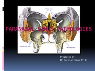
  • 1. PARANASAL SINUS PATHOLOGIES Presented by Dr. Cathrine Diana PG-III
  • 2. contents  Paranasal sinuses  Introduction  Development  Anatomy  Physiology  Examination and investigations  Paranasal sinus pathologies  Classification  Description  Complications  Treatment
  • 3. INTRODUCTION  Sinus (Latin) - fold or pocket.  Paranasal sinuses - four paired, hollow air filled spaces in various cranio-facial bones  Named after the bones in which they are located
  • 4. INTRODUCTION Clinically, Divided into two groups: 1. Anterior group: maxillary, frontal, anterior and middle ethmoidal air cells. They all open in the middle meatus. 2. Posterior group: Posterior ethmoidal and the sphenoid sinus
  • 5. DEVELOPMENT  Excavation of bone by air-filled sacs (pneumatic diverticula) from the nasal cavity.  Begins prenatally - continues through lifetime.
  • 6.
  • 7. ANATOMY– MAXILLARYSINUS  MAXILLARY ANTRECHEA / ANTRUM OF HIGHMORE  Largest  Maxilla - under the eyes, on either side of the nose.  Pyramidal - base toward the lateral wall of nose and apex directed laterally into the zygomatic process.  Capacity of 15 ml (average).
  • 8. FRONTAL SINUS  Situated deep to the inner and outer table of frontal bone  Drain- frontal recess to the middle meatus  Absent on one (15 %) or both sides (5 %)  Drainage pathway – naso- frontal duct- frontal recess - situated at its floor – drains into middle meatus (62%) or ethmoid infundibulum (38%).
  • 9. Ethmoid sinus Thin-walled air cavities in the lateral masses of ethmoid bone, between nose and the eyes. 1. Anterior ethmoidal air cells –3- 11 drain into either the ethmoidal infundibulum or the frontonasal duct. 2. Bullar cells (middle ethmoidal air cells) - usually <3 - open in ethmoidal infundibulum. 3. Posterior group :- Up to 7 - usually drain by a single orifice into the superior meatus.
  • 10. Agar nasi cells  They are the most anterior ethmoidal air cells. .  Its size influence the patency of the frontal recess and the anterior middle meatus.  Haller cells:  Also called infraorbital ethmoid cells.  Present in approx. 20 % pateints. Clinical significance –  Become infected , with potential extension into orbit.  Narrows the maxillary ostium.
  • 11. Onodi cells  These are posterior ethmoidal cells extending into the sphenoid bone ,either adjacent to or impinging upon the optic nerve.  When these Onodi cells abut or surround the optic nerve, the nerve is at risk when surgical excision of these cells is performed.  It is also a potential cause of incomplete sphenoidectomy.
  • 12. SPHENOID SINUS  Body of sphenoid - behind the nose, in the center of the skull.  Rarely symmetrical and separated by a thin bony septum.  Ostium of the sphenoid sinus is situated in the upper part of its anterior wall and drains into sphenoethmoidal recess.  Average size – 2 x 2 x 2 cm.  According to Congdon sphenoid pneumatization can be as follows  Conchal – 5 %  Presellar – 23 %  Post-sellar – 67%
  • 13. ANATOMY- LATERAL NASALWALL  3 projections - superior, middle and inferior concha.  Meatus - space below each concha.  Inferior meatus: nasolacrimal duct  Middle meatus:  Maxillary sinus  Frontal sinus  Anterior ethmoid sinuses  Superior meatus: posterior ethmoid sinuses  Sphenoethmoidal recess: sphenoid sinus
  • 14. OSTEOMEATAL COMPLEX  It is a common channel that links the frontal sinus, anterior and middle ethmoid sinuses and the maxillary sinus to the middle meatus. It is composed of five structures:  Maxillary ostium  Infundibilum  Ethmoidal bulla  Uncinate process  Hiatus semilunaris
  • 15. PHYSIOLOGY– SINUS EPITHELIUM  Respiratory epithelium - ciliated pseudostratified columnar epithelium, goblet cells, and submucosal glands  Produce a protective mucous blanket - traps bacteria and noxious materials, which are carried by ciliary motion to the ostium and into the nose for elimination
  • 16.  Ciliary movements: 50- 300 cilia/ cell; 8-20 beat/ second.  For maximum ciliary activity: Humidity: >85%, Temperature: 18- 40 degree C, pH: 7- 8.  The orientation of the cilia within a given sinus is specific as secretions are propelled towards the natural sinus ostia and from there to the nasopharynx and oropharynx where they are subsequently cleared by swallowing.
  • 17. SINUS HEALTH  Composition of gas content in the maxillary sinus is similar to venous blood, with high CO2 and lower O2 level compared to breathing air.  Sinus health depends on: 1. Mucous secretion of normal viscosity, volume, and composition 2. Normal muco-ciliary flow to prevent mucous stasis and subsequent infection 3. Open sinus ostia to allow adequate drainage and aeration.  Negative factors:  Dryness of air, Cigarette, Temperature variations, hypoxia, hypercapnia, Hypertonic/ hypotonic fluids, Dehydration, pH changes, diseases (like Cystic fibrosis and Primary ciliary dyskinesia), Drugs (phenylephrine, adrenaline, lidocaine, atropine, antihistaminic), Infections, Anatomic obstruction (septal deviation, enlarged or irregular turbinate), Foreign bodies and Nasal polyps.
  • 18. PATHOPHYSIOLOGICALSTAGES OF SINUS DISEASES Initial phase: - reversible Ostium obstruction phase Bacterial phase Chronic phase
  • 19. Osteomeatal complex obstruction ↓ Decreased ventilation of the sinuses ↓ Decreased drainage of the sinuses ↓ pO2 decrease, pCO2 increase, mucous stasis ↓ Inflammation and viscous mucous, ciliary movement slowing ↓ Stasis and proteolytic enzymes ↓ Ciliary damage ↓ Anaerobic microorganisms ↓ More damage
  • 20. FUNCTIONS OF SINUS 1. Reduction of weight of skull 2. Increasing resonance of the voice 3. Providing a buffer against blows to the face. 4. Insulating sensitive structures like dental roots and eyes from rapid temperature fluctuations in the nasal cavity. 5. Humidifying and heating of inhaled air because of slow air turnover in this region. 6. Regulation of intranasal and serum gas pressures 7. Increasing surface area for olfaction 8. Contribute to facial growth
  • 21. EXAMINATION  History and systemic clinical examination:  Check general signs of health  Systemic medical history, history of allergies, drug use and abuse  Occupation history  Examination of and neck for lumps or swollen lymph nodes
  • 22. Examination  Local examination of the nose, face, and neck: 1. Anterior Rhinoscopy: Examination of nose with a nasal speculum to check for abnormal areas, useful in evaluation of nasal obstruction. 2. Posterior rhinoscopy: With a mouth mirror in the nasopharynx
  • 23. Examination - transillumination  Normal transillumination decreases chance of pus in the sinus.  No light reflex suggests mucopurulent material or thickening of nasal mucosa.  Inexpensive screening tool
  • 24. Transillumination of Frontal Sinus Transillumination of Maxillary Sinus 24
  • 25. Examination - endoscopy  Endoscopic examination/ Rhinoscopy:  nasoscope/rhinoscope is a thin, tube-like instrument with a light and a lens for viewing. A special tool on the nasoscope may be used to remove samples of tissue. The tissues samples are viewed under a microscope by a pathologist.
  • 26. EXAMINATION – PLAIN RADIOGRAPHS  Plain radiographs:  to check for Sinus opacifications, Air-fluid level, Mass, Fractures  Caldwell view: PA view/ “forehead-nose” view to evaluate maxilla, maxillary and frontal sinus, ethmoid air cells, lamina papyracea
  • 27.  Water’s view: chin-nose” or “occipito- mental” view for evaluation of the paranasal sinuses. submento-vertical” view to evaluate the sphenoid, the posterior ethmoids, the maxillary and frontal sinuses
  • 28. CT SCANS  CT scans: Excellent views of the sinuses, best for osteomeatal complex and ethmoidal disease  “Limited CT Evaluation” – slice 3-4 mm  CT navigation:  A computer is used to identify the 3- dimensional location of a probe tip placed within the patient's nose or sinuses..  Improves anatomical identification and avoid damage to vital neighbouring structures such as the brain and eyes.
  • 30.
  • 31.
  • 33.
  • 34.
  • 36. EXAMINATION  MRI:  Excellent soft tissue definition - evaluation of neoplastic disease.  MRI (magnetic resonance imaging) with gadolinium: Gadolinium is injected into a vein. The gadolinium collects around the cancer cells so they show up brighter in the picture.
  • 37.  PET scan (positron emission tomography scan): A small amount of radioactive glucose is injected into a vein. The PET scanner rotates around the body and makes a picture of where glucose is being used in the body. Malignant tumor cells show up brighter in the picture because they are more active and take up more glucose than normal cells do.  Stuckensen and colleagues found a sensitivity of 70%, 84%, and 66% and a specificity of 82%, 68%, and 74% for PET, ultrasound, and CT scan in terms of nodal metastasis
  • 38. 9. Histological examination:  Fine-needle aspiration (FNA) biopsy, Incisional biopsy and excisional biopsy are done from pathologic tissues. 10. Culture examination: Correlation of routine nasal culture and sinus culture is poor. Endoscopically guided aspiration of cultures from medial meatus do correlate with sinus culture. Silver stained section showing invasive fungal sinusitis (aspergillus) Allergic mucin of allergic fungal sinusitis
  • 39. Classification  DEVELOPMENTAL VARIATIONS AND ANOMALIES  INFLAMMATORY/INFECTIOUS DISEASES  CYSTS  TUMORS  OTHER SURGICALLY RELEVANT CONDITIONS
  • 40. Developmental variations and anomalies  Paradoxical curvature of middle turbinate  Concha bullosa in middle turbinate  Lateralization and pneumatisation of uncinate process  Variations of ethmoidal roof anatomy  Bulla ethmoidalis – torus ethmoidalis and giant bulla  Others: Agenesis of sphenoid sinus, pneumatisation of greater wings of sphenoid and crista galli.
  • 41. DEVELOPMENTALVARIATIONSAND ANOMALIES Paradoxical curvature: Normally the convexity of the middle turbinate is directed medially toward the nasal septum. When the convexity is directed laterally, it is termed a paradoxical middle turbinate . Most authors agree that the paradoxical middle turbinate can be a contributing factor to sinusitis.
  • 42. DEVELOPMENTALVARIATIONS ANDANOMALIES Concha bullosa: When pneumatization involves the bulbous portion of the middle turbinate it is termed concha bullosa. If only the attachment portion of the middle turbinate is pneumatized, it is termed lamellar concha . A concha bullosa may obstruct the ethmoid infundibulum.
  • 43. Variations of uncinate process The uncinate process may be medialized, lateralized, or pneumatized/bent. Medialization occurs with giant bulla ethmoidalis. Lateralization of the uncinate process may obstruct the infundibulum. Pneumatization (uncinate bulla) can rarely cause obstruction of the infundibulum.
  • 44. Variation of the ethmoidal roof anatomy The ethmoid roof is of critical importance for two reasons.  most vulnerable to iatrogenic cerebrospinal fluid leaks.  anterior ethmoid artery is vulnerable to injury. The depth of the olfactory fossa is determined by the height of the lateral lamella of the cribriform plate. In 1962, Keros classified the depth of the olfactory fossa into three types, that is, Keros type I: <3 mm Keros type II: 4-7 mm Keros type III: 8-16 mm - most vulnerable to iatrogenic injury.
  • 45. Variations of sphenoid sinus … Agenesis of sphenoid sinus  Pneumatisation of other bones  The crista galli is normally bony. When aerated, it may communicate with the frontal recess, causing obstruction of the ostium and thus lead to chronic sinusitis and mucocele formation
  • 46. Bullae ethmoidalis  The bulla ethmoidalis is a prominent anterior ethmoid air cell.  Failure to pneumatise - torus ethmoidalis.  A 'giant bulla' may fill the entire middle meatus and force its way between the uncinate process and the middle turbinate.
  • 47. INFLAMMATORY/ INFECTIOUS CONDITIONS  POLYPS  SINUSITIS  GRANULOMATOUS DISEASES
  • 48. POLYPS  They are fleshy outgrowths of the nasal mucosa that form at the site of dependent edema in the lamina propria of the mucous membrane, usually around the ostia of the maxillary sinuses.  usually start near the ethmoid sinuses and grow into the open areas.  Large polyps can block the sinuses or nasal airway.  Risk factors: Aspirin sensitivity (wheezing), Asthma, Acute and Chronic sinus infections, Cystic fibrosis, Hay fever (allergic rhinitis).
  • 49.  Clinical features:  Nasal obstruction and mouth breathing  Nasal congestion and postnasal drainage  Anosmia, hyposmia  Sneezing, rhinorrhea  Facial pain  Ocular itching  Bleeding polyps occur in rhinosporidiosis  Unilateral polyps occasionally occur in association with or represent benign or malignant tumors of the nose or paranasal sinuses, or in response to a foreign body.  Diagnosis:  physical examination - A developing polyp is teardrop-shaped; when mature, it resembles a peeled seedless grape.  CT scans
  • 50.  Treatment 1. Steroids –may shrink or eliminate polyps  Topical corticosteroid spray - mometasone [30 mcg/spray], beclomethasone [42 mcg/spray], flunisolide - given as 1 or 2 sprays bid in each nasal cavity  1-wk tapered course of oral corticosteroids. 2. Surgery :  FESS  Steroid therapy after surgery - to retard recurrence.  In severe recurrent cases- maxillary sinusotomy or ethmoidectomy, usually done endoscopically. 3. Removal of etiology – control of underlying allergy or infection.
  • 51. SINUSITIS  Definition: Sinusitis is the inflammatory condition of the mucous membrane lining of the sinuses  RHINOSINUSITIS is a better term because:  Allergic or non-allergic rhinitis nearly always precedes sinusitis  Sinusitis without rhinitis is rare  Nasal discharge and congestion are prominent symptoms of sinusitis  Nasal mucosa and sinus mucosa are similar and are contiguous
  • 52. Classifications ACCORDING TO DURATION: 1. Acute :infection lasting 4 weeks, symptoms resolve completely resolved in < 30 days. 2. Subacute :infection lasting between 4 to 12 weeks, yet resolves completely. 3. Recurrent: ≥ 4 discrete acute episodes per year, each completely resolved in < 30 days but recurring in cycles, with at least 10 days between complete resolution of symptoms and initiation of a new episode 4. Chronic: symptoms lasting more than 12 weeks.
  • 53.  ACCORDING TO PATHOGEN: 1. Bacterial: Hospital-acquired acute infections are more often bacterial, typically involving Staphylococcus aureus, Klebsiella pneumoniae, Pseudomonas aeruginosa 2. Viral: In immunocompetent patients - in the community is almost always viral (eg, rhinovirus, influenza, parainfluenza). antibiotics given for:  Mild to moderate sinus symptoms persisting for ≥ 10 days  Severe symptoms (eg, fever ≥ 39°, severe pain) for ≥ 3 to 4 days  Worsening sinus symptoms after initially improving from a typical viral URI ("double sickening" or biphasic illness)
  • 54. 3. Fungal: Usually seen in immunocompromised patients because of poorly controlled diabetes, neutropenia, or HIV infection. It is clinically of 2 types:  Non-invasive fungal sinusitis:  Saprophytic fungal infestation/colonization  Allergic fungal rhinosinusitis (AFRS)  Saprophytic fungus balls (mycetoma)  Invasive fungal sinusitis –  Chronic invasive fungal sinusitis  Granulomatous invasive fungal sinusitis  Acute (fulminant) invasive fungal sinusitis
  • 55. sinusitis  According to source:  Primary  Secondary  According to number of sinuses involved:  Hemisinusitis –all sinuses on one side  Polisinusitis – several sinuses, but not all, are involved  Pansinusitis  According to source of infection:  Rhinogenous  Odontogenic  Traumatic  Hematogenic  Allergic
  • 56. Risk factors: 1. Obstruction to drainage: most important 2. Defect of self-cleaning mechanism of the mucous membrane of sinus – infections causing immobility of the cilia, Increased viscosity of secretions, Immotile cilia syndrome, Prolonged exposure to cigarette smoke 3. Medications  First generation antihistamines (non sedating do not affect)  Anticholinergics, Aspirin, Anesthetic agents, Benzodiazepines 4. Immunodeficiency: Immunoglobulin deficiency (IgA, IgG), diabetes, HIV infection 5. Other factors: prolonged ICU stays, severe burns, cystic fibrosis, and ciliary dyskinesia.
  • 57. Clinical Features: 1. Nasal congestion and discharge 2. Sore throat and postnasal drip 3. Pain or pressure. 4. Oedema of facial tissues. 5. Bad breath or loss of smell (hyposmia/anosmia). 6. Systemic symptoms: Malaise may be present. Fever and chills suggest an extension of the infection beyond the sinuses, rise in temperature, bad appetite, sleep disturbances, changes of the blood (leukocytosis), Productive cough (especially at night) 7. Complications – ocular, neurological, local
  • 58. Diagnosis 1. Diagnosis is clinical; 2. CT and cultures - mainly for chronic, refractory, or atypical cases. 3. X-rays of the apices of the teeth Clinical diagnostic criteria include 2major factors, I major and 2 minor factors or presence of pus in nasal cavity. Major Factors Minor Factors Facial pain/pressure Headache Facial congestion/fullness Maxillary dental pain Nasal drainage/discharge Cough Postnasal drip Halitosis Nasal obstruction/blockage Fatigue Hyposmia/anosmia Ear pain/ pressure/ fullness Fever (acute sinusitis only) Fever Purulence in nasal cavity on examination (diagnostic by itself)
  • 59. TREATMENT OPTIONS MEASURES TO ENHANCE DRAINAGE: 1. Heat therapy: Steam inhalation; hot, wet towels over the affected sinuses; and hot beverages 2. Topical vasoconstrictors/ Nasal decongestants:  Topical nasal sprays (limit use to 3-7 days) - Phenylephrine, Oxymetazoline, Naphthazoline, Tetrahydrozoline, Zylometazoline. Phenylephrine(0.25%) spray q 3 h or oxymetazoline q 8 to 12 h, are effective but should be used for a maximum of 5 days or for a repeating cycle of 3 days on and 3 days off until the sinusitis is resolved  CORTICOSTEROIDS: Corticosteroid nasal sprays can help relieve symptoms but typically take at least 10 days to be effective 3. Systemic vasoconstrictors: Systemic vasoconstrictors, such as pseudoephedrine 30 mg po (for adults) q 4 to 6 h, are less effective.
  • 60. 4. Nasal irrigation:  Commercial buffered sprays, Bulb syringe, waterpik and ceramic irrigators with lavage tip or disposable enema bucket  Washes away irritants and moistens the dry nose.  cumbersome and uncomfortable - better for patients with recurrent sinusitis.  ANTIHISTAMINICS: recommended if allergy present. They can be oral or topical  HYDRATION
  • 61. 5. Mucoactive drugs:  main purpose - increase the ability to expectorate sputum and/or decrease mucus hypersecretion. 1. Expectorants: hypertonic saline, iodine containing compounds, guaifenesin (glyceryl guaiacolate), ion channel modifiers (tricyclic nucleotides) 2. Mucoregulators: carbocysteine, anticholinergic drugs, glucocorticoids, macrolide antibiotics 3. Mucolytics: classic mucolytic (N-Acetyl Cysteine), peptide mucolytic, non destructive mucolytics 4. Mucokinetics: bronchodialators, ambroxol
  • 62. a) Nebulization: best form of physiotherapy. Can be done using compressors or ultrasonic nebulisers. Normal saline solutions are nebulized which has a hydrating effect on the mucous lining. Ultrasonic nebulisers can set the rate according to need. b) Laser therapy: used directly over the sinuses to reduce inflammation c) Ultrasound therapy: sound waves are conducted through a hypoallergenic gel to reduce inflammation and loosen the accumulated mucous. d) Short-wave diathermy e) Rinoflow therapy: new option, basically micronized endotracheal wash. Used in sinusitis, rhinitis, pharyngitis, laryngitis and secretory otitis media.
  • 63. Antibiotics and antifungal drugs:  Amoxicillin 500 mg tid for 10-14 days - First line  Beta-lactanase resistance - Amoxicillin/clavulanate, Cefuroxime, Cefpodoxime, Cefprozil
  • 64.
  • 65. Surgery  Indications:  Sinusitis unresponsive to antibiotic therapy  Necrotic sinusitis  Orbital complications (abscess and phlegmon of orbit)  Intracranial complications (meningitis, brain abscess)  Rhinogenic sepsis  Odontogenic sinusitis combined with maxillary osteomyelitis.  Approaches: 1. Removal of etiology 2. Fess 3. Caldwell-luc procedure 4. Intranasal antrostomy 5. Radical surgeries
  • 66. Removal of etiology:  Treatment of affected tooth.  Caldwell luc approach may be used.  Intra-nasal antrostomy may be needed.
  • 67. Caldwell - luc  Caldwell-Luc is the fenestration of the anterior wall of the maxillary sinus and the surgical drainage of this sinus into the nose via an antrostomy.  a middle meatus antrostomy is being utilized as a more physiologic antrostomy..
  • 69. FESS:  Functional endoscopic sinus surgery (FESS) is the mainstay in the surgical treatment of sinusitis and nasal polyps, including bacterial, fungal, recurrent acute, and chronic sinus problems.  Nasal endoscopes through the nostrils to avoid cutting the skin.  Telescope diameters - 4mm (adult use) and 2.7mm (pediatric use)  Viewing angles - 0 degrees to 30, 45, 70, 90, and 120 degrees  Carry: High definition cameras attached to monitors, tiny articulating instruments - cutting, suction, biopsy, curettage  All the sinuses can be accessed at least to some degree by means of FESS.
  • 70.  Extended approaches: Paranasal sinuses are found to a relatively low- morbidity approach to selected tumors even inside the skull or brain. This can be divided into approaches to: anterior cranial fossa, mid cranial fossa, posterior cranial fossa, infratemporal fossa (incl. pterygopalatine fissure), sella turcica, orbital access, and optic nerve access.  Complications:  Proximity of the sinuses to the eyes, optic nerves, brain and internal carotid arteries  Serious risks are rare occurrences
  • 71. Before FESS 2 months after FESS
  • 72.  Principal element - granuloma formation - a conglomerate of macrophages, epithelioid cells, and multinucleated giant cells. 1. Infectious:  spirochetes (syphilis, yaws)  mycobacteria [tuberculosis, leprosy]  bacterial [rhinoscleroma]  fungus [aspergillus] 2. Inflammatory  Wegener granulomatosis  sarcoidosis,  Churg-Strauss syndrome  cocaine induced midline destructive lesions Granulomatous diseases
  • 73. Cysts  Mucocele and Pyocele  Retention cyst  Pseudocyst  Post-operative maxillary cyst
  • 74. Mucocele and pyocele  Mucocele:  formed when drainage of mucus from one of the paranasal sinuses becomes blocked by obstruction of its ostium.  Contents: clear serous fluid, thick mucoid material, or, if hemorrhage has occurred, thick brown material  Pyocele/mucopyocele:  Infected mucocele  Contents: mixture of mucus and pus from which a causative organism may or may not be seen on smear or subsequently cultured.
  • 75. Pathogenesis:  Blockage of the ostium pressure develops within sinus  expansion of the sinus space erosion, and displacement of bone.  Encroachment of the mucocele upon contiguous structure  Sinus lining – remains normal or becomes attenuated to undergo metaplasia to low cuboidal or squamous cells.
  • 76.  Most commonly: frontal and anterior ethmoid sinuses  Etiology: chronic inflammation, osteomas, fractures, tumors, polyps, scarring, and congenital abnormalities. Management  Decompression by complete removal and curettage  Marsupialisation via endoscopic approach through the middle meatus  Prophylactic Nasofrontal duct obstruction
  • 77.  Retention cyst and Pseudocyst: Together called antral cyst or mucosal cyst  Indistinguishable on radiological or clinical examination.  Quite common.  Mostly - single cysts, but in a few instances - they may be multiple and bilateral.  Clinical features:  Usually asymptomatic – discovered during radiographic examination  Symptoms – similar to chronic sinusitis  Sometimes an antral cyst may produce a swelling
  • 78.  Diagnosis :CT based  Spherical, ovoid or dome-shaped radiopacities that have a smooth and uniform outline  narrow or broad base.  Size from minute to very large  Usually remain static  Many regress spontaneously
  • 79. POST-OPERATIVE MAXILLARYCYST (SURGICALCILIATEDCYSTOFMAXILLA)  Delayed complication arising years after surgery of maxillary sinus.  Causes:  Caldwell–Luc procedure including a nasal antrostomy  Gun- shot injuries  Fractures of the malar–maxillary complex  Mid-face osteotomies.  Clinical features:  Pain, discomfort or swelling in the cheek or face, or intra-orally in the palate or alveolus.  Pus discharge.  Radiographs - well-defined radiolucent area closely related to the maxillary sinus.  Treatment: enucleation.
  • 80. TUMORS  0.2% of all malignancies  80% - maxillary sinus.  Men>> women.  40 and 70 years.  Carcinomas >> sarcomas  Metastases are relatively rare.  Tumours of the sphenoid and frontal sinuses are extremely rare - no standard staging system  Most common - squamous cell carcinoma
  • 81. WHO HISTOLOGICAL CLASSIFICATION OF NASAL AND PNS TUMORS
  • 82. TNM CLASSIFICATION OF CARCINOMAS OF NOSE AND PARANASAL SINUSES .
  • 83. Risk factors: 1. Woodworking (carpentry), Shoemaking, Metal-plating, Flour mill or bakery work. 2. Human papillomavirus (HPV) infection 3. Male 4. Older than 40 years. 5. Smoking.
  • 84. Clinical features  No signs or symptoms in the early stages.  later:  Blocked sinuses that do not clear, or sinus pressure.  Headaches or pain in the sinus areas.  Rhinitis and epistaxis  A lump or sore inside the nose that does not heal.  A lump on the face or roof of the mouth.  Numbness or tingling in the face.  Swelling or other trouble with the eyes, such as double vision or the eyes pointing in different directions.  Pain in the upper teeth, loose teeth, or dentures that no longer fit well.  Pain or pressure in the ear.
  • 85. Treatment 1. Surgery: For all stages of paranasal sinus and nasal cavity cancer.  Transfacial approaches  1. lateral rhinotomy/ weberfergussen incision  2. diffenbech extension  3. Lynch extension with the modification  External ethmoidectomy/frontoehtmoidectomy  Bicoronal with Mid face degloving  Subcranial approach  Acess osteotomies
  • 86.  Trans oral approach:  Caldwell lue  Denker  Janson – horgan approach  Trans nasal:  Transseptal approach  Endoscopic approaches
  • 87. Draw backs of endoscopic approach  -Not indicated in case of extensive involvement  Trans orbital extension  Scar tissue due to previous surgery  in case to reduce the bony fracture
  • 88.  TRANSORAL ROBOTIC SURGERY  New advances in technology facilitate minimal access  To avoid large transcervical or face-splitting incisions.  Transoral robotic surgery allows access to tumors within the posterior oral cavity and oropharynx via multiple robotic arms and a high-definition/ magnification camera.  The operator sits at a separate console and via remote control can operate the various instruments.  Electrocautery, laser, and standard dissection instruments can be used with the robot.  Advantages include surgical access via minimal approaches, resulting in definitive pathologic assessment while minimizing transection and resection of critical swallowing musculature Oral Maxillofacial Surg Clin N Am 24 (2012) 307–316
  • 89. Trans oral Robotic surgery  more-precise movements in narrow spaces and the capability to work around corners.  This result in preservation of maximum amount of healthy muscular and neurovascular tissue which helps the patient swallow on their own sooner and discharged home earlier.
  • 90. 1. Radiation therapy: External radiation therapy, Internal radiation therapy - depending on the type and stage of the cancer being treated.  IMRT:  Intensity-modulated radiation therapy (IMRT) can deliver high doses of radiation with precision while minimizing damage to surrounding tissues.  IMRT can conform to the irregular shape of a tumor, delivering higher doses directly to the tumor cells and potentially destroying more tumor cells.  The technique requires more precise planning due to the sharp dose falloff gradient between the gross tumor and the surrounding normal tissue.  IMRT provides locoregional control (90%) and is well tolerated by patients. Oral Maxillofacial Surg Clin N Am 24 (2012) 307–316
  • 91. Chemotherapy:  Systemic chemotherapy: When chemotherapy drug is given PO, IV or IM the drugs enter the bloodstream and can reach cancer cells throughout the body  Regional chemotherapy: When chemotherapy is placed directly into the cerebrospinal fluid, an organ, or a body cavity, the drugs mainly affect cancer cells in those areas.  Combination chemotherapy is treatment using more than one anticancer drug.  The way the chemotherapy is given depends on the type and stage of the cancer being treated.  Intra arterial cisplastin therapy: As a primary therapy, the complete response rate was 83.3%, The 2-year local control rate was 63.0%, and the 2-year overall survival rate was 75.5%. The 2-year preservation rate of the hard palate was 97.1%, that of the eyeball was 97.2%, and that of visual function was 94.4%. This treatment regimen can contribute to improving the quality of life of patients without reducing the curability of the therapy. Int. J. Oral Maxillofac. Surg. 2015; 44: 697–704.
  • 92. Other surgically relevant conditions  Oro-antral communication/fistula  Antral foreign bodies  Hypertrophied maxillary sinus
  • 93. Oro-Antral Fistula (oaf)  Oro-antral communication: it is an abnormal connection between oral and Antral cavities.  Oro-antral fistula: long standing oro-antral communication when left open, epithelializes to form a patent fistula between the two cavities.  Types:  ALVEOLOSINUSAL  PALATO-SINUSAL  VESTIBULO-SINUSAL Oro-Antral Communications
  • 94. CAUSES:  Extraction – maxillary posterior teeth  Cysts, tumors  Osteomyelitis  Radiation therapy  Trauma  Implant denture CLINICAL FEATURES:  Some patients are asymptomatic  Unpleasant tasting discharge and odor  Reflux of fluids and foods into the nose from mouth  Leakage of air  Difficulty in smoking and blowing air  Development of chronic sinusitis in infected cases
  • 95. Diagnosis:  Valsalva maneauver  Mirror fog test  Cotton wisp test  Nasal regurgitation of fluid  Radiographs: Sinus floor discontinuity, Sinus opacity, Focal alveolar atrophy, Associated periodontal disease Oro-Antral Communications (UL7)
  • 96. Treatment:  Immediate treatment: Primary purpose is closure of defect and prevention of sinusitis through:  Suturing across the defect with/without periodontal pack, warm saline rinses, antibiotic and antihistaminic therapy with decongestants.  Size < 5mm: non-invasive intervention (spontaneous closure by blood clot)  Size > 5mm, and > 48 hours - : surgical closure : small defects – local flaps; large defects – regional/distant flaps.
  • 97.  Obturators  Buccal Advancement Flap most common.  Described by Rehrmann & made popular by Berger.  Trapezoidal sliding flap – Moczair  buccal osteoperiosteal flapOro-Antral Communications
  • 98.  Palatal flaps 1. Palatal rotational advancement flap most common 2. V-shaped palatal flap (kruger) & 3. Split-thickness palatal flap (ito & hara).Oro-Antral Communications
  • 99.  Combination flaps  Inverted periosteal flap  Tongue flap, temporalis flap  BFP closure  PRF membrane coverage  Autologous bone grafts: press-fit technique  Autologous cartilage grafts: auricular cartilage, auricular cartilage  Alloplastic materials  Transplantation of a mature wisdom tooth (followed by root canal treatment of the tooth 5 - 6 weeks later)  Laser bio-stimulation (over 5 days) Oro-Antral Communication
  • 100. ANTRAL FOREIGN BODIES:  Gutta-percha points, tooth roots, impression materials, dental burs, bone pieces , implants etc.. Treatment has been direct explored by Caldwell-Luc approach, with or without nasal antrostomy. FESS may be done for sinusitis. HYPERTROPHIED MAXILLARY SINUS:  Not pathology but causes difficulty in implant supported rehabilitation of posterior maxilla, with the risk of subsequent development of sinus pathologies. The management includes direct and indirect sinus lifting. Oro-Antral Communications (UL8)
  • 101. Complications  Because of the proximity of the paranasal sinuses to the eyes and brain, complications of sinusitis are divided into  orbital,  neurological and  local complications.
  • 102. Orbital complications  highest frequency - in children under 6 years of age.  Infection usually originates from the ethmoids and occurs through: (1) direct extension through the orbital wall (2) retrograde spread through veins between the sinuses and the orbit.  Lymphatic spread – not significant
  • 103. Orbital complications 1. Preseptal cellulitis, or periorbital cellulitis 2. Orbital cellulitis and edema 3. Subperiosteal abscess 4. orbital abscess 5. Cavernous sinus thrombosis: direct extension or retrograde thrombophlebitis (via the ophthalmic vein) of ethmoid or sphenoid infections.  restriction of extra ocular mobility, proptosis, chemosis, and visual loss  cranial neuropathies and signs of meningitis
  • 104. Neurological complications  Less frequently than orbital  Most commonly related to the frontal or sphenoid sinuses.  Via - direct spread or retrograde thrombophlebitis. A – osteomyelitis B - periorbital abscess C – epidural abscess D – subdural abscess E – brain abscess F – meningitis G - septic thrombosis of superior sagittal sinus
  • 105. Local complications  Osteomyelitis - complication of frontal sinusitis.  Tender, doughy, erythematous swelling over the forehead.  Treatment of choice - surgical eradication of the affected bone under antibiotic coverage.
  • 106. REFERENCES 1. References: 2. Peterson’s principles of oral and maxillofacial surgery  DISEASES of the SINUSES Diagnosis and Management- DAVIDW. KENNEDY, MD, FACS 1. WHO classification of head and neck tumors 2. PL Dhingra - Disease of ear, nose & throat 4th edition 3. Rosai and Ackerman’s Surgical Pathology (9th edition) 4. David L. Daniels et.al. The Frontal Sinus Drainage Pathway and Related Structures, AJNR: 24, August 2003. 5. Interactive Atlas. http://uwmsk.org/sinusanatomy2/axial/axial.html 6. (Adapted from Chow AW, Benninger MS, Brook I, et al: IDSA clinical practice guideline for acute bacterial rhinosinusitis in children and adults. Clinical Infectious Diseases 54 (8):1041–5 (2012).) 7. Thompson and Patterson: Fungal disease of the nose and paranasal sinuses:J Allergy Clin Immunol 2012;129:321-6. 8. Multiplanar Sinus CT: A Systematic Approach to Imaging Before Functional Endoscopic Sinus Surgery Jenny K. Hoang, James D. Eastwood, Christopher L. Tebbit, and Christine M. Glastonbury American Journal of Roentgenology 2010 194:6, W527-W536
  • 107.  Raef S. Ahmed, Roger Ove, Jun Duan, Richard Popple, Glenn B. Cobb Intensity-modulated radiotherapy (IMRT) for carcinoma of the maxillary sinus: A comparison of IMRT planning systems Medical Dosimetry, Volume 31, Issue 3, Autumn 2006, Pages 224-232  Management of rhinomaxillary mucormycosis with Posaconazole in immunocompetent patients Sachin Rai *, Shikha Yadav, Dinesh   Kumar, Vijay Kumar, Vidya Rattan Journal of Oral Biology and Craniofacial Research xxx (2016) xxx  Modified transnanal endoscopic maxillectomy: a novel surgery style of maxillary malignant tumor Yonghua Bi1,2, Shuangba He1, Tao Guo1, Jingwu Sun Int J Clin Exp Med 2016;9(6):11361-11366  Modified double-layered flap technique for closure of anoroantral fistula: Surgical procedure and case reportAlberto Merlinia, Joseph Garibaldia, Matteo Piazzaia, Luca Giorgisb, British Journal of Oral and Maxillofacial Surgery 54 (2016) 959–961  Repair of Oroantral Communication by Use of a Combined Surgical Approach Functional Endoscopic Surgery and Buccal Advancement Flap/Buccal Fat Pad Graft Timothy Adams, DDS,* Daniel Taub, DDS, MD,y and Marc Rosen, MD. J Oral Maxillofac Surg 73:1452-1456, 2015    An Update on Squamous Carcinoma of the Oral Cavity,Oropharynx, and Maxillary SinusJoshua E. Lubek, DDS, MDa,*, Lewis Clayman, DMD, MD Oral Maxillofacial Surg Clin N Am 24 (2012) 307–316