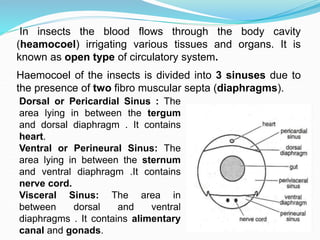
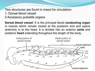
R K PANSE
Assistant Professor
Entomology
In insects the blood flows through the body cavity
(heamocoel) irrigating various tissues and organs. It is
known as open type of circulatory system.
Haemocoel of the insects is divided into 3 sinuses due to
the presence of two fibro muscular septa (diaphragms).
Dorsal or Pericardial Sinus : The
area lying in between the tergum
and dorsal diaphragm . It contains
heart.
Ventral or Perineural Sinus: The
area lying in between the sternum
and ventral diaphragm .It contains
nerve cord.
Visceral Sinus: The area in
between dorsal and ventral
diaphragms . It contains alimentary
canal and gonads.
Two structures are fount in insect for circulation:
1. Dorsal blood vessel
2 Accessory pulsatile organs
Dorsal blood vessel: It is the principal blood conducting organ
in insects which remain closed at the posterior end and opens
anteriorly in to the head. It is divided into an anterior aorta and
posterior heart extending throughout the length of the body.
1. Aorta: It is the anterior part and functions as principal
artery. It is present in the thoracic region and opens in to the
head near the brain. Its attachment with the heart posteriorly
is marked by a aortic valve.
2. Heart: It is the posterior part of dorsal blood vessel
extending up to the terminal end of the abdomen. Heart
remains in position with the help of alary muscles that are
attached to the tergum of the abdomen on one side and to
the dorsal diaphragm on other side. These alary muscles
appear to be distributed fan like over the heart. Heart
consists of number of chambers marked by constrictions
and the presence of the opening called the incurrent ‘ostia’
which allow the entry of blood from pericardial sinus in to the
heart. There may be up to three pairs of thoracic ostia
and nine pairs of abdominal ostia.
Accessory pulsatile organs: Insects
consists of sac like structures called
accessory pulsatile organs, which
are present at the base of the
appendages such as wings, legs and
antenna. They pulsate independently
and supply adequate blood to the
appendages.
Process of blood circulation: Heart mainly function as a
pulsatile organ whose expansion and contraction leads to
blood circulation . It takes place generally in an anti clock
manner starting from posterior end to the anterior end in a
forward direction. Circulation of blood takes place in two
phases due to the action of the alary muscles as well as
the muscles of the walls of the heart. The two phases are:
1. Diastole: During which expansion of heart takes place.
2. Systole : Contraction of heart takes place.
 1. Diastole: Expansion of heart ( diastole) occurs, when the
alary muscles that are spread fan like over the heart and
connected to the tergum get contracted. It results in
increase of volume of heart and decrease in the area of
pericardial sinus. This creates a pressure on the blood in
pericardial sinus forcing the blood to enter into the heart
through the incurrent ostia. These incurrent ostia allow
only the entry of blood from the sinus in to the heart and
prevents its backflow from the heart to the sinus.
 2. Systole : Contraction of heart (systole), is brought about
by the expansion of the alary muscles as well as
contraction of the muscles of the heart wall. This creates
pressure on the blood within the heart leading to its
forward movement in to the aorta. From the aorta blood
enters in to the head and flows back bathing the visceral
organs in the visceral sinus and neural cord in the
perineural sinus.
 In between diastole and systole there will be a short period
of rest which is known as diastasis. During the process of
backward flow of blood, after entering in to the visceral
and perineural sinuses, blood flow to the posterior part of
the body and in to the pericardial sinus, due to the
undulating movements of the dorsal and ventral
diaphragms. During the process of circulation throughout
the body some part of the blood flows into the accessory
pulsatile organ that are present at the base of appendages
like legs, wings and antennae.
 The rate of heart beat (diastole) generally vary with the
body temperature and physiological conditions of the
body which in turn differs between species (or) between
stages of the insects.
Properties of blood:
 1. Blood is colourless (or) green (or) yellowish with
different types of haemocytes and plasma.
 Green colour is due to chlorophyll dissolved in the plasma
2. Blood covers up 5 – 40% of the total body weight that
vary with the sex and stage (or) age of the insect.
 3. Insect blood contain proteins, lipids, sugars, organic
acids, phosphates, pigments, uric acid etc.
 4. The insect blood of phytophagous insect is rich in ‘K’
where as that of carnivores is rich in ‘Na’
 5. Specific gravity of the blood varies from 1.01 to 1.06.
 6. pH of the blood generally varies from 6-7.
 7. The blood sugar of insects is trehalose.
Functions of haemolymph
1. Lubricant
2. Hydraulic medium
3.Transport and storage
4.Protection: It helps in phagocytocis, encapsulation,
detoxification, coagulation, and wound healing.
5. Heat transfer:
6. Maintenance of osmotic pressure:
7. Reflex bleeding:
8. Metabolic medium:

Lec. 18 Circulatory system.ppt

  • 1.
    R K PANSE AssistantProfessor Entomology
  • 2.
    In insects theblood flows through the body cavity (heamocoel) irrigating various tissues and organs. It is known as open type of circulatory system. Haemocoel of the insects is divided into 3 sinuses due to the presence of two fibro muscular septa (diaphragms). Dorsal or Pericardial Sinus : The area lying in between the tergum and dorsal diaphragm . It contains heart. Ventral or Perineural Sinus: The area lying in between the sternum and ventral diaphragm .It contains nerve cord. Visceral Sinus: The area in between dorsal and ventral diaphragms . It contains alimentary canal and gonads.
  • 3.
    Two structures arefount in insect for circulation: 1. Dorsal blood vessel 2 Accessory pulsatile organs Dorsal blood vessel: It is the principal blood conducting organ in insects which remain closed at the posterior end and opens anteriorly in to the head. It is divided into an anterior aorta and posterior heart extending throughout the length of the body.
  • 4.
    1. Aorta: Itis the anterior part and functions as principal artery. It is present in the thoracic region and opens in to the head near the brain. Its attachment with the heart posteriorly is marked by a aortic valve. 2. Heart: It is the posterior part of dorsal blood vessel extending up to the terminal end of the abdomen. Heart remains in position with the help of alary muscles that are attached to the tergum of the abdomen on one side and to the dorsal diaphragm on other side. These alary muscles appear to be distributed fan like over the heart. Heart consists of number of chambers marked by constrictions and the presence of the opening called the incurrent ‘ostia’ which allow the entry of blood from pericardial sinus in to the heart. There may be up to three pairs of thoracic ostia and nine pairs of abdominal ostia.
  • 5.
    Accessory pulsatile organs:Insects consists of sac like structures called accessory pulsatile organs, which are present at the base of the appendages such as wings, legs and antenna. They pulsate independently and supply adequate blood to the appendages.
  • 6.
    Process of bloodcirculation: Heart mainly function as a pulsatile organ whose expansion and contraction leads to blood circulation . It takes place generally in an anti clock manner starting from posterior end to the anterior end in a forward direction. Circulation of blood takes place in two phases due to the action of the alary muscles as well as the muscles of the walls of the heart. The two phases are: 1. Diastole: During which expansion of heart takes place. 2. Systole : Contraction of heart takes place.
  • 7.
     1. Diastole:Expansion of heart ( diastole) occurs, when the alary muscles that are spread fan like over the heart and connected to the tergum get contracted. It results in increase of volume of heart and decrease in the area of pericardial sinus. This creates a pressure on the blood in pericardial sinus forcing the blood to enter into the heart through the incurrent ostia. These incurrent ostia allow only the entry of blood from the sinus in to the heart and prevents its backflow from the heart to the sinus.  2. Systole : Contraction of heart (systole), is brought about by the expansion of the alary muscles as well as contraction of the muscles of the heart wall. This creates pressure on the blood within the heart leading to its forward movement in to the aorta. From the aorta blood enters in to the head and flows back bathing the visceral organs in the visceral sinus and neural cord in the perineural sinus.
  • 8.
     In betweendiastole and systole there will be a short period of rest which is known as diastasis. During the process of backward flow of blood, after entering in to the visceral and perineural sinuses, blood flow to the posterior part of the body and in to the pericardial sinus, due to the undulating movements of the dorsal and ventral diaphragms. During the process of circulation throughout the body some part of the blood flows into the accessory pulsatile organ that are present at the base of appendages like legs, wings and antennae.  The rate of heart beat (diastole) generally vary with the body temperature and physiological conditions of the body which in turn differs between species (or) between stages of the insects.
  • 9.
    Properties of blood: 1. Blood is colourless (or) green (or) yellowish with different types of haemocytes and plasma.  Green colour is due to chlorophyll dissolved in the plasma 2. Blood covers up 5 – 40% of the total body weight that vary with the sex and stage (or) age of the insect.  3. Insect blood contain proteins, lipids, sugars, organic acids, phosphates, pigments, uric acid etc.  4. The insect blood of phytophagous insect is rich in ‘K’ where as that of carnivores is rich in ‘Na’  5. Specific gravity of the blood varies from 1.01 to 1.06.  6. pH of the blood generally varies from 6-7.  7. The blood sugar of insects is trehalose.
  • 10.
    Functions of haemolymph 1.Lubricant 2. Hydraulic medium 3.Transport and storage 4.Protection: It helps in phagocytocis, encapsulation, detoxification, coagulation, and wound healing. 5. Heat transfer: 6. Maintenance of osmotic pressure: 7. Reflex bleeding: 8. Metabolic medium: