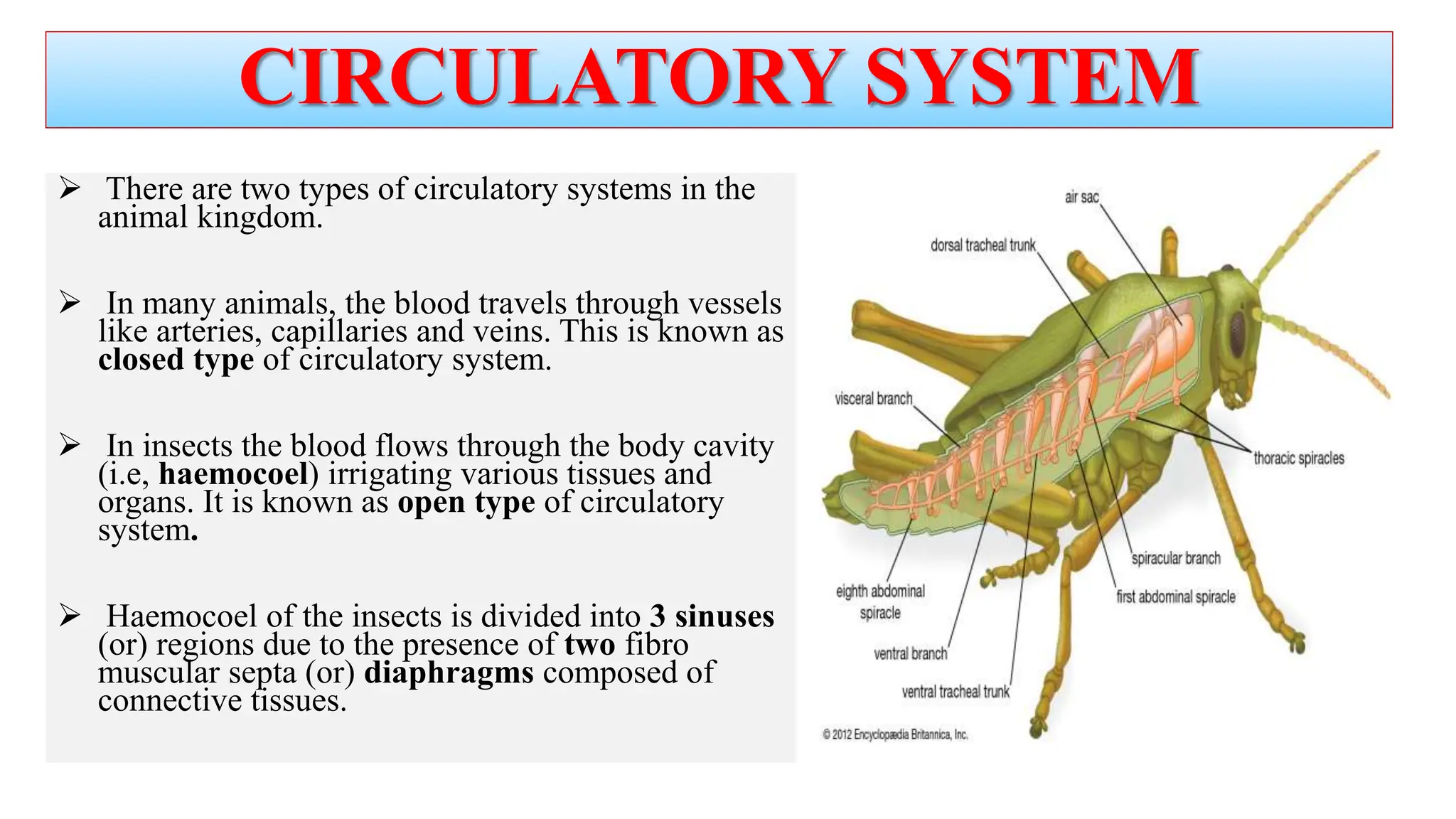
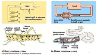
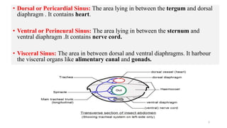
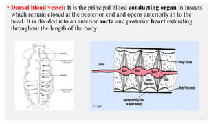
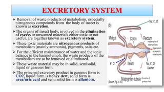
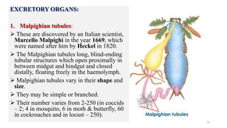
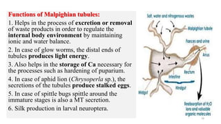
CIRCULATORY SYSTEM
 There are two types of circulatory systems in the
animal kingdom.
 In many animals, the blood travels through vessels
like arteries, capillaries and veins. This is known as
closed type of circulatory system.
 In insects the blood flows through the body cavity
(i.e, haemocoel) irrigating various tissues and
organs. It is known as open type of circulatory
system.
 Haemocoel of the insects is divided into 3 sinuses
(or) regions due to the presence of two fibro
muscular septa (or) diaphragms composed of
connective tissues.
2
• Dorsal or Pericardial Sinus: The area lying in between the tergum and dorsal
diaphragm . It contains heart.
• Ventral or Perineural Sinus: The area lying in between the sternum and
ventral diaphragm .It contains nerve cord.
• Visceral Sinus: The area in between dorsal and ventral diaphragms. It harbour
the visceral organs like alimentary canal and gonads.
3
• Dorsal blood vessel: It is the principal blood conducting organ in insects
which remain closed at the posterior end and opens anteriorly in to the
head. It is divided into an anterior aorta and posterior heart extending
throughout the length of the body.
4
1. Aorta:
 It is the anterior part of the dorsal
blood vessel and functions as
principal artery.
 It is present in the thoracic region
and opens in to the head near the
brain.
 Its attachment with the heart
posteriorly is marked by a aortic
valve.
 Anteriorly sometimes it gets divided
into 2 (or) more cephalic arteries in
the head.
5
2. Heart:
 It is the posterior part of dorsal blood vessel extending up to the terminal
end of the abdomen.
 Heart remains in position with the help of alary muscles that are attached to
the tergum of the abdomen on one side and to the dorsal diaphragm on other
side.
 These alary muscles appear to be distributed fan like over the heart.
 Heart consists of number of chambers marked by constrictions and the
presence of the opening called the incurrent ‘ostia’ which allow the entry
of blood from pericardial sinus in to the heart.
 The number of ostia depends upon the number of heart chambers which
will be usually 9.
 The wall of heart also consists of muscles.
 Heart mainly functions as the pumping organ into the aorta.
6
• Accessory pulsatile organs:
 Insects consists of sac like structures called accessory pulsatile
organs, which are present at the base of the appendages such as wings,
legs and antenna.
 They pulsate independently and supply adequate blood to the
appendages.
7
• Process of blood circulation:
 Heart mainly function as a pulsatile organ whose expansion and
contraction leads to blood circulation.
 It takes place generally in an anti clock manner starting from posterior
end to the anterior end in a forward direction.
 Circulation of blood takes place in two phases due to the action of the
alary muscles as well as the muscles of the walls of the heart.
 The two phases are
1. Diastole: During which expansion of heart takes place.
2. Systole: Contraction of heart takes place.
8
1. Diastole:
 Expansion of heart (diastole) occurs, when the alary muscles that are
spread fan like over the heart and connected to the tergum get
contracted.
 It results in increase of volume of heart and decrease in the area of
pericardial sinus.
 This creates a pressure on the blood in pericardial sinus forcing the
blood to enter into the heart through the incurrent ostia.
 This incurrent ostium allows only the entry of blood from the sinus in
to the heart and prevents its backflow from the heart to the sinus.
9
2. Systole:
 Contraction of heart (systole), is brought about by the expansion of the alary muscles
as well as contraction of the muscles of the heart wall.
 This creates pressure on the blood within the heart leading to its forward movement
in to the aorta.
 From the aorta blood enters in to the head and flows back bathing the visceral organs
in the visceral sinus and neural cord in the perineural sinus.
 In between diastole and systole there will be a short period of rest which is known as
diastasis.
 During the process of backward flow of blood, after entering in to the visceral and
perineural sinuses, blood flow to the posterior part of the body and in to the pericardial
sinus, due to the undulating movements of the dorsal and ventral diaphragms.
 During the process of circulation throughout the body some part of the blood flows
into the accessory pulsatile organ that are present at the base of appendages like legs,
wings and antennae.
 The rate of heart beat (diastole) generally vary with the body temperature and
physiological conditions of the body which in turn differs between species (or)
between stages of the insects.
10
Properties of blood:
1. Blood is colourless (or) green (or) yellowish with different types of
haemocytes and plasma. Green colour is due to chlorophyll dissolved in the
plasma and red colour is due to haemoglobin in Chironomus midge.
2. Blood covers up 5 – 40% of the total body weight that vary with the sex and
stage (or) age of the insect.
Plasma is an aqueous solution of inorganic ions, lipids,sugars, amino acids,
protein.
3. Insect blood contains proteins, lipids, sugars, organic acids, phosphates,
pigments, uric acid etc.
4. The insect blood of phytophagous insect is rich in ‘Mg’ and ‘K’ where as
that of carnivores is rich in ‘Na’
5. Specific gravity of the blood varies from 1.01 to 1.06.
Water content is 84-92 per cent.
11
12
Major proteins are lipoprotein, glycoprotein and enzymes. In high altitude insects glycerol is present which ast as an
antifreezing compound.
6. pH of the blood generally varies from 6-7.
7. The blood sugar of insects is trehalose.
8. Blood lacks vitamin ‘K’
Haemocytes :
The blood cells or haemocytes are several types and all are nucleate.
1. Prohaemocyte : smallest of all cells with largest nucleous.
2. Plasmatocyte (Phagocyte) aids in phagocytocis.
3. Granular haemocyte: contain large number of cytoplasmic inclusions.
4. Spherule cell: Cytoplasmic inclusion obscure the nucleous.
5. Cytocyte(Coagulocyte): Play an important role in blood coagulation and plasma precipitation.
6. Oenocytoids : Large cells with ecentris nucleus.
7. Adipo haemocytes: Round with distinct fat droplets.
8. Podocyte: Large flattened cells with number of protoplasmic projections.
Functions of blood:
1. Transport and storage : Digested nutrients, minerals or food materials and gases (Chironomid
larvae) were transported.
2. Lubricant : Blood stores water for the tissues.
3. Encapsulation: to protect from the large metazoan parasites, the haemocytes of blood, become
aggregated around the foreign body forming a capsule of 2-3 layers. This leads to the death of the
foreign bodies due to lack of O2 supply.
4. Phagocytosis: to get protection from micro organisms like bacteria, viruses and fungi, the
haemocytes completely engulf the foreign body and gets autolysed (this is the principal function
of haemocytes)
5. Immunity and Maintainance of Osmotic pressure: blood gives immunity by producing
antibodies to restrict further infections. Ions and amino acids present in blood balanced osmotic
pressure.
6. Hydraulic medium: hydraulic pressure developed due to blood pumping is useful in the
Ecydysis, wing expansion in adults, maintenance of body shape in caterpillars.
13
7. Connective tissue formation: blood provides lipoproteins that are
necessary for the formation of the connective tissue.
8. Wound healing (or) coagulation : haemocytes extend pseudopodia which
forms a cellular network over the wounded site (or) plasmtocytes coagulate
forming a plug over the wound (or) haemocytes are arranged in to multi
layered sheaths over the wounded site, thus helping in wound healing.
9. Detoxification: as the haemocytes are capable of detoxifying the toxic
chemicals, insects get the ability to resist the toxic effects of chemicals.
10. Reflex bleeding: it is a phenomenon where emission of blood occurs
through the pores (or) slits of the cuticle which mainly helps the insects for
getting protection from their natural enemies. E.g. Aphids.
14
 Removal of waste products of metabolism, especially
nitrogenous compounds from the body of insect is
known as excretion.
The organs of insect body, involved in the elimination
of excess or unwanted materials either toxic or not
useful, are together known as excretory system.
 These toxic materials are nitrogenous products of
metabolism (mainly ammonia), pigments, salts etc.
 For the efficient maintenance of water and the ionic
balance in the haemolymph, the waste products of the
metabolism are to be removed or eliminated.
 These waste material may be in solid, semisolid,
liquid or gaseous form.
 The principal excretory product in gaseous form is
CO2, liquid form is honey dew, solid form is
urea/uric acid and semi solid form is allantoin.
EXCRETORY SYSTEM
15
EXCRETORY ORGANS:
1. Malpighian tubules:
 These are discovered by an Italian scientist,
Marcello Malpighi in the year 1669, which
were named after him by Heckel in 1820.
 The Malpighian tubules long, blind-ending
tubular structures which open proximally in
between midgut and hindgut and closed
distally, floating freely in the haemolymph.
 Malpighian tubules vary in their shape and
size.
 They may be simple or branched.
 Their number varies from 2-250 (in coccids
– 2; 4 in mosquito, 6 in moth & butterfly, 60
in cockroaches and in locust – 250).
16
 The shape of tubules may be sac like, papillae like or
branched.
 Malpighian tubules are absent in aphids and Collembola.
 In some of the insects such as caterpillars and coleopterans,
the distal ends of the Malpighian tubules get reattached to
the alimentary canal by opening in to the rectum of hindgut.
This condition is called ‘crypto nephridial condition’.
 The crypto nephridial arrangement is concerned with re
absorption of water from rectum.( In adult coleoptera, larval
Lepidoptera)
 The cells of Malpighian tubules also produce enzymes,
acid and alkaline phosphatases, dehydrogenase (succinic),
lipases, vitamins like thiamine, ascorbic acid etc.
17
Functions of Malpighian tubules:
1. Helps in the process of excretion or removal
of waste products in order to regulate the
internal body environment by maintaining
ionic and water balance.
2. In case of glow worms, the distal ends of
tubules produces light energy.
3. Also helps in the storage of Ca necessary for
the processes such as hardening of puparium.
4. In case of aphid lion (Chrysoperla sp.), the
secretions of the tubules produce stalked eggs.
5. In case of spittle bugs spittle around the
immature stages is also a MT secretion.
6. Silk production in larval neuroptera.
18
2. Integument:
 Through the process of moulting, insects remove the waste
nitrogenous products, i.e. they are deposited in the form of exuviae.
 In some insects, where respiration occurs through body wall, CO2 is
removed through integument as waste product (cutaneous
respiration).
3. Tracheal system:
 The respiratory tubes, the trachea which are distributed throughout
the body, function in elimination of CO2 through spiracles.
4. Alimentary canal:
 The gut of the insects also play a major role in excretion by
removing the unwanted material, dead cells formed during enzyme
secretion (holocrine) and intima layer during moulting.
 Rectum plays an important role in excretion by reabsorbing the water
from faeces.
19
5. Nephrocytes: These are the special cells that are distributed in the
body cavity and scattered. Nephrocytes are cells that take up foreign
chemicals of relatively high molecular weight which Malpighian
tubules may be incapable of dealing with.
They are of two types
1. Dorsal or pericardial nephrocytes on either side of the heart in
pericardial sinus, present in immature and adult stages of most of the
insects.
2. Ventral nephrocytes, arranged as a chain below the foregut and
attached by its two ends to the salivary glands. e.g.: dipterous larvae.
 Nephrocytes helps in the removal of ammonia, chlorides, dyes,
colloidal particles etc.
20
6. Oenocytes:
 These are large cells and are usually present near the abdominal spiracles.
 They arise from the ectoderm or hypodermis.
 These cells are thought to secrete cuticulin layer of the epicuticle and in
cockroach, surface grease which covers the integument is believed to be
involved in excretion.
7. Urate cells:
 Some of the fat body cells which store urea or uric acid in the form of granules
are known as urate cells.
 Preserved uric acid can be utilized subsequently.
 These are present when Malpighian tubules are absent or may become non-
functional.
 In some of the insects such as cockroach, the waste material in the form of urea
or uric acid is stored throughout its life in the fat body cells without any harmful
effect. 21
Storage excretion:
This phenomenon of storage of urea / uric acid in the fat body cells is called ‘storage excretion’ which is
useful for supply of nitrogen, when insect feeds on nitrogen deficient food.
Uric acid is stored in body wall , give white color in Red cotton bug
Uric acid is stored in the wing scales giving white color in Pierid butterflies.
Waste products of pupal metabolism (Meconium) is stored and released during adult emergence.
Outer coat of spermatophore.
8. Labial glands:
 These are seen in Collembola, Diplura, Thysanura.
 They consists of a sac like structures called ampulla that leads to a long coiled labyrinth that open at the
base of labium in the head.
 These glands help to remove ammonia.
9. Chloride cells:
 These are the cells distributed on the body of aquatic insects such as larva of mayfly or stone fly.
 They absorb ions from salt water (body) and then excrete in to surrounding medium to compensate the
changes in the ionic concentration of haemolymph.
22
23
Physiology of excretion system:
The malpighian tubules produce a filtrate ( the primary urine) which is isomotic but ionically dissimilar to
haemolmph and reabsorbs water and solutes, but eliminate others.
The malpighion tubules produces an isosmotic filtrate which is high in K and low in Na with Cl as major anion.
Sugars and most amino acids are also passively filtered from the haemolomph via junction the tubules .sugar is
reabsorbed from lumen and returned to haemolymph.
The continuous secretory activity of each malpighian tubule leads to a flow of primary urine from its lumen
towards and into the gut.
In the rectum the urine is modified by removal of solutes and water to maintain fluid and ionic homeostasis of
the body.
Type of excretory waste:
Uricotelism:Terrestrial insects excrete waste products as uric acid or its salts called urates, which were water
insoluble and require less amount of water for waste product removal.
Ammonotelism: In aquatic insects ammonia is the waste product, which is freely soluble in water and requires
more amount of water for removal.

insect circulatory and excretory systems

  • 1.
    CIRCULATORY SYSTEM  Thereare two types of circulatory systems in the animal kingdom.  In many animals, the blood travels through vessels like arteries, capillaries and veins. This is known as closed type of circulatory system.  In insects the blood flows through the body cavity (i.e, haemocoel) irrigating various tissues and organs. It is known as open type of circulatory system.  Haemocoel of the insects is divided into 3 sinuses (or) regions due to the presence of two fibro muscular septa (or) diaphragms composed of connective tissues.
  • 2.
  • 3.
    • Dorsal orPericardial Sinus: The area lying in between the tergum and dorsal diaphragm . It contains heart. • Ventral or Perineural Sinus: The area lying in between the sternum and ventral diaphragm .It contains nerve cord. • Visceral Sinus: The area in between dorsal and ventral diaphragms. It harbour the visceral organs like alimentary canal and gonads. 3
  • 4.
    • Dorsal bloodvessel: It is the principal blood conducting organ in insects which remain closed at the posterior end and opens anteriorly in to the head. It is divided into an anterior aorta and posterior heart extending throughout the length of the body. 4
  • 5.
    1. Aorta:  Itis the anterior part of the dorsal blood vessel and functions as principal artery.  It is present in the thoracic region and opens in to the head near the brain.  Its attachment with the heart posteriorly is marked by a aortic valve.  Anteriorly sometimes it gets divided into 2 (or) more cephalic arteries in the head. 5
  • 6.
    2. Heart:  Itis the posterior part of dorsal blood vessel extending up to the terminal end of the abdomen.  Heart remains in position with the help of alary muscles that are attached to the tergum of the abdomen on one side and to the dorsal diaphragm on other side.  These alary muscles appear to be distributed fan like over the heart.  Heart consists of number of chambers marked by constrictions and the presence of the opening called the incurrent ‘ostia’ which allow the entry of blood from pericardial sinus in to the heart.  The number of ostia depends upon the number of heart chambers which will be usually 9.  The wall of heart also consists of muscles.  Heart mainly functions as the pumping organ into the aorta. 6
  • 7.
    • Accessory pulsatileorgans:  Insects consists of sac like structures called accessory pulsatile organs, which are present at the base of the appendages such as wings, legs and antenna.  They pulsate independently and supply adequate blood to the appendages. 7
  • 8.
    • Process ofblood circulation:  Heart mainly function as a pulsatile organ whose expansion and contraction leads to blood circulation.  It takes place generally in an anti clock manner starting from posterior end to the anterior end in a forward direction.  Circulation of blood takes place in two phases due to the action of the alary muscles as well as the muscles of the walls of the heart.  The two phases are 1. Diastole: During which expansion of heart takes place. 2. Systole: Contraction of heart takes place. 8
  • 9.
    1. Diastole:  Expansionof heart (diastole) occurs, when the alary muscles that are spread fan like over the heart and connected to the tergum get contracted.  It results in increase of volume of heart and decrease in the area of pericardial sinus.  This creates a pressure on the blood in pericardial sinus forcing the blood to enter into the heart through the incurrent ostia.  This incurrent ostium allows only the entry of blood from the sinus in to the heart and prevents its backflow from the heart to the sinus. 9
  • 10.
    2. Systole:  Contractionof heart (systole), is brought about by the expansion of the alary muscles as well as contraction of the muscles of the heart wall.  This creates pressure on the blood within the heart leading to its forward movement in to the aorta.  From the aorta blood enters in to the head and flows back bathing the visceral organs in the visceral sinus and neural cord in the perineural sinus.  In between diastole and systole there will be a short period of rest which is known as diastasis.  During the process of backward flow of blood, after entering in to the visceral and perineural sinuses, blood flow to the posterior part of the body and in to the pericardial sinus, due to the undulating movements of the dorsal and ventral diaphragms.  During the process of circulation throughout the body some part of the blood flows into the accessory pulsatile organ that are present at the base of appendages like legs, wings and antennae.  The rate of heart beat (diastole) generally vary with the body temperature and physiological conditions of the body which in turn differs between species (or) between stages of the insects. 10
  • 11.
    Properties of blood: 1.Blood is colourless (or) green (or) yellowish with different types of haemocytes and plasma. Green colour is due to chlorophyll dissolved in the plasma and red colour is due to haemoglobin in Chironomus midge. 2. Blood covers up 5 – 40% of the total body weight that vary with the sex and stage (or) age of the insect. Plasma is an aqueous solution of inorganic ions, lipids,sugars, amino acids, protein. 3. Insect blood contains proteins, lipids, sugars, organic acids, phosphates, pigments, uric acid etc. 4. The insect blood of phytophagous insect is rich in ‘Mg’ and ‘K’ where as that of carnivores is rich in ‘Na’ 5. Specific gravity of the blood varies from 1.01 to 1.06. Water content is 84-92 per cent. 11
  • 12.
    12 Major proteins arelipoprotein, glycoprotein and enzymes. In high altitude insects glycerol is present which ast as an antifreezing compound. 6. pH of the blood generally varies from 6-7. 7. The blood sugar of insects is trehalose. 8. Blood lacks vitamin ‘K’ Haemocytes : The blood cells or haemocytes are several types and all are nucleate. 1. Prohaemocyte : smallest of all cells with largest nucleous. 2. Plasmatocyte (Phagocyte) aids in phagocytocis. 3. Granular haemocyte: contain large number of cytoplasmic inclusions. 4. Spherule cell: Cytoplasmic inclusion obscure the nucleous. 5. Cytocyte(Coagulocyte): Play an important role in blood coagulation and plasma precipitation. 6. Oenocytoids : Large cells with ecentris nucleus. 7. Adipo haemocytes: Round with distinct fat droplets. 8. Podocyte: Large flattened cells with number of protoplasmic projections.
  • 13.
    Functions of blood: 1.Transport and storage : Digested nutrients, minerals or food materials and gases (Chironomid larvae) were transported. 2. Lubricant : Blood stores water for the tissues. 3. Encapsulation: to protect from the large metazoan parasites, the haemocytes of blood, become aggregated around the foreign body forming a capsule of 2-3 layers. This leads to the death of the foreign bodies due to lack of O2 supply. 4. Phagocytosis: to get protection from micro organisms like bacteria, viruses and fungi, the haemocytes completely engulf the foreign body and gets autolysed (this is the principal function of haemocytes) 5. Immunity and Maintainance of Osmotic pressure: blood gives immunity by producing antibodies to restrict further infections. Ions and amino acids present in blood balanced osmotic pressure. 6. Hydraulic medium: hydraulic pressure developed due to blood pumping is useful in the Ecydysis, wing expansion in adults, maintenance of body shape in caterpillars. 13
  • 14.
    7. Connective tissueformation: blood provides lipoproteins that are necessary for the formation of the connective tissue. 8. Wound healing (or) coagulation : haemocytes extend pseudopodia which forms a cellular network over the wounded site (or) plasmtocytes coagulate forming a plug over the wound (or) haemocytes are arranged in to multi layered sheaths over the wounded site, thus helping in wound healing. 9. Detoxification: as the haemocytes are capable of detoxifying the toxic chemicals, insects get the ability to resist the toxic effects of chemicals. 10. Reflex bleeding: it is a phenomenon where emission of blood occurs through the pores (or) slits of the cuticle which mainly helps the insects for getting protection from their natural enemies. E.g. Aphids. 14
  • 15.
     Removal ofwaste products of metabolism, especially nitrogenous compounds from the body of insect is known as excretion. The organs of insect body, involved in the elimination of excess or unwanted materials either toxic or not useful, are together known as excretory system.  These toxic materials are nitrogenous products of metabolism (mainly ammonia), pigments, salts etc.  For the efficient maintenance of water and the ionic balance in the haemolymph, the waste products of the metabolism are to be removed or eliminated.  These waste material may be in solid, semisolid, liquid or gaseous form.  The principal excretory product in gaseous form is CO2, liquid form is honey dew, solid form is urea/uric acid and semi solid form is allantoin. EXCRETORY SYSTEM 15
  • 16.
    EXCRETORY ORGANS: 1. Malpighiantubules:  These are discovered by an Italian scientist, Marcello Malpighi in the year 1669, which were named after him by Heckel in 1820.  The Malpighian tubules long, blind-ending tubular structures which open proximally in between midgut and hindgut and closed distally, floating freely in the haemolymph.  Malpighian tubules vary in their shape and size.  They may be simple or branched.  Their number varies from 2-250 (in coccids – 2; 4 in mosquito, 6 in moth & butterfly, 60 in cockroaches and in locust – 250). 16
  • 17.
     The shapeof tubules may be sac like, papillae like or branched.  Malpighian tubules are absent in aphids and Collembola.  In some of the insects such as caterpillars and coleopterans, the distal ends of the Malpighian tubules get reattached to the alimentary canal by opening in to the rectum of hindgut. This condition is called ‘crypto nephridial condition’.  The crypto nephridial arrangement is concerned with re absorption of water from rectum.( In adult coleoptera, larval Lepidoptera)  The cells of Malpighian tubules also produce enzymes, acid and alkaline phosphatases, dehydrogenase (succinic), lipases, vitamins like thiamine, ascorbic acid etc. 17
  • 18.
    Functions of Malpighiantubules: 1. Helps in the process of excretion or removal of waste products in order to regulate the internal body environment by maintaining ionic and water balance. 2. In case of glow worms, the distal ends of tubules produces light energy. 3. Also helps in the storage of Ca necessary for the processes such as hardening of puparium. 4. In case of aphid lion (Chrysoperla sp.), the secretions of the tubules produce stalked eggs. 5. In case of spittle bugs spittle around the immature stages is also a MT secretion. 6. Silk production in larval neuroptera. 18
  • 19.
    2. Integument:  Throughthe process of moulting, insects remove the waste nitrogenous products, i.e. they are deposited in the form of exuviae.  In some insects, where respiration occurs through body wall, CO2 is removed through integument as waste product (cutaneous respiration). 3. Tracheal system:  The respiratory tubes, the trachea which are distributed throughout the body, function in elimination of CO2 through spiracles. 4. Alimentary canal:  The gut of the insects also play a major role in excretion by removing the unwanted material, dead cells formed during enzyme secretion (holocrine) and intima layer during moulting.  Rectum plays an important role in excretion by reabsorbing the water from faeces. 19
  • 20.
    5. Nephrocytes: Theseare the special cells that are distributed in the body cavity and scattered. Nephrocytes are cells that take up foreign chemicals of relatively high molecular weight which Malpighian tubules may be incapable of dealing with. They are of two types 1. Dorsal or pericardial nephrocytes on either side of the heart in pericardial sinus, present in immature and adult stages of most of the insects. 2. Ventral nephrocytes, arranged as a chain below the foregut and attached by its two ends to the salivary glands. e.g.: dipterous larvae.  Nephrocytes helps in the removal of ammonia, chlorides, dyes, colloidal particles etc. 20
  • 21.
    6. Oenocytes:  Theseare large cells and are usually present near the abdominal spiracles.  They arise from the ectoderm or hypodermis.  These cells are thought to secrete cuticulin layer of the epicuticle and in cockroach, surface grease which covers the integument is believed to be involved in excretion. 7. Urate cells:  Some of the fat body cells which store urea or uric acid in the form of granules are known as urate cells.  Preserved uric acid can be utilized subsequently.  These are present when Malpighian tubules are absent or may become non- functional.  In some of the insects such as cockroach, the waste material in the form of urea or uric acid is stored throughout its life in the fat body cells without any harmful effect. 21
  • 22.
    Storage excretion: This phenomenonof storage of urea / uric acid in the fat body cells is called ‘storage excretion’ which is useful for supply of nitrogen, when insect feeds on nitrogen deficient food. Uric acid is stored in body wall , give white color in Red cotton bug Uric acid is stored in the wing scales giving white color in Pierid butterflies. Waste products of pupal metabolism (Meconium) is stored and released during adult emergence. Outer coat of spermatophore. 8. Labial glands:  These are seen in Collembola, Diplura, Thysanura.  They consists of a sac like structures called ampulla that leads to a long coiled labyrinth that open at the base of labium in the head.  These glands help to remove ammonia. 9. Chloride cells:  These are the cells distributed on the body of aquatic insects such as larva of mayfly or stone fly.  They absorb ions from salt water (body) and then excrete in to surrounding medium to compensate the changes in the ionic concentration of haemolymph. 22
  • 23.
    23 Physiology of excretionsystem: The malpighian tubules produce a filtrate ( the primary urine) which is isomotic but ionically dissimilar to haemolmph and reabsorbs water and solutes, but eliminate others. The malpighion tubules produces an isosmotic filtrate which is high in K and low in Na with Cl as major anion. Sugars and most amino acids are also passively filtered from the haemolomph via junction the tubules .sugar is reabsorbed from lumen and returned to haemolymph. The continuous secretory activity of each malpighian tubule leads to a flow of primary urine from its lumen towards and into the gut. In the rectum the urine is modified by removal of solutes and water to maintain fluid and ionic homeostasis of the body. Type of excretory waste: Uricotelism:Terrestrial insects excrete waste products as uric acid or its salts called urates, which were water insoluble and require less amount of water for waste product removal. Ammonotelism: In aquatic insects ammonia is the waste product, which is freely soluble in water and requires more amount of water for removal.