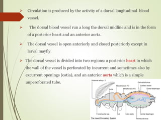
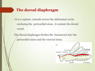
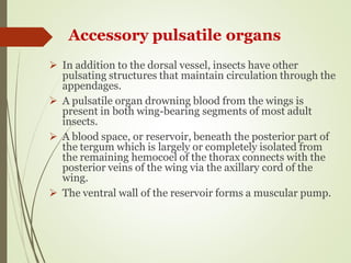
Zoo514
Dr. Reem Alajmi
Insect
Circulatory System
Circulatory system & Circulation
in insects
 Insects have an open circulatory system , the blood
(hemolymph) occupy all the body cavity (haemocoel ) , the
internal organs and tissues are exposed freely to it.
 The haemocoel is divided into three major sinuses by
fibro– muscular septa or diaphragms; a dorsal pericardia
sinus, a perivisceral sinus and a ventral perineural sinus.
 Circulation is produced by the activity of a dorsal longitudinal blood
vessel.
 The dorsal blood vessel run a long the dorsal midline and is in the form
of a posterior heart and an anterior aorta.
 The dorsal vessel is open anteriorly and closed posteriorly except in
larval mayfly.
 The dorsal vessel is divided into two regions: a posterior heart in which
the wall of the vessel is perforated by incurrent and sometimes also by
excurrent openings (ostia), and an anterior aorta which is a simple
unperoforated tube.
- It is a septum :extends across the abdominal cavity
enclosing the pericardial sinus , it contain the dorsal
vessel.
-The dorsal diaphragm divides the haemocoel into the
pericardial sinus and the visceral sinus.
The dorsal diaphragm
-The ventral diaphragm forms a
continuous ventral sheath .
- It extends from the prothorax to
the end of the body .
- It encloses the perineural sinus.
- The perineural sinus encloses the
nerve cord.
Ventral diaphragm
 It runs along the dorsal midline , for the whole of the body.
 It leaves the alimentary canal , just above the esophagus.
 It is divided into a posterior heart in which the wall is perforated and
an anterior aorta which is a simple unperforated tube.
 It collects blood from the abdominal cavity and discharges it in the
head .
 It is open anteriorly but closed posteriorly.
 The wall of the dorsal vessel is contractile. It consists of a single layer
of circular and spiral muscle fibrils.
Dorsal vessel
 It is often restricted to the abdomen or it may
extend as far as the prothorax .
 It is often a continuous tube that is not divided
into chambers, except in Orthoptera it is slightly
enlarged into ampullae, these are the points where
the ostia pierce the wall.
 The heart may be directly bound to the dorsal wall
or suspended from it by elastic filaments.
 A pair of alary musclesare attached laterally to the
walls of each chamber
The heart
Incurrent ostia
 They are vertical, slit-like openings in the lateral wall of
the heart.
 The maximum number may exist are 12 pairs, 9
abdominal and 3 thoracic.
 The anterior and posterior lips of each ostium are
reflexed into the heart to form a valve which permits the
flow of blood into the heart at diastole (expansion of the
heart), but prevents its outward passage at systole.
 These are ventro- lateral openings in the wall of the heart.
 No internal valves.
 Externally each opening is surrounded by a papilla of a
spongy multinucleate cells which expand during systole,
so the hemolymph is forced out, and contracts during
diastole, so that entry of blood is prevented.
Excurrent Ostia
Segmental vessels
 Most Blattodea and Mantodea have
no excurrent ostia, but the blood
leaves the heart via segmental vessels
that extend laterally.
The Aorta
 It is slender anterior prolongation of the heart, it
carries the blood from the heart .
 In addition to the dorsal vessel, insects have other
pulsating structures that maintain circulation through the
appendages.
 A pulsatile organ drowning blood from the wings is
present in both wing-bearing segments of most adult
insects.
 A blood space, or reservoir, beneath the posterior part of
the tergum which is largely or completely isolated from
the remaining hemocoel of the thorax connects with the
posterior veins of the wing via the axillary cord of the
wing.
 The ventral wall of the reservoir forms a muscular pump.
Accessory pulsatile organs
A pulsatile organ is also found at the base of each
antenna.
It consists of an ampulla from which a fine tube
extends almost to the tip of the antenna.
In most insects , the ampullae have dilator muscles.
Compression drives hemolymph into the antenna.
 Most insects have a longitudinal septum in the legs which
divides the lumen into two sinuses and permits a bidirectional
flow of blood within the leg.
 These are muscles that are closely associated
with the heart.
 In Orthoptera ten abdominal and two
thoracic pairs are present.
 In other insect species the number is
reduced.
 They form integral part of the dorsal
diaphragm which spreads between them as
connective tissue membrane .
Alary muscles
 Systole
- It is the contraction phase of the heartbeat.
-It results from the contraction of the muscles in the heart wall which starts
posteriorly and spreads forward as a Wave.
 Diastole
- It is the relaxation phase of the heartbeat.
- It results from relaxation of the muscles assisted by the elastic filaments
supporting the heart.
- After diastole there is a third phase in the heart cycle known as diastasis in which
the heart remains in the expanded stat.
- Frequency with which the heart contracts varies from 14beats/ minute to 150
beats/minute
Heartbeat
At systole
 The blood is pumped forwards through the heart, entering the
perivisceral sinus through the anterior opening of the aorta in the
head and through the excurrent ostia where these exist.
 The valves on the incurrent ostia prevent the escape of blood
through these openings.
 The force of blood leaving the aorta anteriorly tends to push blood
backwards in the perivisceral sinus.
 The backwards flow is aided by the movements of the dorsal
diaphragm and by the inflow of blood into the heart, through the
incurrent ostia, at diastole.
 Movements of the ventral diaphragm presumably help to maintain
the supply of blood to the ventral nerve cord.
Circulation
 Normal circulation
 Blood is drawn into the heart through the ostia.
At diastole
Circulation through the wings
 In most cases, blood is drawn in and out of the wings by the thoracic pulsatile
organs. Specific veins are contributed to this function.
 In Lepidotera, blood enters the wings along all the veins and go out again
during heartbeat reversal.
 The movement of blood out of the veins causes the tracheae inside them to
expand so that air is drawn into the wing.
 When heartbeat starts beating forwards again, the negative pressure on the
wing tracheae is removed and their elasticity causes them to contract.
 This produces a negative pressure in the hemolymph space outside the trachea
and blood flows in.
 In insects lacking leg pulsatile organs, the flow of blood
through the legs is thought to be maintained by pressure
differences at the base.
 Age and stage. Heartbeat is faster in adult than immature stages.
 Environmental factors such as temperature. Activity usually
stops above 45-50 ˚C and below 1-5 ˚C. Within this range, the rate is higher at
higher temperatures.
 Strong movement of the gut ( slow it or stop it). It is common for the heart to stop
beating sometimes for a few seconds.
 It is also common for the heartbeat to undergo periodic reversal, with waves of
contraction starting at the front. When this occurs, blood is forced out of the
incurrent ostia.
Factors affecting the rate of the heartbeat
 No nerve cells are associated with the heart , and the heart
muscles are the seat of automation ( Myogenic beat ). Although
the myogenic pattern maybe modulated naturally or hormonally.
 The alary muscles are activated by the contraction of the heart
and they exert tension that modifies the heartbeat.
 The direction of a beat, from back to front or vice versa, maybe
related to the distribution of blood pressure. Also it may be
related to the availability of oxygen. In the absence of a good
oxygen supply, the rate of heartbeat is strongly reduced.
Control of heartbeat
Hemolymph
 Insect blood is known as hemolymph.
 It circulate round the body, bathing the tissue directly.
 It consists of a fluid plasma in which blood cells,
hemocytes, are suspended.
 Making up usually less than 25% of an insect’s body weight.
 Hemolymph water comprises 20-25% of the total body
water in adult insects, but in caterpillars, the figure is close
to 50%. This reflects the important hydrostatic functions of
the hemolymph in these larval forms
 It transports hormones, nutrients and wastes and has a
role in, osmoregulation, temperature control, Immunity,
storage (water, carbohydrates and fats) and skeletal
function.
 It also plays an essential part in the moulting process
(McGavin 2001, Triplehorn 2005 ). An additional role of
the hemolymph in some orders, can be that of predatory
defence. It can contain unpalatable and odorous
chemicals that will act as a deterrent to predators
(Gullan 2005 ).
- In Plasma Inorganic & Organic Constituents
 Inorganic
 anion
1- Chloride is the most abundant inorganic anion in insect blood.
- Conc. high ( hemimetabolous )
- low ( holometabolous ) lower than 10% of total osmolar
concentration.
2- Phosohates & carbonates
Constituents of the plasma
 Cations
1- Sodium Na + is the most abundant cation.
- Conc. Varies with taxonomic position & diet.
- phytophagous : low
- predators : high
2- Potassium K & Magnisium Mg
- high in phytophagous
- low in carnivorous
- Blood –sucking e.g., Cimex & Stomoxys high Na + low K ++
• Na + and K ++: in the ionized form.
• Ca & Mg : are bound to macromolecules
 e.g., in locust : K conc, increases before moulting.
 These changes affect the behaviour of insect , since ; neuromuscular
junctions are directly exposed to Hl. & low concentration in K ++ raises
the muscle resting potential.
 Mg : concentration is high in phytophagous insects ( as a result of its
high conc. In the diet since it is a constituent of chlorophyll .
 Mg conc. Is low in Lepidoptera when the larvae stop feeding.
 It’s conc. Is low in adult than the larvae
 In aquatic insects, the ionic composition of hemolymph affected by the
composition of water.
Variation in cation concentration are wide
- Copper ( A constituent of tyrosinase)
- Iron
- Zinc
- Manganese
- These are found bound to organic complex & not found
as free ions .
Metallic traces
1- Free Amino Acids (A.A.)
- Very high level of free a.a. which constituting 33 –65 %
about ( 0.29 –2.43 gm) , most of the known amino
acids are present.
- They vary both quantitatively & qualitatively from one
species to another & in different stages of the same sp.
- They depend on those available in food.
- Changes in A.A. concentration at different stages in the
life cycle.
Organic constituent
- Numerous proteins are present in the hemolymph with a variety of
functions.
- They are not all present at the same time but, there are changes
through the life cycle.
- Proteins present in insect haemolymph are grouped as: storage
proteins, lipid transport proteins, vitelogenins, enzymes,
proteinase inhibitors, chromoproteins and a range of proteins
involved in the immune response of insects.
2-proteins
- Uric acid , urea and ammonia.
- Trehalose, a disaccharide, is the most characteristics sugar found
in insect hemolymph. Activity and starvation affect its conc. As it
is the main source of energy
- Glucose is also often present and other carbohydrates.
- Lipids. Marked increase may occur during development or flight ,
where lipid in fat body are mobilized as fuel for the flight.
- Organic acids
i ) e.g., Citrate : It is present in high concentration ( vary with
species and with development ).
ii ) Organic phosphates.
3- Other organic constituents
- Pigments
Respiratory pigments do not occur in insect blood. Except for :
Chironomus larvae : Haemoglobin is present in solution in plasma
- The inorganic and organic solutes present in the hemolymph contribute to its osmotic pressure. Variation in
plasma osmotic component in different orders. Three categories are present:
1- Na & Cl form most of the osmolar conc.
Basic type of insect blood & most of the arthropods.
e.g., Odonata. Ephemeroptera , Homoptera, Orthoptera.
2- Cl is low relative to Na which constitute 21-48 % of total osmolar conc, and A. A. are in high conc.
e.g., Diptera , Neuroptera& most Coleoptera.
3 – A.A. accounts for about 40% of the total osmolar conc. The other substance never exceed 10 % .
e.g., Lepidoptera & Hymenoptera.
Properties of the plasma
1- Osmotic pressure
2- Hemolymph pH
 Definition: pH is defined as the negative logarithm of hydrogen
ion concentration of a solution.
 pH= - log H+
 In most insects the blood is slightly acidic, pH 6.4 – 6.8, although
slightly alkaline values have been recorded in a dragonfly larva
and in the larva of midge Chironomus.
 Most enzymes work within a limited pH range so its control is
important .
 During normal activity the blood become acidic due to liberation
of acid metabolites (CO2), this tendency is buffered by
substances in the blood.
 The buffering capacity of insect blood (its ability to prevent
change in pH) is low in the normal physiological range, but it
increases sharply on either side of this range.
 Bicarbonates and phosphates are the most important buffers.
40
1- Nutritive materials are carried out from alimentary canal and
storage tissues to the sites at which they are metabolized.
2- Carrying excretory products from the place of origin to the
malpighian tubules.
3- Transporting hormones from endocrine organs to the site of action.
4- The plasma contains much more CO2 than O2, CO2 in solution
accounts for about 20% of the total blood but the remainder is
bound as bicarbonate. the greater affinity of blood to CO2 than to
O2 is important for the cyclic release of CO2 which occurs in some
insects.
5 – Store for some substances e.g., Trehalose as a source of energy and
its supply is achieved rapidly from fat body.
41 Functions of Plasma
6- Water storage:
- An insect can maintain the level of its cell fluids if the food is dry.
- The water itself functions as a hydrostatic skeleton in forms such
as larvae , where the cuticle is soft.
7- At molting the hydrostatic properties are used in expansion of
appendages. ( The amount of fluid present at this time influences
the ultimate size of the insect. e.g., larvae of Lucilia with more
moisture produce bigger adult).
8- Reflex bleeding results from the increase in hydrostatic pressure (
plasma is forced through weak spots or pores in the cuticle . In
species with this phenomena the blood contain some repellant
substances.
42
43 9- As a result of muscular activity; there is an increase in
hydrostatic properties ( this is responsible for the eversion of
the penis in male insects and the osmeterium ( Fleshy bifurcate
defensive device on the prothoracic segment in the larvae of
Papilinoidae) .
Osmeterium of Papilio xuthus Larva.
 The blood or hemolymph of insects consists of a
fluid plasma in which nucleated cells are
suspended , These cells are sessile. Several
different types of blood cells occur . Their functions
include: encapsulation , nodule formation and
phagocytosis which are considered as cellular
immunity , wound healing and intermediate
metabolism .
44
Insect Blood Cells (Hemocytes)
 Rowley and Ratcliffe (1981) and Gupta (1997, 1985, 1991)
grouped insect hemocytes into six cell types:
1- Prohemocytes.
2- Plasmatocytes.
3- Granulocytes (which are probably the same as cystocytes
or coagulocytes)
4- Spherulocytes.
5- Oenocytoids.
6- Adipohemocytes.
45
 Another group, Brehelin and Zachary (1986) make
the distinction among nine types of hemocytes:
prohemocytes, plasmatocytes, oenocytoids,
spherule cells, thrombocytoids, and four granular
hemocytes labeled GH1, GH2, GH3, and GH4.
46
47
48
 The previous figure (A–H) Light microscopy semithin sections (A–F) and
transmission electron microscopy (G–H) of C. aeruginosa hemocytes. (A)
Prohemocyte showing a high nucleus/cell ratio. (B) Granulocyte filled with the
typical granules. (C) Plasmatocyte showing fine, poorly stained, granulations.
(D)Coagulocyte showing nucleus with a heterochromatic aspect and the
perinuclear area lacking fine granules. (E) Oenocytoid with the typical large
andround profile with the central nucleus and the homogeneously stained
cytoplasm with few granules. (F) Spherule cells filled with the typical large
round granules. (G) Prohemocyte showing the large nucleus with an evident
nucleolus (nu), rough endoplasmic reticulum (r), small mitochondria (m) and
electron dense vesicles (v). (H) Granulocyte presenting a lobated nucleus (n)
with an evident nucleolus (nu). The cytoplasm show small mitochondria (m),
rough endoplasmic reticulum (r), Golgi complex (g), glycogen granules (gl)
and round electron lucent vesicles (asterisks). A microtubule ring (mr) just
beneath the plasma membrane and pseudopodia (p) are present. (I)
Contribution of trans Golgi vesicles (tgv) fusing towards the large immature
granules (white arrows). Glycogen aggregates are present (black arrows
49
Fig: Scanning electron microscopy of
Rhynchophorus ferrugineus larvae hemocytes
1- Prohemocytes
 The prohemocytes are very variable in shape but usually
small round cells . Nucleus fills almost the entire
cytoplasm. They are characterized by undeveloped
intracellular organelles .
 They rarely comprise more than 5% of the total hemocyte
population.
 They are the stem cells from which most other hemocyte
types are formed.
51
Types of Hemocytes
( Ultrastructure identification)
52
An illustration of the most common types of hemocytes from insect hemolymph. PR,
prohemocyte; PL, plasmatocyte; GR, granulocyte; SP, spherulocyte; CO, coagulocyte.
Several different shapes of plasmatocytes are shown in A, B, and C. The arrows
indicate transformations of cells that are believed to occur (according to Woodring,
1985).
 Plasmatocytes are variable in shape usually round and oval .
 They are the most abundant cell types. with a centrally placed,
spherical nucleus surrounded by well vacuolated cytoplasm.
 Plasmatocytes are characterized by large nucleus containing a
large nucleolus.
 They possess well developed Golgi apparatus and endoplasmic
reticulum as well as many lysosomes.
 The cells are capable of amoeboid movement and are
phagocytic.
 They are involved in phagocytosis and encapsulation of
foreign organisms invading insect so they are responsible
for most of the cellular immunity of the insect .
53
2 ) Plasmatocytes (phagocytes)
 Round or disc-shaped, with a relatively small nucleus
surrounded by cytoplasm, well developed granular rough
endoplasmic reticulum and Golgi apparatus.
 They discharge their contents on the surfaces of intruding
organisms as early part of the defense response.
 They probably derived from plasmatocytes.
54 3 ) Granulocytes
4) Spherulocytes
They are characterized by the large,
refractile spherules which may occupy 90%
of the cytoplasm.
They are not common although they are
found in most of the studied species.
Their function is unknown.
55
5- Oenocytoids
Mainly occur in Lepidoptera.
They exhibit little development of rough
endoplasmic reticulum or Golgi complexes,
but they have a complex array of microtubules.
Their function is unknown.
56
6- Adipohemocytes
They are characterized by containing lipid
droplets.
They contain well developed endoplasmic
reticulum and Golgi complexes.
57
Many other hemocyte types are present and they
vary according to the different insect species and
also to the type of investigation
58
59
Functions of Hemocytes
1- Phagocytosis
 The most common function of hemocytes is phagocytosis of
foreign particles, microorganisms and tissue debris that are
comparatively smaller in size than the cell.
 Many cells are capable of phagocytosis but plasmatocytes and
granular hemocyets are the most important.
 Endocytosis, especially phagocytosis, is important in both
metamorphosis and defense against disease, as well as in
routine cleaning up of dead or damaged cells.
60
 The hemocytes collect at the site of injuries,
forming a plug which helps to seal the wound,
proliferating and removing the dead tissues and
discard it.
 The blood of various insects when collected in
glass capillaries, usually clots within 10 – 15
minutes.
61 2- Wound healing and Coagulation
 On the other hand the blood of other insect species never clots or
coagulates.
 In Cockroaches , the apparent coagulum is merely a clump of
hemocytes; the blood cells become round, develop thread – like
pseudopodia, and agglutinate to form a plug which is the essential
factor of the clot. When blood cells coagulates due to the presence of
hyaline hemocytes, these cells extrude thread – like pseudopodial
expansions which fuse to form a meshwork in which the other cells
are entangled.
62
 This occurs in response to invasion of the body by large quantities of
particulate matter (especially bacterial aggregates) that cannot be
removed effectively by phagocytosis.
 This process includes two phases:
First: Within minutes of the foreign matter arriving in the body cavity,
it is surrounded either by granulocytes or by specific plasma proteins
and by material discharged from coagulocytes.
63 3- Nodule formation
- This initial layer, which is often melanized includes
“plasmatocyte-spreading peptide” that induces
plasmatocytes to aggregate and adhere to a foreign
surface
 In the second phase, which takes several hours to
complete, plasmatocytes surround the melanized
core, become flattened, and form a multicellular
sheath comparable to that seen after encapsulation
(Strand, 2002).
64
 Encapsulation is essentially nodule formation on a larger
scale.
 After the invader is initially coated with a thin layer of
granulocytes or plasma proteins, layers of plasmatocytes
surround it (Nappi, 1975; Lavine and Strand, 2001, 2002)
 This reaction on the part of hemocytes forms an insect’s
most important defense mechanism against metazoan
endoparasites such as nematodes and insects.
65 4- Encapsulation
 An encapsulated organism almost always dies, as a
result of starvation, asphyxiation, and perhaps
poisoning by quinones, antibacterial peptides,
hydrogen peroxide and nitric oxide.
66
 All tissues and organs of insects are separated from
the body cavity by connective tissue membranes and
basement membranes.
 Blood cells usually collect around foreign bodies and
form capsules. Usually the cellular character of these
capsules persists, but sometimes the nuclei disappear
and a capsule of connective tissue is produced. The
hemocytes are concerned in connective tissue
formation.
67 5- Connective tissue formation
6- Metabolic and Homeostatic Functions
 Hemocytes have been implicated in a variety of
metabolic and homeostatic functions.
 Cells have been shown to contain, for example,
glycogen, mucopolysaccharide, lipid, and protein, it
has been suggested that hemocytes are important in
storage of nutrients and their distribution to growing
tissues, formation of connective tissue, synthesis of
chitin, and maintenance of hemolymph sugar level.
68
 Cells might be important in hemolymph amino
acid homeostasis.
 Certain hemocytes (spherule cells in Diptera,
oenocytoids in other insects) contain enzymes for
metabolism of tyrosine, derivatives of which are
important in tanning and/or darkening of cuticle.
69
Numbers of Hemocytes
 Small insects have many fewer hemocytes than
large insects.
 Number of hemocytes are variable between species
and in different life stage in the same species.
 Count of the number of cells per unit volume of
hemolymph (usually called total hemocyte count)
may not reflect the total number of hemocytes in
circulation because the blood volume varies.
70
Hemocyte profile
 The relative abundance of different types of
hemocytes (called the hemocyte profile or
differential hemocyte count) is not constant.
 Plasmatocytes and granulocytes are usually the
most abundant, often comprising about 80% of the
total hemocyte population.
 There number varied again with different life stage
and physiological state.
71

insect_circulatory_system................

  • 1.
  • 2.
    Circulatory system &Circulation in insects  Insects have an open circulatory system , the blood (hemolymph) occupy all the body cavity (haemocoel ) , the internal organs and tissues are exposed freely to it.  The haemocoel is divided into three major sinuses by fibro– muscular septa or diaphragms; a dorsal pericardia sinus, a perivisceral sinus and a ventral perineural sinus.
  • 3.
     Circulation isproduced by the activity of a dorsal longitudinal blood vessel.  The dorsal blood vessel run a long the dorsal midline and is in the form of a posterior heart and an anterior aorta.  The dorsal vessel is open anteriorly and closed posteriorly except in larval mayfly.  The dorsal vessel is divided into two regions: a posterior heart in which the wall of the vessel is perforated by incurrent and sometimes also by excurrent openings (ostia), and an anterior aorta which is a simple unperoforated tube.
  • 5.
    - It isa septum :extends across the abdominal cavity enclosing the pericardial sinus , it contain the dorsal vessel. -The dorsal diaphragm divides the haemocoel into the pericardial sinus and the visceral sinus. The dorsal diaphragm
  • 6.
    -The ventral diaphragmforms a continuous ventral sheath . - It extends from the prothorax to the end of the body . - It encloses the perineural sinus. - The perineural sinus encloses the nerve cord. Ventral diaphragm
  • 8.
     It runsalong the dorsal midline , for the whole of the body.  It leaves the alimentary canal , just above the esophagus.  It is divided into a posterior heart in which the wall is perforated and an anterior aorta which is a simple unperforated tube.  It collects blood from the abdominal cavity and discharges it in the head .  It is open anteriorly but closed posteriorly.  The wall of the dorsal vessel is contractile. It consists of a single layer of circular and spiral muscle fibrils. Dorsal vessel
  • 9.
     It isoften restricted to the abdomen or it may extend as far as the prothorax .  It is often a continuous tube that is not divided into chambers, except in Orthoptera it is slightly enlarged into ampullae, these are the points where the ostia pierce the wall.  The heart may be directly bound to the dorsal wall or suspended from it by elastic filaments.  A pair of alary musclesare attached laterally to the walls of each chamber The heart
  • 10.
    Incurrent ostia  Theyare vertical, slit-like openings in the lateral wall of the heart.  The maximum number may exist are 12 pairs, 9 abdominal and 3 thoracic.  The anterior and posterior lips of each ostium are reflexed into the heart to form a valve which permits the flow of blood into the heart at diastole (expansion of the heart), but prevents its outward passage at systole.
  • 11.
     These areventro- lateral openings in the wall of the heart.  No internal valves.  Externally each opening is surrounded by a papilla of a spongy multinucleate cells which expand during systole, so the hemolymph is forced out, and contracts during diastole, so that entry of blood is prevented. Excurrent Ostia
  • 12.
    Segmental vessels  MostBlattodea and Mantodea have no excurrent ostia, but the blood leaves the heart via segmental vessels that extend laterally.
  • 13.
    The Aorta  Itis slender anterior prolongation of the heart, it carries the blood from the heart .
  • 14.
     In additionto the dorsal vessel, insects have other pulsating structures that maintain circulation through the appendages.  A pulsatile organ drowning blood from the wings is present in both wing-bearing segments of most adult insects.  A blood space, or reservoir, beneath the posterior part of the tergum which is largely or completely isolated from the remaining hemocoel of the thorax connects with the posterior veins of the wing via the axillary cord of the wing.  The ventral wall of the reservoir forms a muscular pump. Accessory pulsatile organs
  • 16.
    A pulsatile organis also found at the base of each antenna. It consists of an ampulla from which a fine tube extends almost to the tip of the antenna. In most insects , the ampullae have dilator muscles. Compression drives hemolymph into the antenna.
  • 17.
     Most insectshave a longitudinal septum in the legs which divides the lumen into two sinuses and permits a bidirectional flow of blood within the leg.
  • 18.
     These aremuscles that are closely associated with the heart.  In Orthoptera ten abdominal and two thoracic pairs are present.  In other insect species the number is reduced.  They form integral part of the dorsal diaphragm which spreads between them as connective tissue membrane . Alary muscles
  • 19.
     Systole - Itis the contraction phase of the heartbeat. -It results from the contraction of the muscles in the heart wall which starts posteriorly and spreads forward as a Wave.  Diastole - It is the relaxation phase of the heartbeat. - It results from relaxation of the muscles assisted by the elastic filaments supporting the heart. - After diastole there is a third phase in the heart cycle known as diastasis in which the heart remains in the expanded stat. - Frequency with which the heart contracts varies from 14beats/ minute to 150 beats/minute Heartbeat
  • 20.
    At systole  Theblood is pumped forwards through the heart, entering the perivisceral sinus through the anterior opening of the aorta in the head and through the excurrent ostia where these exist.  The valves on the incurrent ostia prevent the escape of blood through these openings.  The force of blood leaving the aorta anteriorly tends to push blood backwards in the perivisceral sinus.  The backwards flow is aided by the movements of the dorsal diaphragm and by the inflow of blood into the heart, through the incurrent ostia, at diastole.  Movements of the ventral diaphragm presumably help to maintain the supply of blood to the ventral nerve cord. Circulation  Normal circulation
  • 21.
     Blood isdrawn into the heart through the ostia. At diastole Circulation through the wings  In most cases, blood is drawn in and out of the wings by the thoracic pulsatile organs. Specific veins are contributed to this function.  In Lepidotera, blood enters the wings along all the veins and go out again during heartbeat reversal.  The movement of blood out of the veins causes the tracheae inside them to expand so that air is drawn into the wing.  When heartbeat starts beating forwards again, the negative pressure on the wing tracheae is removed and their elasticity causes them to contract.  This produces a negative pressure in the hemolymph space outside the trachea and blood flows in.
  • 22.
     In insectslacking leg pulsatile organs, the flow of blood through the legs is thought to be maintained by pressure differences at the base.
  • 24.
     Age andstage. Heartbeat is faster in adult than immature stages.  Environmental factors such as temperature. Activity usually stops above 45-50 ˚C and below 1-5 ˚C. Within this range, the rate is higher at higher temperatures.  Strong movement of the gut ( slow it or stop it). It is common for the heart to stop beating sometimes for a few seconds.  It is also common for the heartbeat to undergo periodic reversal, with waves of contraction starting at the front. When this occurs, blood is forced out of the incurrent ostia. Factors affecting the rate of the heartbeat
  • 25.
     No nervecells are associated with the heart , and the heart muscles are the seat of automation ( Myogenic beat ). Although the myogenic pattern maybe modulated naturally or hormonally.  The alary muscles are activated by the contraction of the heart and they exert tension that modifies the heartbeat.  The direction of a beat, from back to front or vice versa, maybe related to the distribution of blood pressure. Also it may be related to the availability of oxygen. In the absence of a good oxygen supply, the rate of heartbeat is strongly reduced. Control of heartbeat
  • 26.
    Hemolymph  Insect bloodis known as hemolymph.  It circulate round the body, bathing the tissue directly.  It consists of a fluid plasma in which blood cells, hemocytes, are suspended.  Making up usually less than 25% of an insect’s body weight.  Hemolymph water comprises 20-25% of the total body water in adult insects, but in caterpillars, the figure is close to 50%. This reflects the important hydrostatic functions of the hemolymph in these larval forms
  • 27.
     It transportshormones, nutrients and wastes and has a role in, osmoregulation, temperature control, Immunity, storage (water, carbohydrates and fats) and skeletal function.  It also plays an essential part in the moulting process (McGavin 2001, Triplehorn 2005 ). An additional role of the hemolymph in some orders, can be that of predatory defence. It can contain unpalatable and odorous chemicals that will act as a deterrent to predators (Gullan 2005 ).
  • 29.
    - In PlasmaInorganic & Organic Constituents  Inorganic  anion 1- Chloride is the most abundant inorganic anion in insect blood. - Conc. high ( hemimetabolous ) - low ( holometabolous ) lower than 10% of total osmolar concentration. 2- Phosohates & carbonates Constituents of the plasma
  • 30.
     Cations 1- SodiumNa + is the most abundant cation. - Conc. Varies with taxonomic position & diet. - phytophagous : low - predators : high 2- Potassium K & Magnisium Mg - high in phytophagous - low in carnivorous - Blood –sucking e.g., Cimex & Stomoxys high Na + low K ++ • Na + and K ++: in the ionized form. • Ca & Mg : are bound to macromolecules
  • 31.
     e.g., inlocust : K conc, increases before moulting.  These changes affect the behaviour of insect , since ; neuromuscular junctions are directly exposed to Hl. & low concentration in K ++ raises the muscle resting potential.  Mg : concentration is high in phytophagous insects ( as a result of its high conc. In the diet since it is a constituent of chlorophyll .  Mg conc. Is low in Lepidoptera when the larvae stop feeding.  It’s conc. Is low in adult than the larvae  In aquatic insects, the ionic composition of hemolymph affected by the composition of water. Variation in cation concentration are wide
  • 32.
    - Copper (A constituent of tyrosinase) - Iron - Zinc - Manganese - These are found bound to organic complex & not found as free ions . Metallic traces
  • 33.
    1- Free AminoAcids (A.A.) - Very high level of free a.a. which constituting 33 –65 % about ( 0.29 –2.43 gm) , most of the known amino acids are present. - They vary both quantitatively & qualitatively from one species to another & in different stages of the same sp. - They depend on those available in food. - Changes in A.A. concentration at different stages in the life cycle. Organic constituent
  • 34.
    - Numerous proteinsare present in the hemolymph with a variety of functions. - They are not all present at the same time but, there are changes through the life cycle. - Proteins present in insect haemolymph are grouped as: storage proteins, lipid transport proteins, vitelogenins, enzymes, proteinase inhibitors, chromoproteins and a range of proteins involved in the immune response of insects. 2-proteins
  • 35.
    - Uric acid, urea and ammonia. - Trehalose, a disaccharide, is the most characteristics sugar found in insect hemolymph. Activity and starvation affect its conc. As it is the main source of energy - Glucose is also often present and other carbohydrates. - Lipids. Marked increase may occur during development or flight , where lipid in fat body are mobilized as fuel for the flight. - Organic acids i ) e.g., Citrate : It is present in high concentration ( vary with species and with development ). ii ) Organic phosphates. 3- Other organic constituents
  • 36.
    - Pigments Respiratory pigmentsdo not occur in insect blood. Except for : Chironomus larvae : Haemoglobin is present in solution in plasma
  • 37.
    - The inorganicand organic solutes present in the hemolymph contribute to its osmotic pressure. Variation in plasma osmotic component in different orders. Three categories are present: 1- Na & Cl form most of the osmolar conc. Basic type of insect blood & most of the arthropods. e.g., Odonata. Ephemeroptera , Homoptera, Orthoptera. 2- Cl is low relative to Na which constitute 21-48 % of total osmolar conc, and A. A. are in high conc. e.g., Diptera , Neuroptera& most Coleoptera. 3 – A.A. accounts for about 40% of the total osmolar conc. The other substance never exceed 10 % . e.g., Lepidoptera & Hymenoptera. Properties of the plasma 1- Osmotic pressure
  • 39.
    2- Hemolymph pH Definition: pH is defined as the negative logarithm of hydrogen ion concentration of a solution.  pH= - log H+  In most insects the blood is slightly acidic, pH 6.4 – 6.8, although slightly alkaline values have been recorded in a dragonfly larva and in the larva of midge Chironomus.  Most enzymes work within a limited pH range so its control is important .  During normal activity the blood become acidic due to liberation of acid metabolites (CO2), this tendency is buffered by substances in the blood.
  • 40.
     The bufferingcapacity of insect blood (its ability to prevent change in pH) is low in the normal physiological range, but it increases sharply on either side of this range.  Bicarbonates and phosphates are the most important buffers. 40
  • 41.
    1- Nutritive materialsare carried out from alimentary canal and storage tissues to the sites at which they are metabolized. 2- Carrying excretory products from the place of origin to the malpighian tubules. 3- Transporting hormones from endocrine organs to the site of action. 4- The plasma contains much more CO2 than O2, CO2 in solution accounts for about 20% of the total blood but the remainder is bound as bicarbonate. the greater affinity of blood to CO2 than to O2 is important for the cyclic release of CO2 which occurs in some insects. 5 – Store for some substances e.g., Trehalose as a source of energy and its supply is achieved rapidly from fat body. 41 Functions of Plasma
  • 42.
    6- Water storage: -An insect can maintain the level of its cell fluids if the food is dry. - The water itself functions as a hydrostatic skeleton in forms such as larvae , where the cuticle is soft. 7- At molting the hydrostatic properties are used in expansion of appendages. ( The amount of fluid present at this time influences the ultimate size of the insect. e.g., larvae of Lucilia with more moisture produce bigger adult). 8- Reflex bleeding results from the increase in hydrostatic pressure ( plasma is forced through weak spots or pores in the cuticle . In species with this phenomena the blood contain some repellant substances. 42
  • 43.
    43 9- Asa result of muscular activity; there is an increase in hydrostatic properties ( this is responsible for the eversion of the penis in male insects and the osmeterium ( Fleshy bifurcate defensive device on the prothoracic segment in the larvae of Papilinoidae) . Osmeterium of Papilio xuthus Larva.
  • 44.
     The bloodor hemolymph of insects consists of a fluid plasma in which nucleated cells are suspended , These cells are sessile. Several different types of blood cells occur . Their functions include: encapsulation , nodule formation and phagocytosis which are considered as cellular immunity , wound healing and intermediate metabolism . 44 Insect Blood Cells (Hemocytes)
  • 45.
     Rowley andRatcliffe (1981) and Gupta (1997, 1985, 1991) grouped insect hemocytes into six cell types: 1- Prohemocytes. 2- Plasmatocytes. 3- Granulocytes (which are probably the same as cystocytes or coagulocytes) 4- Spherulocytes. 5- Oenocytoids. 6- Adipohemocytes. 45
  • 46.
     Another group,Brehelin and Zachary (1986) make the distinction among nine types of hemocytes: prohemocytes, plasmatocytes, oenocytoids, spherule cells, thrombocytoids, and four granular hemocytes labeled GH1, GH2, GH3, and GH4. 46
  • 47.
  • 48.
  • 49.
     The previousfigure (A–H) Light microscopy semithin sections (A–F) and transmission electron microscopy (G–H) of C. aeruginosa hemocytes. (A) Prohemocyte showing a high nucleus/cell ratio. (B) Granulocyte filled with the typical granules. (C) Plasmatocyte showing fine, poorly stained, granulations. (D)Coagulocyte showing nucleus with a heterochromatic aspect and the perinuclear area lacking fine granules. (E) Oenocytoid with the typical large andround profile with the central nucleus and the homogeneously stained cytoplasm with few granules. (F) Spherule cells filled with the typical large round granules. (G) Prohemocyte showing the large nucleus with an evident nucleolus (nu), rough endoplasmic reticulum (r), small mitochondria (m) and electron dense vesicles (v). (H) Granulocyte presenting a lobated nucleus (n) with an evident nucleolus (nu). The cytoplasm show small mitochondria (m), rough endoplasmic reticulum (r), Golgi complex (g), glycogen granules (gl) and round electron lucent vesicles (asterisks). A microtubule ring (mr) just beneath the plasma membrane and pseudopodia (p) are present. (I) Contribution of trans Golgi vesicles (tgv) fusing towards the large immature granules (white arrows). Glycogen aggregates are present (black arrows 49
  • 50.
    Fig: Scanning electronmicroscopy of Rhynchophorus ferrugineus larvae hemocytes
  • 51.
    1- Prohemocytes  Theprohemocytes are very variable in shape but usually small round cells . Nucleus fills almost the entire cytoplasm. They are characterized by undeveloped intracellular organelles .  They rarely comprise more than 5% of the total hemocyte population.  They are the stem cells from which most other hemocyte types are formed. 51 Types of Hemocytes ( Ultrastructure identification)
  • 52.
    52 An illustration ofthe most common types of hemocytes from insect hemolymph. PR, prohemocyte; PL, plasmatocyte; GR, granulocyte; SP, spherulocyte; CO, coagulocyte. Several different shapes of plasmatocytes are shown in A, B, and C. The arrows indicate transformations of cells that are believed to occur (according to Woodring, 1985).
  • 53.
     Plasmatocytes arevariable in shape usually round and oval .  They are the most abundant cell types. with a centrally placed, spherical nucleus surrounded by well vacuolated cytoplasm.  Plasmatocytes are characterized by large nucleus containing a large nucleolus.  They possess well developed Golgi apparatus and endoplasmic reticulum as well as many lysosomes.  The cells are capable of amoeboid movement and are phagocytic.  They are involved in phagocytosis and encapsulation of foreign organisms invading insect so they are responsible for most of the cellular immunity of the insect . 53 2 ) Plasmatocytes (phagocytes)
  • 54.
     Round ordisc-shaped, with a relatively small nucleus surrounded by cytoplasm, well developed granular rough endoplasmic reticulum and Golgi apparatus.  They discharge their contents on the surfaces of intruding organisms as early part of the defense response.  They probably derived from plasmatocytes. 54 3 ) Granulocytes
  • 55.
    4) Spherulocytes They arecharacterized by the large, refractile spherules which may occupy 90% of the cytoplasm. They are not common although they are found in most of the studied species. Their function is unknown. 55
  • 56.
    5- Oenocytoids Mainly occurin Lepidoptera. They exhibit little development of rough endoplasmic reticulum or Golgi complexes, but they have a complex array of microtubules. Their function is unknown. 56
  • 57.
    6- Adipohemocytes They arecharacterized by containing lipid droplets. They contain well developed endoplasmic reticulum and Golgi complexes. 57
  • 58.
    Many other hemocytetypes are present and they vary according to the different insect species and also to the type of investigation 58
  • 59.
  • 60.
    Functions of Hemocytes 1-Phagocytosis  The most common function of hemocytes is phagocytosis of foreign particles, microorganisms and tissue debris that are comparatively smaller in size than the cell.  Many cells are capable of phagocytosis but plasmatocytes and granular hemocyets are the most important.  Endocytosis, especially phagocytosis, is important in both metamorphosis and defense against disease, as well as in routine cleaning up of dead or damaged cells. 60
  • 61.
     The hemocytescollect at the site of injuries, forming a plug which helps to seal the wound, proliferating and removing the dead tissues and discard it.  The blood of various insects when collected in glass capillaries, usually clots within 10 – 15 minutes. 61 2- Wound healing and Coagulation
  • 62.
     On theother hand the blood of other insect species never clots or coagulates.  In Cockroaches , the apparent coagulum is merely a clump of hemocytes; the blood cells become round, develop thread – like pseudopodia, and agglutinate to form a plug which is the essential factor of the clot. When blood cells coagulates due to the presence of hyaline hemocytes, these cells extrude thread – like pseudopodial expansions which fuse to form a meshwork in which the other cells are entangled. 62
  • 63.
     This occursin response to invasion of the body by large quantities of particulate matter (especially bacterial aggregates) that cannot be removed effectively by phagocytosis.  This process includes two phases: First: Within minutes of the foreign matter arriving in the body cavity, it is surrounded either by granulocytes or by specific plasma proteins and by material discharged from coagulocytes. 63 3- Nodule formation
  • 64.
    - This initiallayer, which is often melanized includes “plasmatocyte-spreading peptide” that induces plasmatocytes to aggregate and adhere to a foreign surface  In the second phase, which takes several hours to complete, plasmatocytes surround the melanized core, become flattened, and form a multicellular sheath comparable to that seen after encapsulation (Strand, 2002). 64
  • 65.
     Encapsulation isessentially nodule formation on a larger scale.  After the invader is initially coated with a thin layer of granulocytes or plasma proteins, layers of plasmatocytes surround it (Nappi, 1975; Lavine and Strand, 2001, 2002)  This reaction on the part of hemocytes forms an insect’s most important defense mechanism against metazoan endoparasites such as nematodes and insects. 65 4- Encapsulation
  • 66.
     An encapsulatedorganism almost always dies, as a result of starvation, asphyxiation, and perhaps poisoning by quinones, antibacterial peptides, hydrogen peroxide and nitric oxide. 66
  • 67.
     All tissuesand organs of insects are separated from the body cavity by connective tissue membranes and basement membranes.  Blood cells usually collect around foreign bodies and form capsules. Usually the cellular character of these capsules persists, but sometimes the nuclei disappear and a capsule of connective tissue is produced. The hemocytes are concerned in connective tissue formation. 67 5- Connective tissue formation
  • 68.
    6- Metabolic andHomeostatic Functions  Hemocytes have been implicated in a variety of metabolic and homeostatic functions.  Cells have been shown to contain, for example, glycogen, mucopolysaccharide, lipid, and protein, it has been suggested that hemocytes are important in storage of nutrients and their distribution to growing tissues, formation of connective tissue, synthesis of chitin, and maintenance of hemolymph sugar level. 68
  • 69.
     Cells mightbe important in hemolymph amino acid homeostasis.  Certain hemocytes (spherule cells in Diptera, oenocytoids in other insects) contain enzymes for metabolism of tyrosine, derivatives of which are important in tanning and/or darkening of cuticle. 69
  • 70.
    Numbers of Hemocytes Small insects have many fewer hemocytes than large insects.  Number of hemocytes are variable between species and in different life stage in the same species.  Count of the number of cells per unit volume of hemolymph (usually called total hemocyte count) may not reflect the total number of hemocytes in circulation because the blood volume varies. 70
  • 71.
    Hemocyte profile  Therelative abundance of different types of hemocytes (called the hemocyte profile or differential hemocyte count) is not constant.  Plasmatocytes and granulocytes are usually the most abundant, often comprising about 80% of the total hemocyte population.  There number varied again with different life stage and physiological state. 71