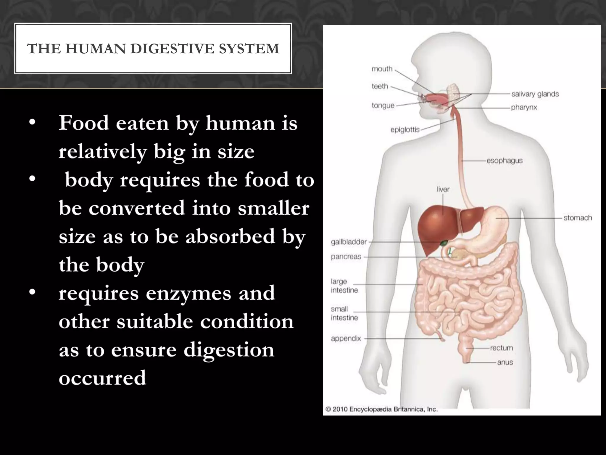
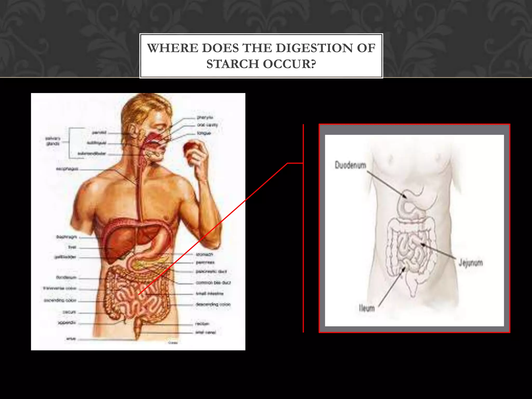
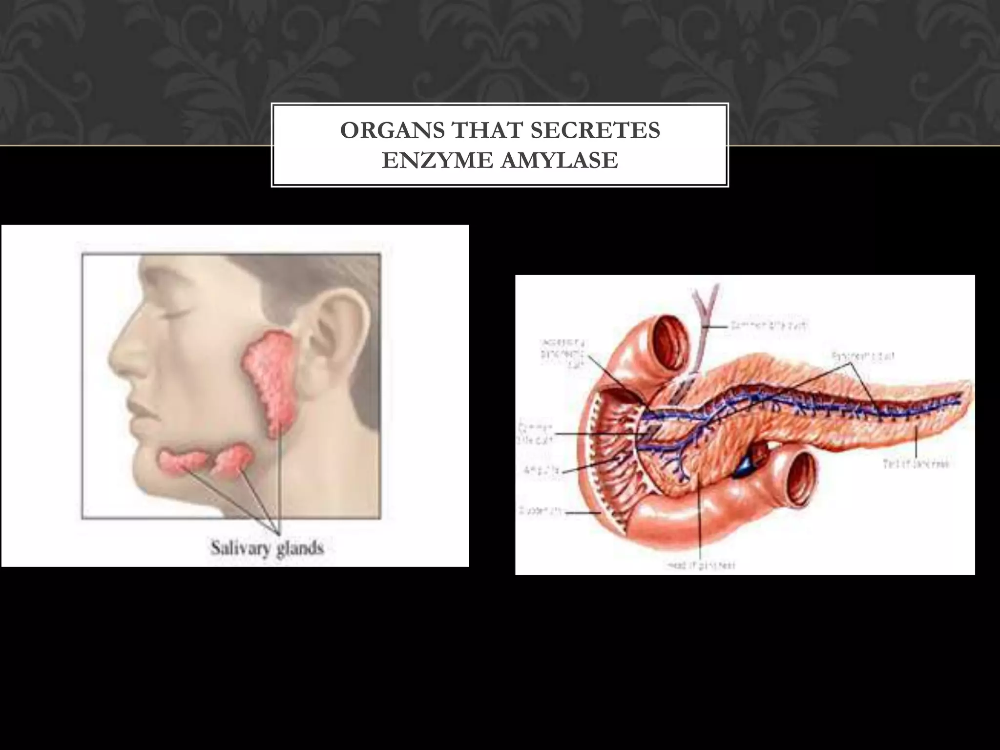
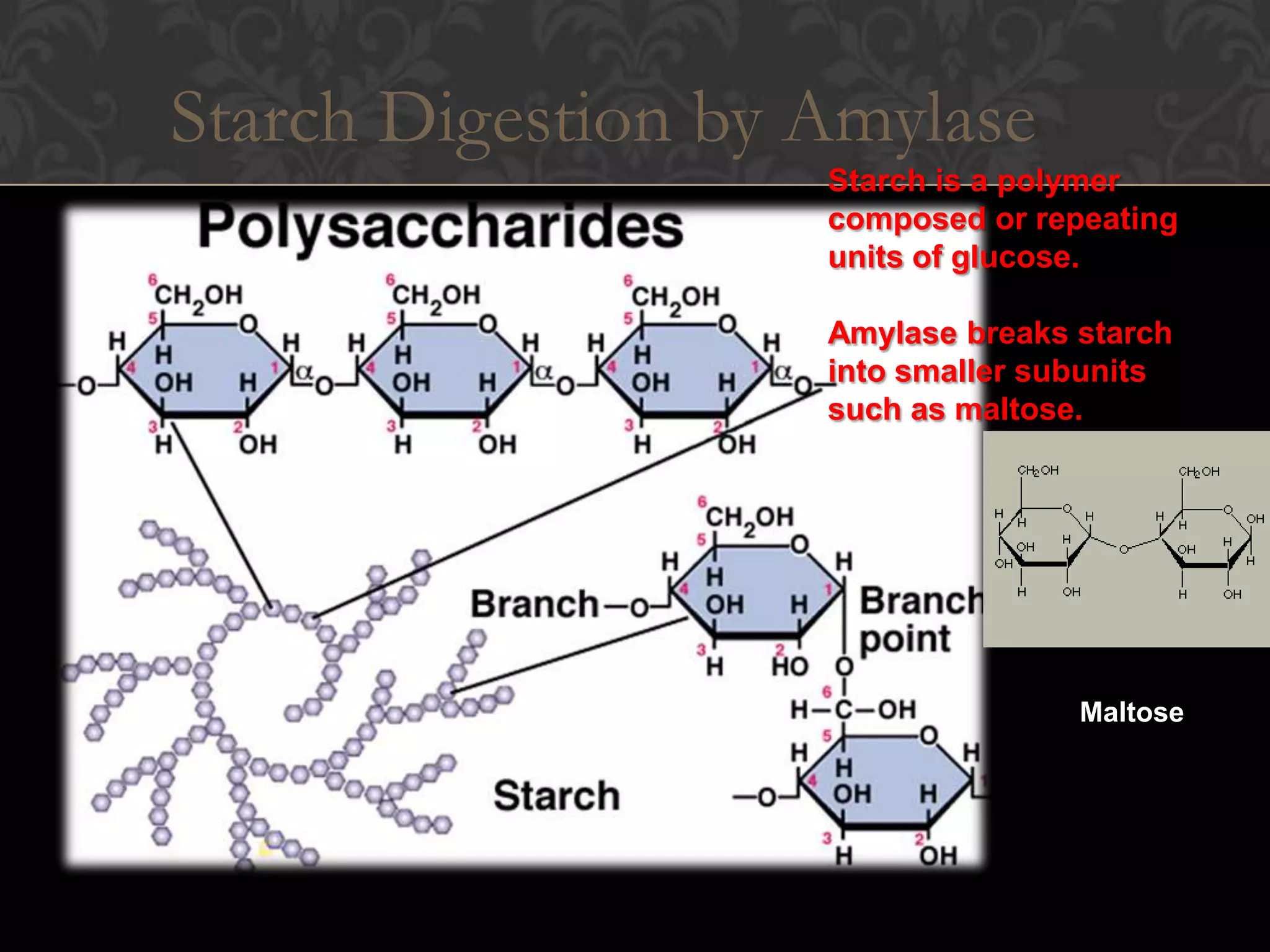
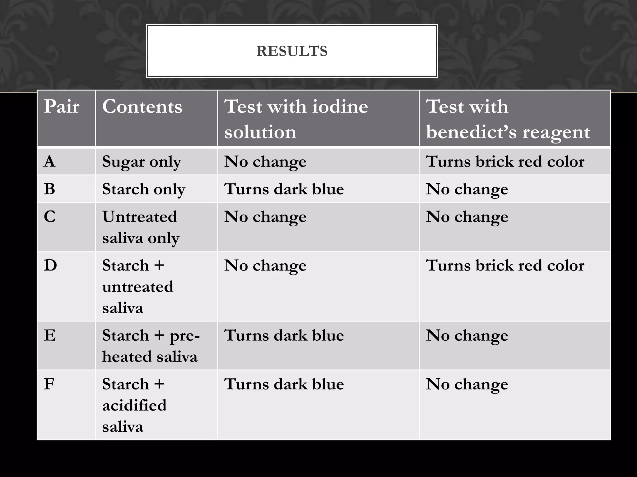
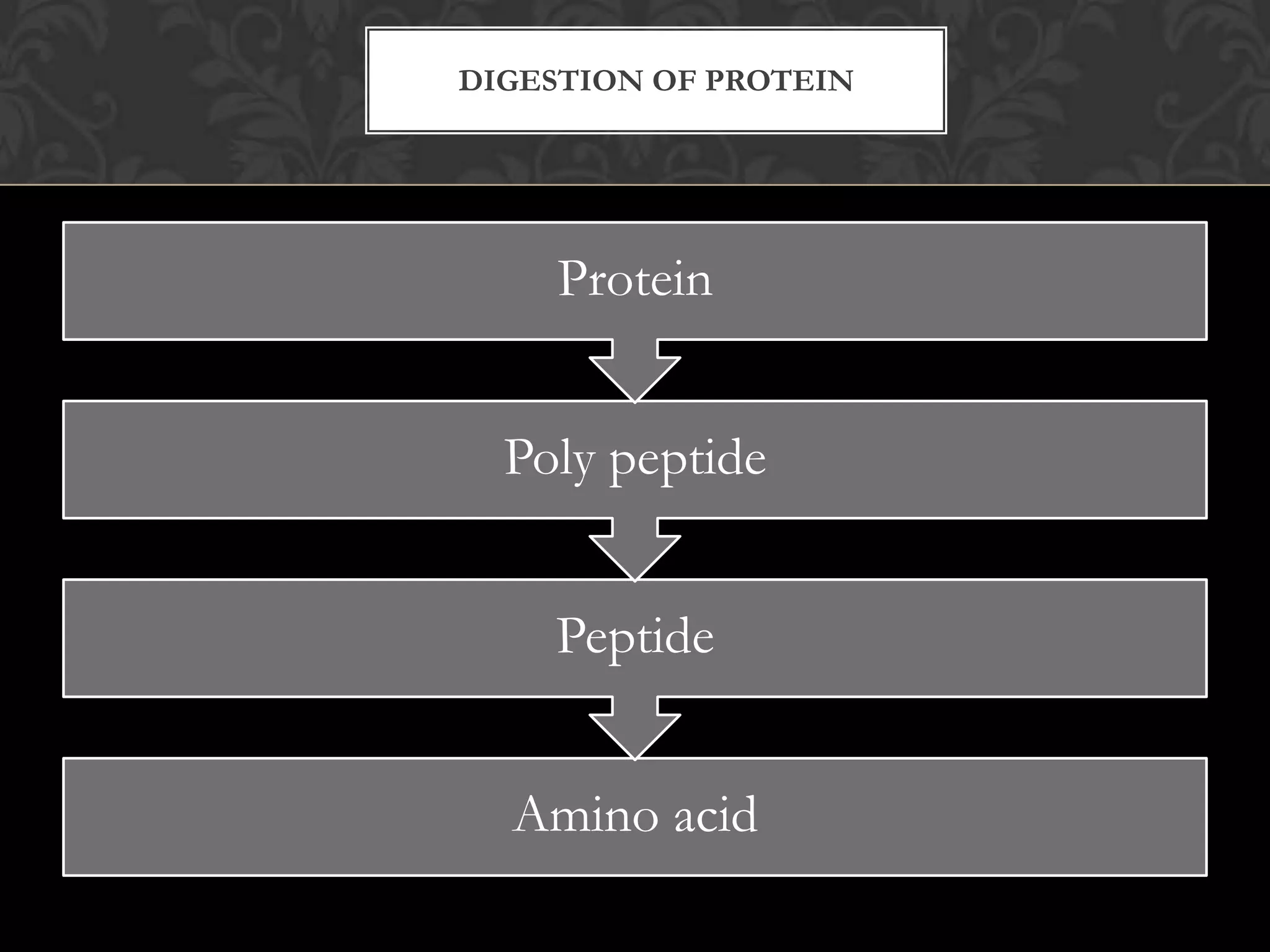
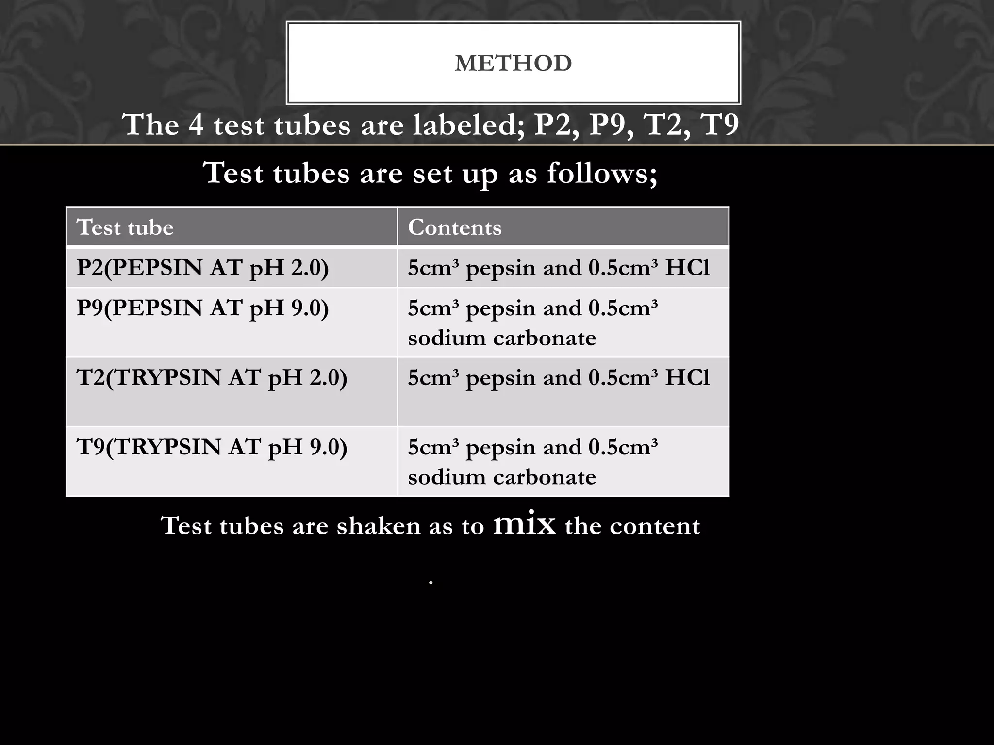
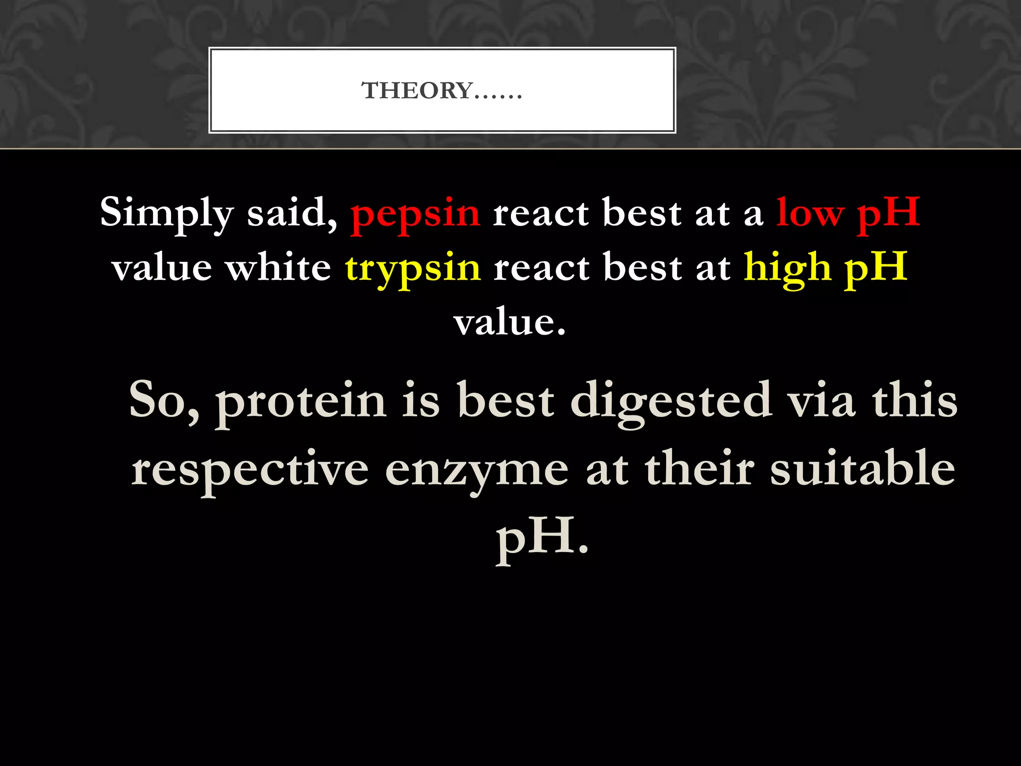
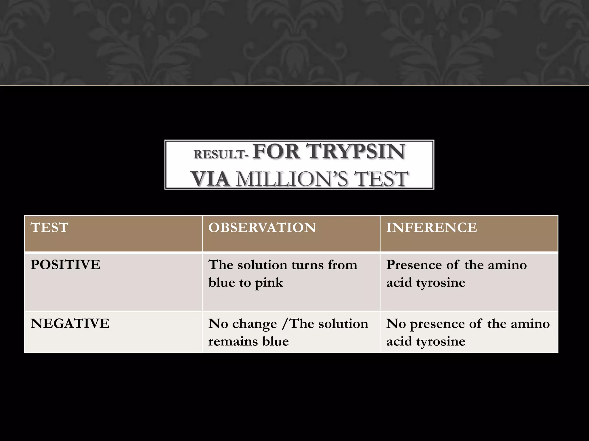
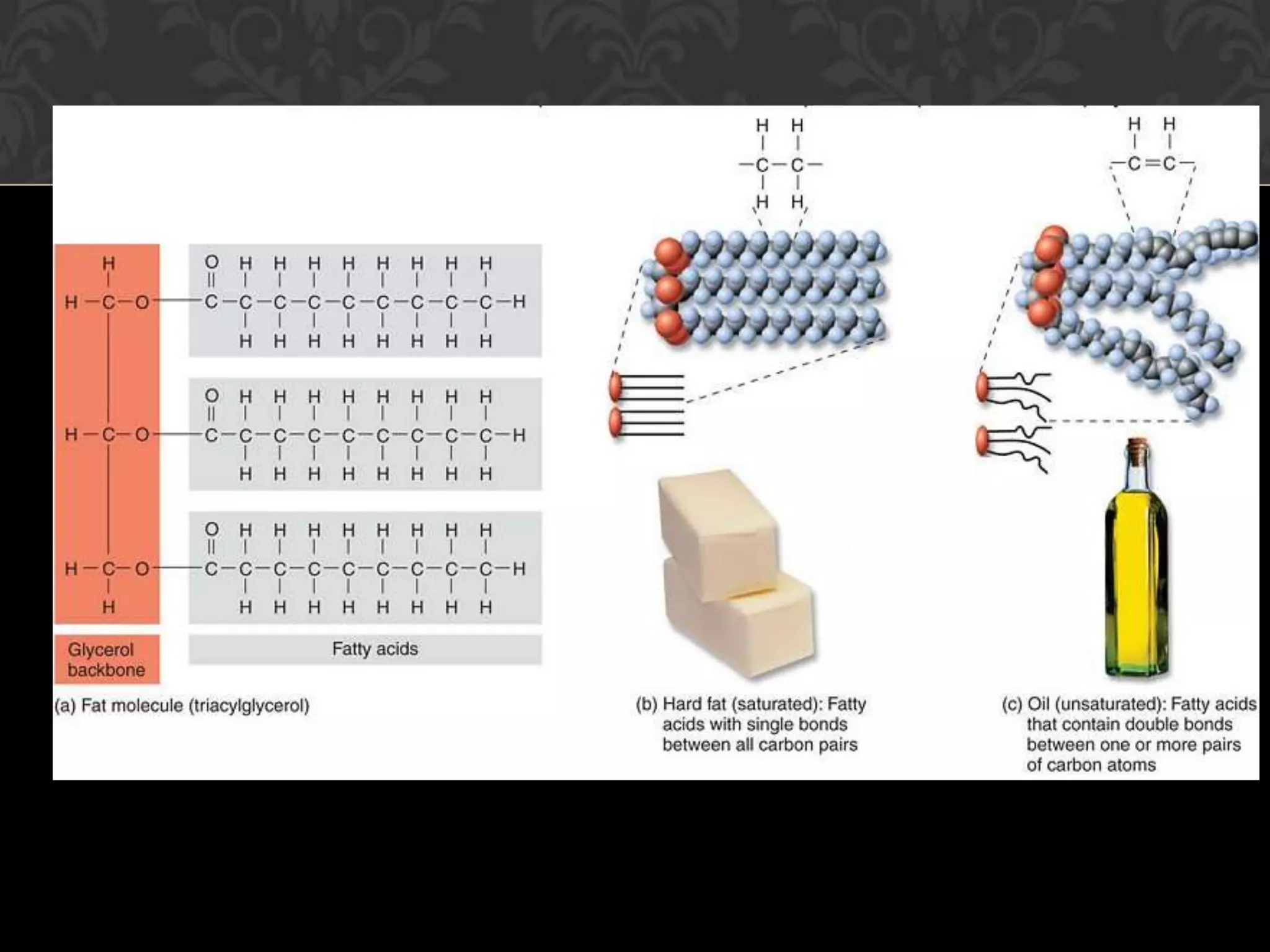
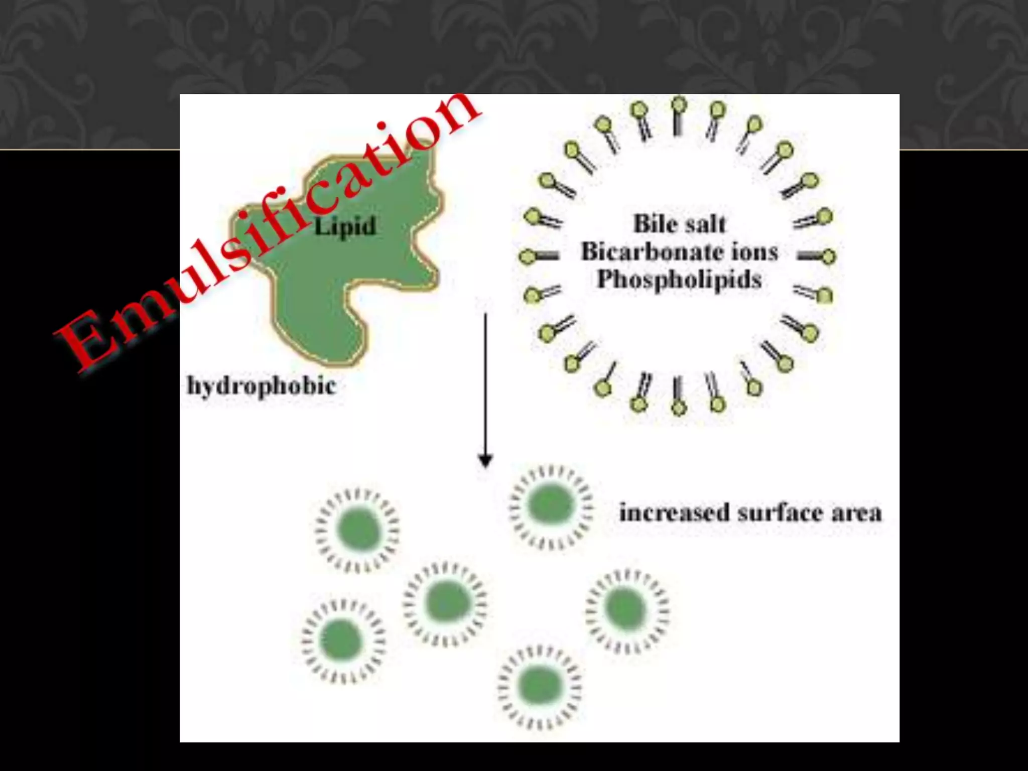
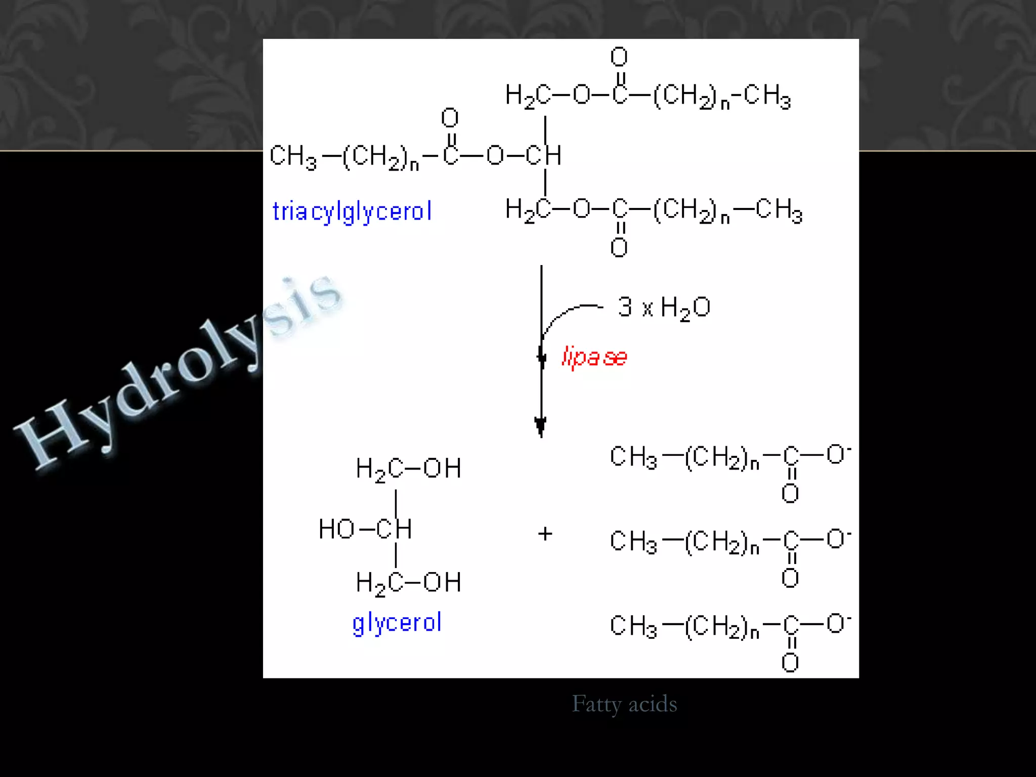
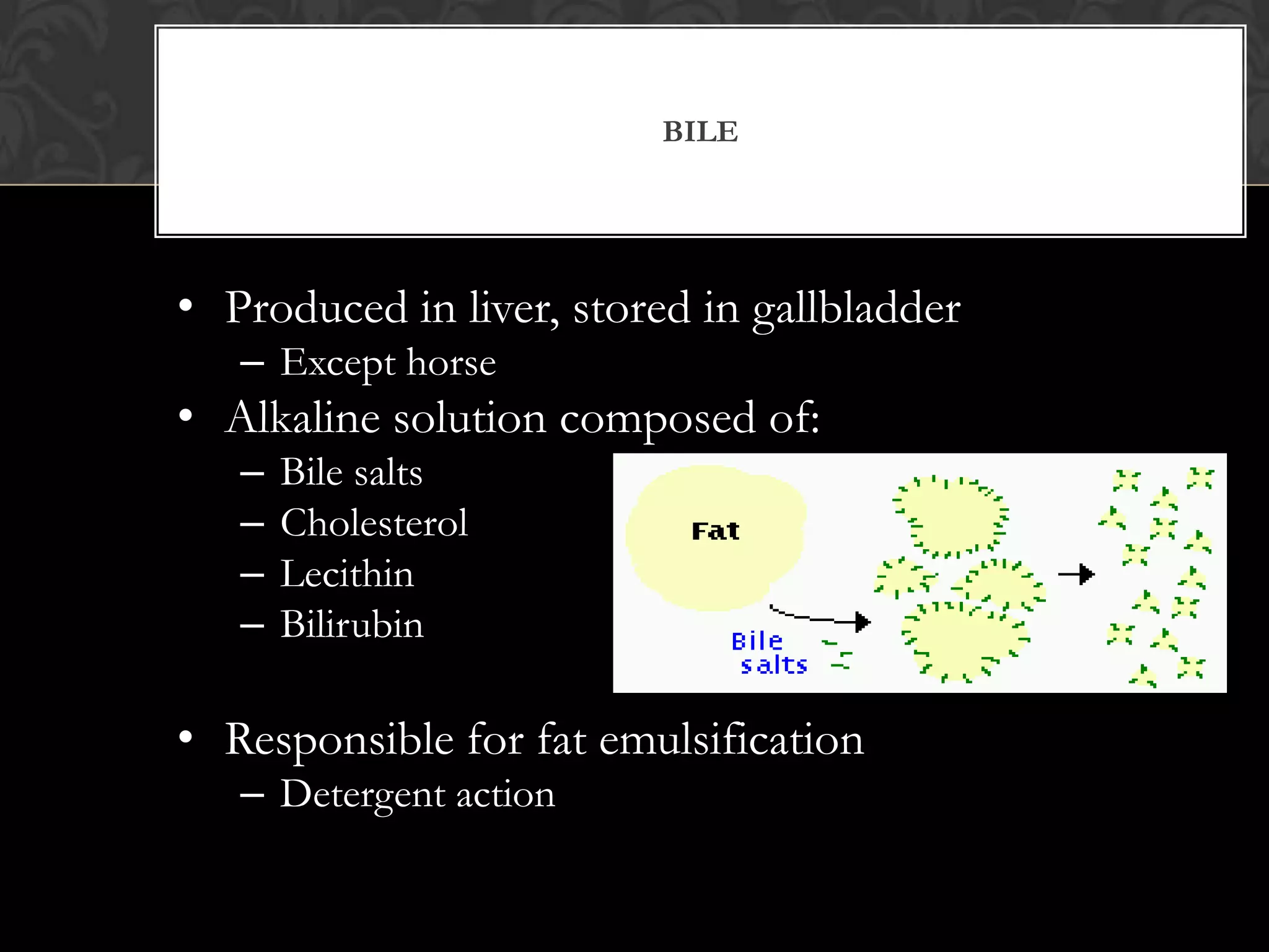
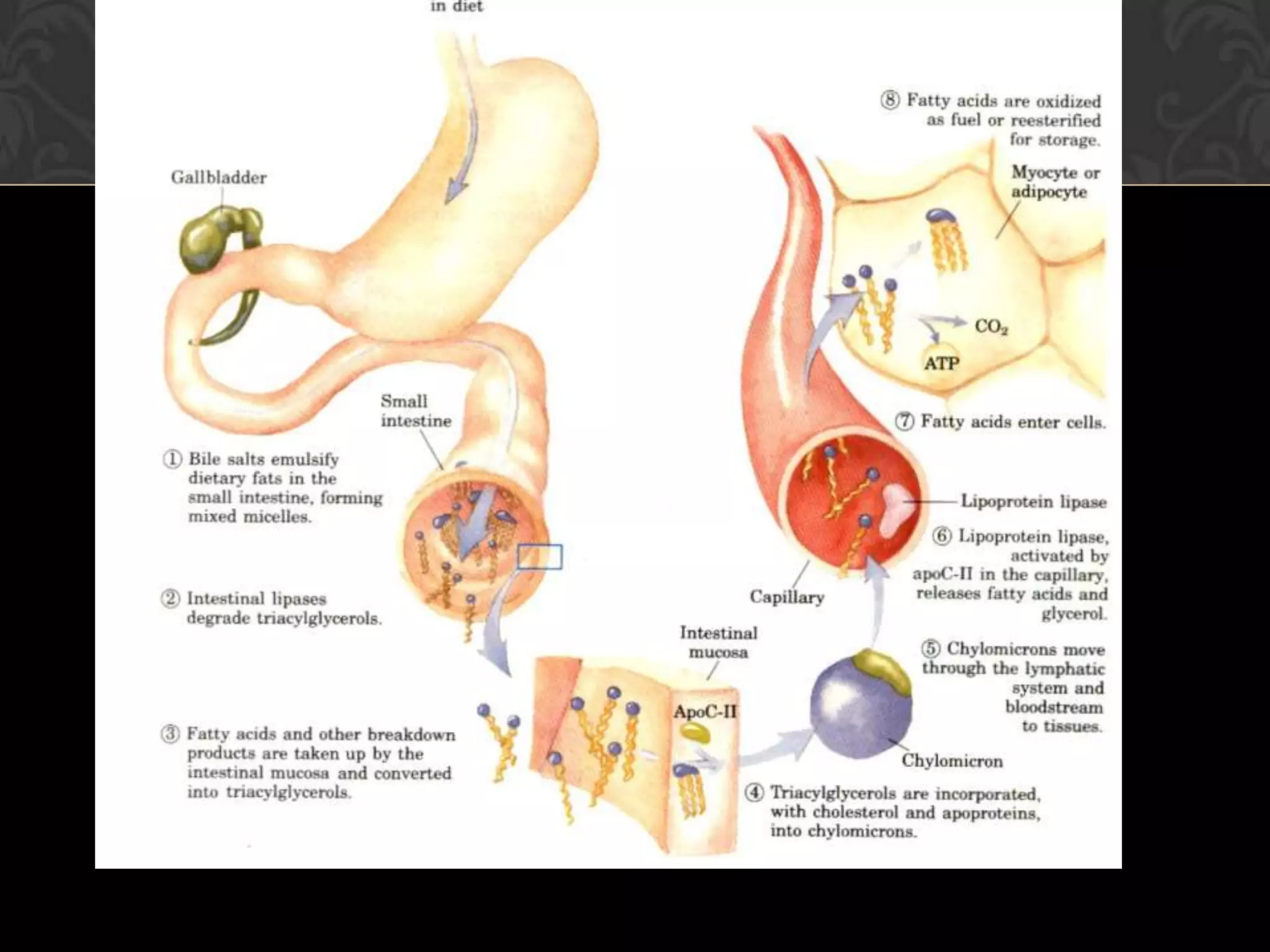
LAB 6: DIGESTION OF STARCH, PROTEIN, AND FATS IN HUMAN


         BY: WAN ISKANDAR FIRDAUS B SHAHARIMAN
               MUHAMMAD IZWAN BIN NORMAN
                  ANTHONEO JAY AUGUSTINE
THE HUMAN DIGESTIVE SYSTEM
DIGESTION OF STARCH
WHERE DOES THE DIGESTION OF
      STARCH OCCUR?
ORGANS THAT SECRETES
  ENZYME AMYLASE
Starch Digestion by Amylase
                    Starch is a polymer
                    composed or repeating
                    units of glucose.

                    Amylase breaks starch
                    into smaller subunits
                    such as maltose.


                                   Maltose
METHODS


                          Transfer one
      20cm3 of                                 Transfer a further
                       quarter of the saliva
 uncontaminated                                 quarter of the
                       into a test tube and
saliva was collected
  in a measuring
                       place this test tube       saliva into
                       in boiling water for    another test tube
       cylinder
                            15 minutes


                         Set up
Leave the test
 tubes for at
                        six pairs               Add four drops
                                               of hydrochloric
   least 10              of test                 acid, mix well
                                                and leave for at
   minutes                tubes                least 15 minutes
Starch +
                      Starch only
Sugar only                            untreated
                                        saliva


                                       Starch +
Untreated saliva      Starch +        pre-heated
     only          acidified saliva      saliva
TESTS


Iodine test
                      Benedict’s test
RESULTS


Pair   Contents        Test with iodine   Test with
                       solution           benedict’s reagent
A      Sugar only      No change          Turns brick red color
B      Starch only     Turns dark blue    No change
C      Untreated       No change          No change
       saliva only
D      Starch +        No change          Turns brick red color
       untreated
       saliva
E      Starch + pre-   Turns dark blue    No change
       heated saliva
F      Starch +        Turns dark blue    No change
       acidified
       saliva
LAB 6:
DIGESTION
OF PROTEIN
 IN HUMAN
DIGESTION OF PROTEIN


    Protein


  Poly peptide


    Peptide


  Amino acid
MATERIALS USED


  4 test tubes          Incubator (37’c)
Test tube rack         1% pepsin solution
 2 syringes or         1% trypsin solution
pipettes (10 ml)           0.1 M HCL
 2 syringes or           0.2 M sodium
pipettes (1 ml)             carbonate
     Label                 Egg white
   pH paper
METHOD

    The 4 test tubes are labeled; P2, P9, T2, T9
         Test tubes are set up as follows;
Test tube                 Contents
P2(PEPSIN AT pH 2.0)      5cm³ pepsin and 0.5cm³ HCl
P9(PEPSIN AT pH 9.0)      5cm³ pepsin and 0.5cm³
                          sodium carbonate
T2(TRYPSIN AT pH 2.0)     5cm³ pepsin and 0.5cm³ HCl

T9(TRYPSIN AT pH 9.0)     5cm³ pepsin and 0.5cm³
                          sodium carbonate

       Test tubes are shaken as to mix the content
                            .
A small strip of egg white is added to each of the
                      test tube
Then, test tubes are placed in an incubator at 37 C
             and then leave for 2 hours
The contents are examined and the appearance of
  the egg white and it’s surrounding is observed.
                Results are recorded.
THEORY……


Simply said, pepsin react best at a low pH
 value white trypsin react best at high pH
                   value.
 So, protein is best digested via this
 respective enzyme at their suitable
                 pH.
RESULT- FOR
                    PEPSIN VIA
               BIURET TEST

TEST          OBSERVATION         INFERENCE

POSITIVE      The solution turns Presence of
              from blue to violet( peptide bonds in
              purple)              protein


NEGATIVE      No change /The      No presence of
              solution remains    peptide bonds in
              blue                protein
FOR TRYPSIN
           RESULT-

           VIA MILLION’S TEST

TEST        OBSERVATION               INFERENCE

POSITIVE    The solution turns from   Presence of the amino
            blue to pink              acid tyrosine

NEGATIVE    No change /The solution   No presence of the amino
            remains blue              acid tyrosine
IF POSITIVE….
DIGESTION OF FATS
METHOD


1) The first 3 test tubes are labeled and
   marked at 3cm and 6 cm levels. 1% to
    2% pancreatic solutions is added to
    test tube 1-3 to the 3cm mark. 4 test
             tubes are prepared
               Test tube 1:
 1% bile (until 6cm mark) + 2/3 drops
phenolphthalein + 0.2% NaOH drop by
 drop until pink + 3 drops vegetable oil
 Shake and incubate at 37 C for 1 hour
Test tube 2:
Distilled water (until 6cm mark) +
2/3 drops phenolpthalein + 0.2%
NaOH drop by drop until pink + 3
           drops of oil
 Shake and incubate at 37 C for 1
               hour
Test tube 3:
 1% bile (until 6cm mark) + 2/3
  drops phenolphthalein + 0.2%
NaOH drop by drop until pink + 3
           drops of oil
Shake and allow to remain at room
         temp. for 1 hour
Test tube 1:
        PinkColourless
Fats are emulsified and digested
           Test tube 2:
          Pink remains
     Fats are not emulsified
           Test tube 3:
          Pink remains
     Condition not suitable
        Low temperature

              RESULT
FATS

» Fats are made of carbon, hydrogen, and oxygen.
» Fats supply your body with:
    Energy
    Form your cells
    Maintain body temperature
    Protect your nerves
UNSATURATED FATS

» Unsaturated Fats have at least one unsaturated
  bond in a place where hydrogen can be added to
  the molecule.
» Unsaturated fats are usually liquid at room
  temperature.

» Unsaturated fats are classified as either
  monounsaturated fats or polyunsaturated fats.
SATURATED FATS

» Fats that have all the hydrogen the carbon atoms
  can hold are called saturated fats.
» Saturated fats are usually solid at room
  temperature.

» Too much saturated fat in your diet can lead to
  heart disease.
FAT BREAKDOWN IN THE
         SMALL INTESTINE
» Fats are digested by mixing the fat with the digestive enzyme
  lipase (from pancreatic juice), which enters the duodenum
  from the pancreas - the main source of enzymes for
  digesting fats and proteins.
» Lipase chops up lipid molecules into fatty acid and glycerol.
» Fat does not dissolve in water.
» To overcome this problem the digestive system uses a
  substance called bile, produced in the liver but stored in
  the gallbladder, which enters the duodenum via the bile duct.
» Bile emulsifies fats - meaning, it disperses them into small
  droplets which then become suspended in the watery
  contents of the digestive tract.
» Emulsification allows lipase to gain easier access to the fat
  molecules and thus accelerates their breakdown and
  digestion.
+
Water soluble
Fatty acids
» Absorption of fat into the body occurs in the villi - the millions of
  finger-like projections which cover the walls of the small intestine.
» Inside each villus is a series of lymph vessels (lacteals) and blood
  vessels (capillaries).
» The lacteals absorb the fatty acids and glycerol into the lymphatic
  system which eventually drains into the bloodstream.
» The fatty acids are transported via the bloodstream to the membranes
  of adipose cells or muscle cells, where they are stored or oxidized for
  energy.
» Fats are only converted to glucose for energy supply during extreme
  condition.
» The glycerol part is absorbed by the liver and is either converted into
  glucose (gluconeogenesis), and/or used to help breakdown glucose
  into energy (glycolysis).

      HOW FAT IS ABSORBED INTO
        THE BLOODSTREAM
BILE


• Produced in liver, stored in gallbladder
   – Except horse
• Alkaline solution composed of:
   –   Bile salts
   –   Cholesterol
   –   Lecithin
   –   Bilirubin

• Responsible for fat emulsification
   – Detergent action
Fatty acid +
           glycerol
Lacteal


          Lymph

Lab 6

  • 1.
    LAB 6: DIGESTIONOF STARCH, PROTEIN, AND FATS IN HUMAN BY: WAN ISKANDAR FIRDAUS B SHAHARIMAN MUHAMMAD IZWAN BIN NORMAN ANTHONEO JAY AUGUSTINE
  • 2.
  • 3.
  • 4.
    WHERE DOES THEDIGESTION OF STARCH OCCUR?
  • 5.
    ORGANS THAT SECRETES ENZYME AMYLASE
  • 6.
    Starch Digestion byAmylase Starch is a polymer composed or repeating units of glucose. Amylase breaks starch into smaller subunits such as maltose. Maltose
  • 7.
    METHODS Transfer one 20cm3 of Transfer a further quarter of the saliva uncontaminated quarter of the into a test tube and saliva was collected in a measuring place this test tube saliva into in boiling water for another test tube cylinder 15 minutes Set up Leave the test tubes for at six pairs Add four drops of hydrochloric least 10 of test acid, mix well and leave for at minutes tubes least 15 minutes
  • 8.
    Starch + Starch only Sugar only untreated saliva Starch + Untreated saliva Starch + pre-heated only acidified saliva saliva
  • 9.
    TESTS Iodine test Benedict’s test
  • 10.
    RESULTS Pair Contents Test with iodine Test with solution benedict’s reagent A Sugar only No change Turns brick red color B Starch only Turns dark blue No change C Untreated No change No change saliva only D Starch + No change Turns brick red color untreated saliva E Starch + pre- Turns dark blue No change heated saliva F Starch + Turns dark blue No change acidified saliva
  • 11.
  • 12.
    DIGESTION OF PROTEIN Protein Poly peptide Peptide Amino acid
  • 13.
    MATERIALS USED 4 test tubes Incubator (37’c) Test tube rack 1% pepsin solution 2 syringes or 1% trypsin solution pipettes (10 ml) 0.1 M HCL 2 syringes or 0.2 M sodium pipettes (1 ml) carbonate Label Egg white pH paper
  • 14.
    METHOD The 4 test tubes are labeled; P2, P9, T2, T9 Test tubes are set up as follows; Test tube Contents P2(PEPSIN AT pH 2.0) 5cm³ pepsin and 0.5cm³ HCl P9(PEPSIN AT pH 9.0) 5cm³ pepsin and 0.5cm³ sodium carbonate T2(TRYPSIN AT pH 2.0) 5cm³ pepsin and 0.5cm³ HCl T9(TRYPSIN AT pH 9.0) 5cm³ pepsin and 0.5cm³ sodium carbonate Test tubes are shaken as to mix the content .
  • 15.
    A small stripof egg white is added to each of the test tube Then, test tubes are placed in an incubator at 37 C and then leave for 2 hours The contents are examined and the appearance of the egg white and it’s surrounding is observed. Results are recorded.
  • 16.
    THEORY…… Simply said, pepsinreact best at a low pH value white trypsin react best at high pH value. So, protein is best digested via this respective enzyme at their suitable pH.
  • 17.
    RESULT- FOR PEPSIN VIA BIURET TEST TEST OBSERVATION INFERENCE POSITIVE The solution turns Presence of from blue to violet( peptide bonds in purple) protein NEGATIVE No change /The No presence of solution remains peptide bonds in blue protein
  • 19.
    FOR TRYPSIN RESULT- VIA MILLION’S TEST TEST OBSERVATION INFERENCE POSITIVE The solution turns from Presence of the amino blue to pink acid tyrosine NEGATIVE No change /The solution No presence of the amino remains blue acid tyrosine
  • 20.
  • 21.
  • 22.
    METHOD 1) The first3 test tubes are labeled and marked at 3cm and 6 cm levels. 1% to 2% pancreatic solutions is added to test tube 1-3 to the 3cm mark. 4 test tubes are prepared Test tube 1: 1% bile (until 6cm mark) + 2/3 drops phenolphthalein + 0.2% NaOH drop by drop until pink + 3 drops vegetable oil Shake and incubate at 37 C for 1 hour
  • 23.
    Test tube 2: Distilledwater (until 6cm mark) + 2/3 drops phenolpthalein + 0.2% NaOH drop by drop until pink + 3 drops of oil Shake and incubate at 37 C for 1 hour
  • 24.
    Test tube 3: 1% bile (until 6cm mark) + 2/3 drops phenolphthalein + 0.2% NaOH drop by drop until pink + 3 drops of oil Shake and allow to remain at room temp. for 1 hour
  • 25.
    Test tube 1: PinkColourless Fats are emulsified and digested Test tube 2: Pink remains Fats are not emulsified Test tube 3: Pink remains Condition not suitable Low temperature RESULT
  • 26.
    FATS » Fats aremade of carbon, hydrogen, and oxygen. » Fats supply your body with:  Energy  Form your cells  Maintain body temperature  Protect your nerves
  • 28.
    UNSATURATED FATS » UnsaturatedFats have at least one unsaturated bond in a place where hydrogen can be added to the molecule. » Unsaturated fats are usually liquid at room temperature. » Unsaturated fats are classified as either monounsaturated fats or polyunsaturated fats.
  • 29.
    SATURATED FATS » Fatsthat have all the hydrogen the carbon atoms can hold are called saturated fats. » Saturated fats are usually solid at room temperature. » Too much saturated fat in your diet can lead to heart disease.
  • 30.
    FAT BREAKDOWN INTHE SMALL INTESTINE » Fats are digested by mixing the fat with the digestive enzyme lipase (from pancreatic juice), which enters the duodenum from the pancreas - the main source of enzymes for digesting fats and proteins. » Lipase chops up lipid molecules into fatty acid and glycerol. » Fat does not dissolve in water. » To overcome this problem the digestive system uses a substance called bile, produced in the liver but stored in the gallbladder, which enters the duodenum via the bile duct. » Bile emulsifies fats - meaning, it disperses them into small droplets which then become suspended in the watery contents of the digestive tract. » Emulsification allows lipase to gain easier access to the fat molecules and thus accelerates their breakdown and digestion.
  • 31.
  • 32.
  • 33.
    » Absorption offat into the body occurs in the villi - the millions of finger-like projections which cover the walls of the small intestine. » Inside each villus is a series of lymph vessels (lacteals) and blood vessels (capillaries). » The lacteals absorb the fatty acids and glycerol into the lymphatic system which eventually drains into the bloodstream. » The fatty acids are transported via the bloodstream to the membranes of adipose cells or muscle cells, where they are stored or oxidized for energy. » Fats are only converted to glucose for energy supply during extreme condition. » The glycerol part is absorbed by the liver and is either converted into glucose (gluconeogenesis), and/or used to help breakdown glucose into energy (glycolysis). HOW FAT IS ABSORBED INTO THE BLOODSTREAM
  • 34.
    BILE • Produced inliver, stored in gallbladder – Except horse • Alkaline solution composed of: – Bile salts – Cholesterol – Lecithin – Bilirubin • Responsible for fat emulsification – Detergent action
  • 36.
    Fatty acid + glycerol Lacteal Lymph

Editor's Notes

  • #11 In pair A, there are no change in both test. This is because sugar is not a form of starch or reducing sugarIn B, iodine changes from yellowish brown to dark blue if added to starch. Starch is not a reducing sugarIn pair C, there are no change in both test. This is because untreated saliva is not a form of starch or reducing sugarIn pair D, starch had been hydrolyze to maltose, a simple sugar. Thus there is no change in iodine test but the mixture turned brick red color with Benedict’s testIn pair E, Starch is not hydrolyzed by pre-heated saliva so when tested with iodine it turns dark blue. This is cause by the damage on the salivary amylase enzyme structure that is responsible in hydrolyzing starch to maltose.In pair F, the salivary amylase also damaged too since the lower pH caused the structure of the enzyme to alter.
  • #26 Test tube 1: fats are hydrolyzedTest tube 2: no bile, enzyme absentTest tube 3: low temp. not body temp