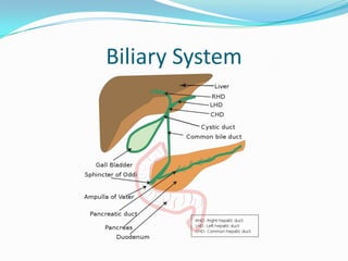
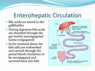
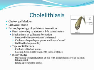
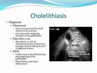
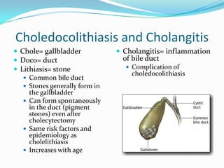
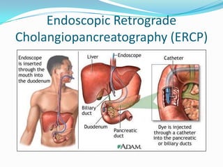
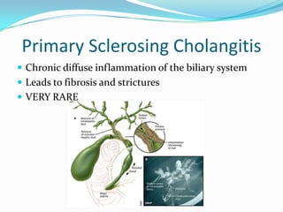
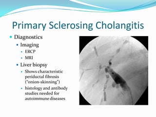
Biliary DiseaseElizabeth Bunting, MS, PA-CFebruary 11, 2011
ObjectivesDefine bile, bile salts, and bile acids, and state the function of each.Indicate which bile salts are the “primary” bile salts.Describe bile acid metabolism and the enterohepatic circulation.Define cholestasis.Describe the diagnostic approach to the patient with cholestasis.Describe the causes of cholestasis.Define gallstone.Describe the pathophysiology of gallstone formation.Describe the clinical features of gallstones.Discuss the clinical features, lab tests and imaging studies useful in the diagnosis of gallstone disease.Discuss the treatment of gallstone disease.Define cholesterolosis.Define biliarydyskinesia.
Biliary System
What is Bile? Why do we need salts and acids?Bile= bile acids + phospholipids + cholesterolPrimary Bile Acids= (cholic acid and chenodeoxycholic) acid which are formed by cholesterol from the liver along with amino acidsSecondary Bile Acids= bacterial metabolites (deoxycholate and lithocholate) of primary bile acids formed in the colonWhat do they do? They help excrete cholesterol, aid in fat digestion and absorption of fat, cholesterol and fat-soluble vitamins in the intestinesBile forms micelles (bound to cholesterol and fat) and aids in their absorption through micellar transport mechanism
Enterohepatic CirculationBile acids are stored in the gallbladderDuring digestion bile acids are absorbed through the gut mostly uncongugated (some congugated)In the terminal ileum the bile salts are reabsorbed and carried through the portal blood circulation to be reconjugated and secreted back into bile
CholestasisChole= gallbladder or its ductsStasis= stuck!Cholestasis is the obstruction of the secretion of bileDifferent forms of Cholestasis: Cholelithiasis (gallstones), Choledocholithiasis (gallstones in the common bile duct)What else can cause Cholestasis? Tumors, cysts, pancreatic problems blocking the duct (pancreatitis), liver disease
Patient with CholestasisRUQ pain may be intermittent if blockage is intermittentJaundiceDark urineLight stoolsWeight loss
CholestasisLabs and ImagingASL and ALT may be elevatedBlood in stool suggests cancer etiologyAlkaline Phosphatase will be elevatedUltrasound needed to look for stones or tumorsCT may be needed to clarify obstructionMRI can be used to further evaluate liver disease
Cholelithiasis
CholelithiasisChole= gallbladderLithiasis= stonePathophysiology of gallstone formationForm secondary to abnormal bile constituentsMechanisms of gallstone formationIncreased biliary secretion of cholesterolCholesterol crystals precipitate and form a “stone”Gallbladder hypomotilityTypes of GallstonesCholesterol 80% of stonesCalcium bilirubinate (pigment) <20% of stonesBiliary sludge Mucus like (supersaturation of bile with either cholesterol or calcium bilirubinate)Likely a precursor to stones
Cholesterol Gallstones
CholelithiasisEpidemiologyWomen > MenIncreases with ageNative AmericansRisk factors (4 F’s)ObesityRapid weight loss (bariatric surgery)Increasing ageHigh calorie (specifically fat) dietPregnancy/ OCPPrimary Biliary cirrhosisClofibrate, octreotide, ceftriaxone therapyChronic Hemolysis (sickle cell andemia)Chronic biliary tract infection - men(hepatitis)Diabetes and insulin resistanceCrohn’sdiesase
CholelithiasisClinical presentationMany times asymptomaticGallbladder pain presents as intermittent severe RUQ pain radiation to the scapula (biliary colic)Pain happens in 15-25% of gallstonesCan be epigastric pain radiating to the RUQOnset of pain is generally sudden and can last from 30 min to 5 hours.N/v generally accompany painTend to be postprandial, especially a high fat mealMore common at night
CholelithiasisDiagnosisUltrasoundVery accurate even for small stones (2mm and up)Can assess the emptying function of the gallbladderPlain film x-rayMay detect 10-15% of cholesterol stones if they have enough calcium and up to 50% of pigment stonesHIDALooks more at the functioning and emptying of the gallbladderDetermines cystic duct obstruction
CholelithiasisTreatment of gallstonesONLY FOR SYMPTOMATIC PATIENTSLaparoscopic cholecystectomyGenerally outpt surgery with quick recovery (generally <1 week)LithotripsyIn combo with bile salt therapy for a single radiolucent stone (<20mm)Infrequently used in the US anymoreChenodeoxycholic and Ursodeoxycholic Acid (7mg/kg/day of each or just UCSA 8-13mg/kg/day in divided doses)Bile salts that can be given orally to dissolve stones over 2 year period of timeFor patients who refuse surgery and have a functioning gallbladder (visualized by oral cholecystography)Most gallstones recur within 5 yearsFew patients are candidates for this
Acute CholecystitisChole=gallbladder  Cystitis= inflammation of the cyst wallCaused by gallstone obstruction 90% of the timeBiliary pain that progressively worsensMost patients (>50%) have experienced attacks before that resolved spontaneously
Acute CholecystitisPersistant severe RUQ pain Generally after a fatty mealFeverN/VAnorexiaRUQ TTP with +Murphy’s signGuarding/reboundEnlarged gallbladder palpable in 15% of patientsDistended abdomen with hypoactive bowel soundsJaundice in 25% of patients
Acute CholecystitisLabsElevated WBC (12,000-15,000)Elevated bili possibleAST/ALT often elevatedAlkaline phosphatase can be high especially in ascending cholangitisAmylase can be highImagingPlain film x-ray show stones in 15% of casesHIDA shows cystic duct obstruction (most common cause of acute cholecystitis)RUQ U/s shows stones, but does not as good at showing acute cholecystitis.Usually done first to show stones and then progress to HIDA
Acute CholecystitisDifferential DiagnosisPUDPancreatitisAppendicitisPerforated colonic carcinoma or diverticulumLiver abscessHepatitisPneumoniaRarely myocardial ischemia
Acute CholecystitisComplicationsGangrene	Leading to perforation and possible abscessEmphasematouscholecystitisEmpyemaChronic CholecystitisRepeated acute cholecystitisCholesterolosis- gallbladderpolypoid enlargement due to cholesterol deposits (AKA strawberry gallbladder)CholangitisHydropsAcute cholecystitis resolves but obstruction persists causing mucoid fluid to collect in the gallbladderPoreclain gallbladder Increased risk of ca
Acute CholecystitisTreatmentLap CholecystectomyConservative treatment (not able to have surgery or waiting for stabilization before surgery)NPO, IV fluids, analgesia, IV Abx (3rd gen cephalosporin + Flagyl, severe cases may need Fluoroquinolone + Flagyl75% will see remission of sx in 2-7 daysMany recurrencesMeperidine (Demerol) for pain control.  Morphine can cause spasm of the sphincter of Oddi and increase painPrognosisLow mortality rate (<0.2%)Surgery consistent with resolution of sx
Choledocolithiasis and CholangitisChole= gallbladderDoco= ductLithiasis= stoneCommon bile ductStones generally form in the gallbladderCan form spontaneously in the duct (pigment stones) even after cholecytectomySame risk factors and epidemiology as cholelithiasisIncreases with ageCholangitis= inflammation of bile ductComplication of choledocolithiasis
Choledocolithiasis and CholangitisSigns and symptoms of CholangitisCHARCOT’S TRIAD- fever jaundice, severe RUQ painPruritisDark urineAcholic stools (light colored)Signs and symptoms of Acute Supportive CholangitisREYNOLD PENTAD- Charcot’s Triad + AMS +hypotensionENDOSCOPIC EMERGENCY
Choledocolithiasis and CholangitisLabsVery elevated ALT/AST from obstruction (often >1000)Elevated BiliAlkphos rises slowlyAmylase can be elevated when comorbid pancreatitisElevated WBC in cholangitisImagingU/s and CT show dilitation of ductsEndoscopic U/s, helical CT and MRI used if low suspicionERCP is gold standardTherapeutic as well- can do sphincterotomy with stone extraction or stent placement
Endoscopic Retrograde Cholangiopancreatography (ERCP)
Choledocolithiasis and CholangitisDifferential DiagnosisCancer- pancreas, ampulla of Vater, bile duct, gallbladderCompression from metastatic ca (GI tract of breast)Chronic liver diseasePrimary biliary cirrhosisSclerosingcholangitisDrug-induced liver diseaseTreatmentEndoscopic sphincterotomy and stone extraction (usually by ERCP)Lap chole usually follows thisAntibiotics if bile cultures or blood cultures are positive Ampicillin+ Gentamicin+ Cipro or Flagyl
Primary SclerosingCholangitisChronic diffuse inflammation of the biliary systemLeads to fibrosis and stricturesVERY RARE
Primary SclerosingCholangitisEpidemiologyMen>womenAges 20-50Risk FactorsUlcerative  Colitis significantly increases riskCrohn’s some increased riskHLA- B8, DR-3, DR-41st degree family member
Primary SclerosingChloangitisClinical PresentationProgressive obstructive jaundiceFatiguePruritisAnorexiaindigestionLabs Elevated AlkphosLow serum albumin (secondary to malabsorption)Possibly low platelet count
Primary SclerosingCholangitisDiagnosticsImagingERCPMRILiver biopsyShows characteristic periductal fibrosis (“onion-skinning”)histology and antibody studies needed for autoimmune diseases
Primary SclerosingCholangitisComplicationsCholangiocarcinoma in 20% of casesCheck CA 19-9Annual imaging with U/s, CT or MRI with MRCPGallstonesCholecystitisGallbladder polypsGallbladder carcinoma
Primary SclerosingCholangitisTreatmentAcute bacterialCipro 750mg bidChronicBalloon dilitation or stentingMay increase risk of complications or cholangitisResection of dominant bile duct strictureMay lead to longer survival and decreased risk of cholangiocarcinomaIn pt with UC, colorectal surveillance is necessary to reduce colorectal caUrsodeoxycholic acid could reduce colorectal dysplasiaFor pt with cirrhosis and primary sclerosingcholangitis liver transplantation will likely be needed
Primary SclerosingCholangitisPrognosisSurvival 12-17 yearsLiver transplant increases survival if before cholangiocarcinomaAdverse prognostic factors	Older age at dxHigher bilirubin/ ASTLower albuminVariceal bleedingDominant bile duct strictureExtrahepatic changes
Biliary Stricture95% of the time are secondary surgery (anastomosis or injury)Other causes: Blunt trauma to the abdomenPancreatitisErosion of the duct by gallstonePrior endoscopic sphincterotomy
Biliary StrictureUnless complete obstruction, may not be apparent immediately after surgeryErosion causes Biloma (collection of bile) which leads to infection and likely fibrous strictureMost common complication is CholangitisNeeds ERCP for repair, stent or dilitation
BiliaryDyskinesiaGallbladder dysfunction (AKA chronic acalculouscholecystitis)Most common in women <50 years of ageClinical presentationEpisodic RUQ painIntolerance to fatty foodsMild nauseaImagingPt have already had U/s, x-ray, EGD, upper GI without findingsERCP diagnostic with <35% ejection fraction at 20  minutesERCP with injection of sincalide reproduces RUQ painTreatmentLap Cholecystectomy cures 85-90% of patientsPathology report shows chronic cholecystitis
THE END!QUESTIONS?????
ReferencesCurrent Medical Diagnosis and Treatment 2010Wikipedia and Google for imagesHarrison’s Principles of Internal Medicine for content and imagesImages:http://www.uninet.edu/cin2001-old/conf/moreno/moreno2.htmlhttp://www.meddean.luc.edu/lumen/meded/Radio/curriculum/Surgery/cholecystitis_list2.htmhttp://www.gallstoneremove.info/recognizing-the-signs-of-gallstones/http://www.gallstones.com/
ReferencesImages cont:http://curezone.com/gallstones/default.asphttp://commons.wikimedia.org/wiki/File:Biliary_system_new.svghttp://www.bmj.com/content/323/7322/1170.fullhttp://healthcaretips-fact-guide.blogspot.com/2009/07/health-tips-facts-jaundice-symptomshome.htmlwww.hpb.org.ukwww.drjoycerichards.comwww.emedicine.medscape.com
ReferencesMore images!www.hopkins-gi.orgwww.disease-picture.comwww.s00.middlebury.eduwww.radiographics.rsna.orgFinal picture is by my adorable husband, Bill Bunting

Biliary Disease

  • 1.
    Biliary DiseaseElizabeth Bunting,MS, PA-CFebruary 11, 2011
  • 2.
    ObjectivesDefine bile, bilesalts, and bile acids, and state the function of each.Indicate which bile salts are the “primary” bile salts.Describe bile acid metabolism and the enterohepatic circulation.Define cholestasis.Describe the diagnostic approach to the patient with cholestasis.Describe the causes of cholestasis.Define gallstone.Describe the pathophysiology of gallstone formation.Describe the clinical features of gallstones.Discuss the clinical features, lab tests and imaging studies useful in the diagnosis of gallstone disease.Discuss the treatment of gallstone disease.Define cholesterolosis.Define biliarydyskinesia.
  • 3.
  • 4.
    What is Bile?Why do we need salts and acids?Bile= bile acids + phospholipids + cholesterolPrimary Bile Acids= (cholic acid and chenodeoxycholic) acid which are formed by cholesterol from the liver along with amino acidsSecondary Bile Acids= bacterial metabolites (deoxycholate and lithocholate) of primary bile acids formed in the colonWhat do they do? They help excrete cholesterol, aid in fat digestion and absorption of fat, cholesterol and fat-soluble vitamins in the intestinesBile forms micelles (bound to cholesterol and fat) and aids in their absorption through micellar transport mechanism
  • 5.
    Enterohepatic CirculationBile acidsare stored in the gallbladderDuring digestion bile acids are absorbed through the gut mostly uncongugated (some congugated)In the terminal ileum the bile salts are reabsorbed and carried through the portal blood circulation to be reconjugated and secreted back into bile
  • 6.
    CholestasisChole= gallbladder orits ductsStasis= stuck!Cholestasis is the obstruction of the secretion of bileDifferent forms of Cholestasis: Cholelithiasis (gallstones), Choledocholithiasis (gallstones in the common bile duct)What else can cause Cholestasis? Tumors, cysts, pancreatic problems blocking the duct (pancreatitis), liver disease
  • 7.
    Patient with CholestasisRUQpain may be intermittent if blockage is intermittentJaundiceDark urineLight stoolsWeight loss
  • 8.
    CholestasisLabs and ImagingASLand ALT may be elevatedBlood in stool suggests cancer etiologyAlkaline Phosphatase will be elevatedUltrasound needed to look for stones or tumorsCT may be needed to clarify obstructionMRI can be used to further evaluate liver disease
  • 9.
  • 10.
    CholelithiasisChole= gallbladderLithiasis= stonePathophysiologyof gallstone formationForm secondary to abnormal bile constituentsMechanisms of gallstone formationIncreased biliary secretion of cholesterolCholesterol crystals precipitate and form a “stone”Gallbladder hypomotilityTypes of GallstonesCholesterol 80% of stonesCalcium bilirubinate (pigment) <20% of stonesBiliary sludge Mucus like (supersaturation of bile with either cholesterol or calcium bilirubinate)Likely a precursor to stones
  • 11.
  • 12.
    CholelithiasisEpidemiologyWomen > MenIncreaseswith ageNative AmericansRisk factors (4 F’s)ObesityRapid weight loss (bariatric surgery)Increasing ageHigh calorie (specifically fat) dietPregnancy/ OCPPrimary Biliary cirrhosisClofibrate, octreotide, ceftriaxone therapyChronic Hemolysis (sickle cell andemia)Chronic biliary tract infection - men(hepatitis)Diabetes and insulin resistanceCrohn’sdiesase
  • 13.
    CholelithiasisClinical presentationMany timesasymptomaticGallbladder pain presents as intermittent severe RUQ pain radiation to the scapula (biliary colic)Pain happens in 15-25% of gallstonesCan be epigastric pain radiating to the RUQOnset of pain is generally sudden and can last from 30 min to 5 hours.N/v generally accompany painTend to be postprandial, especially a high fat mealMore common at night
  • 14.
    CholelithiasisDiagnosisUltrasoundVery accurate evenfor small stones (2mm and up)Can assess the emptying function of the gallbladderPlain film x-rayMay detect 10-15% of cholesterol stones if they have enough calcium and up to 50% of pigment stonesHIDALooks more at the functioning and emptying of the gallbladderDetermines cystic duct obstruction
  • 15.
    CholelithiasisTreatment of gallstonesONLYFOR SYMPTOMATIC PATIENTSLaparoscopic cholecystectomyGenerally outpt surgery with quick recovery (generally <1 week)LithotripsyIn combo with bile salt therapy for a single radiolucent stone (<20mm)Infrequently used in the US anymoreChenodeoxycholic and Ursodeoxycholic Acid (7mg/kg/day of each or just UCSA 8-13mg/kg/day in divided doses)Bile salts that can be given orally to dissolve stones over 2 year period of timeFor patients who refuse surgery and have a functioning gallbladder (visualized by oral cholecystography)Most gallstones recur within 5 yearsFew patients are candidates for this
  • 16.
    Acute CholecystitisChole=gallbladder Cystitis= inflammation of the cyst wallCaused by gallstone obstruction 90% of the timeBiliary pain that progressively worsensMost patients (>50%) have experienced attacks before that resolved spontaneously
  • 17.
    Acute CholecystitisPersistant severeRUQ pain Generally after a fatty mealFeverN/VAnorexiaRUQ TTP with +Murphy’s signGuarding/reboundEnlarged gallbladder palpable in 15% of patientsDistended abdomen with hypoactive bowel soundsJaundice in 25% of patients
  • 18.
    Acute CholecystitisLabsElevated WBC(12,000-15,000)Elevated bili possibleAST/ALT often elevatedAlkaline phosphatase can be high especially in ascending cholangitisAmylase can be highImagingPlain film x-ray show stones in 15% of casesHIDA shows cystic duct obstruction (most common cause of acute cholecystitis)RUQ U/s shows stones, but does not as good at showing acute cholecystitis.Usually done first to show stones and then progress to HIDA
  • 19.
    Acute CholecystitisDifferential DiagnosisPUDPancreatitisAppendicitisPerforatedcolonic carcinoma or diverticulumLiver abscessHepatitisPneumoniaRarely myocardial ischemia
  • 20.
    Acute CholecystitisComplicationsGangrene Leading toperforation and possible abscessEmphasematouscholecystitisEmpyemaChronic CholecystitisRepeated acute cholecystitisCholesterolosis- gallbladderpolypoid enlargement due to cholesterol deposits (AKA strawberry gallbladder)CholangitisHydropsAcute cholecystitis resolves but obstruction persists causing mucoid fluid to collect in the gallbladderPoreclain gallbladder Increased risk of ca
  • 21.
    Acute CholecystitisTreatmentLap CholecystectomyConservativetreatment (not able to have surgery or waiting for stabilization before surgery)NPO, IV fluids, analgesia, IV Abx (3rd gen cephalosporin + Flagyl, severe cases may need Fluoroquinolone + Flagyl75% will see remission of sx in 2-7 daysMany recurrencesMeperidine (Demerol) for pain control. Morphine can cause spasm of the sphincter of Oddi and increase painPrognosisLow mortality rate (<0.2%)Surgery consistent with resolution of sx
  • 22.
    Choledocolithiasis and CholangitisChole=gallbladderDoco= ductLithiasis= stoneCommon bile ductStones generally form in the gallbladderCan form spontaneously in the duct (pigment stones) even after cholecytectomySame risk factors and epidemiology as cholelithiasisIncreases with ageCholangitis= inflammation of bile ductComplication of choledocolithiasis
  • 23.
    Choledocolithiasis and CholangitisSignsand symptoms of CholangitisCHARCOT’S TRIAD- fever jaundice, severe RUQ painPruritisDark urineAcholic stools (light colored)Signs and symptoms of Acute Supportive CholangitisREYNOLD PENTAD- Charcot’s Triad + AMS +hypotensionENDOSCOPIC EMERGENCY
  • 24.
    Choledocolithiasis and CholangitisLabsVeryelevated ALT/AST from obstruction (often >1000)Elevated BiliAlkphos rises slowlyAmylase can be elevated when comorbid pancreatitisElevated WBC in cholangitisImagingU/s and CT show dilitation of ductsEndoscopic U/s, helical CT and MRI used if low suspicionERCP is gold standardTherapeutic as well- can do sphincterotomy with stone extraction or stent placement
  • 25.
  • 26.
    Choledocolithiasis and CholangitisDifferentialDiagnosisCancer- pancreas, ampulla of Vater, bile duct, gallbladderCompression from metastatic ca (GI tract of breast)Chronic liver diseasePrimary biliary cirrhosisSclerosingcholangitisDrug-induced liver diseaseTreatmentEndoscopic sphincterotomy and stone extraction (usually by ERCP)Lap chole usually follows thisAntibiotics if bile cultures or blood cultures are positive Ampicillin+ Gentamicin+ Cipro or Flagyl
  • 27.
    Primary SclerosingCholangitisChronic diffuseinflammation of the biliary systemLeads to fibrosis and stricturesVERY RARE
  • 28.
    Primary SclerosingCholangitisEpidemiologyMen>womenAges 20-50RiskFactorsUlcerative Colitis significantly increases riskCrohn’s some increased riskHLA- B8, DR-3, DR-41st degree family member
  • 29.
    Primary SclerosingChloangitisClinical PresentationProgressiveobstructive jaundiceFatiguePruritisAnorexiaindigestionLabs Elevated AlkphosLow serum albumin (secondary to malabsorption)Possibly low platelet count
  • 30.
    Primary SclerosingCholangitisDiagnosticsImagingERCPMRILiver biopsyShowscharacteristic periductal fibrosis (“onion-skinning”)histology and antibody studies needed for autoimmune diseases
  • 31.
    Primary SclerosingCholangitisComplicationsCholangiocarcinoma in20% of casesCheck CA 19-9Annual imaging with U/s, CT or MRI with MRCPGallstonesCholecystitisGallbladder polypsGallbladder carcinoma
  • 32.
    Primary SclerosingCholangitisTreatmentAcute bacterialCipro750mg bidChronicBalloon dilitation or stentingMay increase risk of complications or cholangitisResection of dominant bile duct strictureMay lead to longer survival and decreased risk of cholangiocarcinomaIn pt with UC, colorectal surveillance is necessary to reduce colorectal caUrsodeoxycholic acid could reduce colorectal dysplasiaFor pt with cirrhosis and primary sclerosingcholangitis liver transplantation will likely be needed
  • 33.
    Primary SclerosingCholangitisPrognosisSurvival 12-17yearsLiver transplant increases survival if before cholangiocarcinomaAdverse prognostic factors Older age at dxHigher bilirubin/ ASTLower albuminVariceal bleedingDominant bile duct strictureExtrahepatic changes
  • 34.
    Biliary Stricture95% ofthe time are secondary surgery (anastomosis or injury)Other causes: Blunt trauma to the abdomenPancreatitisErosion of the duct by gallstonePrior endoscopic sphincterotomy
  • 35.
    Biliary StrictureUnless completeobstruction, may not be apparent immediately after surgeryErosion causes Biloma (collection of bile) which leads to infection and likely fibrous strictureMost common complication is CholangitisNeeds ERCP for repair, stent or dilitation
  • 36.
    BiliaryDyskinesiaGallbladder dysfunction (AKAchronic acalculouscholecystitis)Most common in women <50 years of ageClinical presentationEpisodic RUQ painIntolerance to fatty foodsMild nauseaImagingPt have already had U/s, x-ray, EGD, upper GI without findingsERCP diagnostic with <35% ejection fraction at 20 minutesERCP with injection of sincalide reproduces RUQ painTreatmentLap Cholecystectomy cures 85-90% of patientsPathology report shows chronic cholecystitis
  • 37.
  • 38.
    ReferencesCurrent Medical Diagnosisand Treatment 2010Wikipedia and Google for imagesHarrison’s Principles of Internal Medicine for content and imagesImages:http://www.uninet.edu/cin2001-old/conf/moreno/moreno2.htmlhttp://www.meddean.luc.edu/lumen/meded/Radio/curriculum/Surgery/cholecystitis_list2.htmhttp://www.gallstoneremove.info/recognizing-the-signs-of-gallstones/http://www.gallstones.com/
  • 39.
  • 40.

Editor's Notes

  • #3 Objectives are not complete. Follow these notes and coordinate readings in your book!