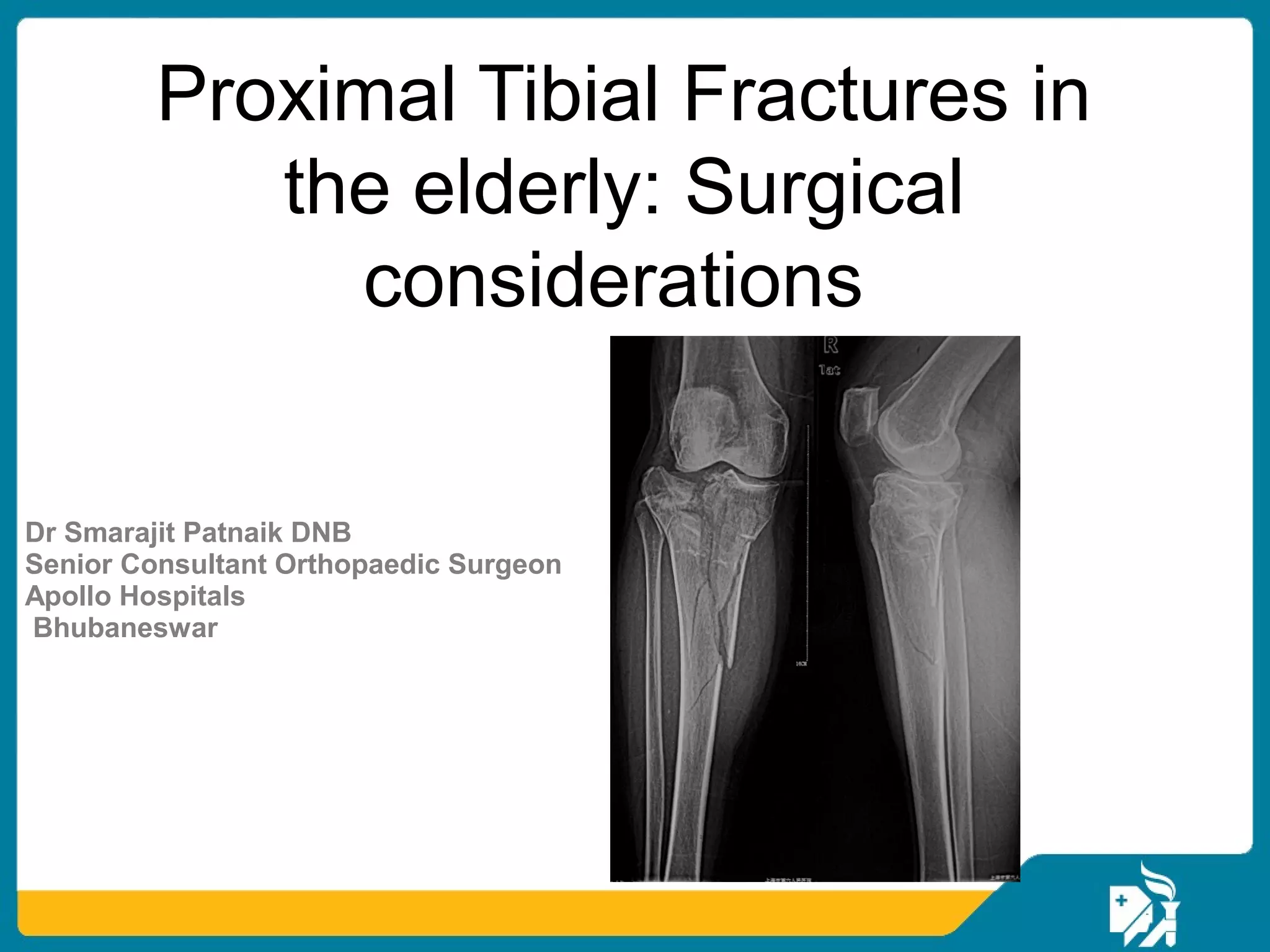
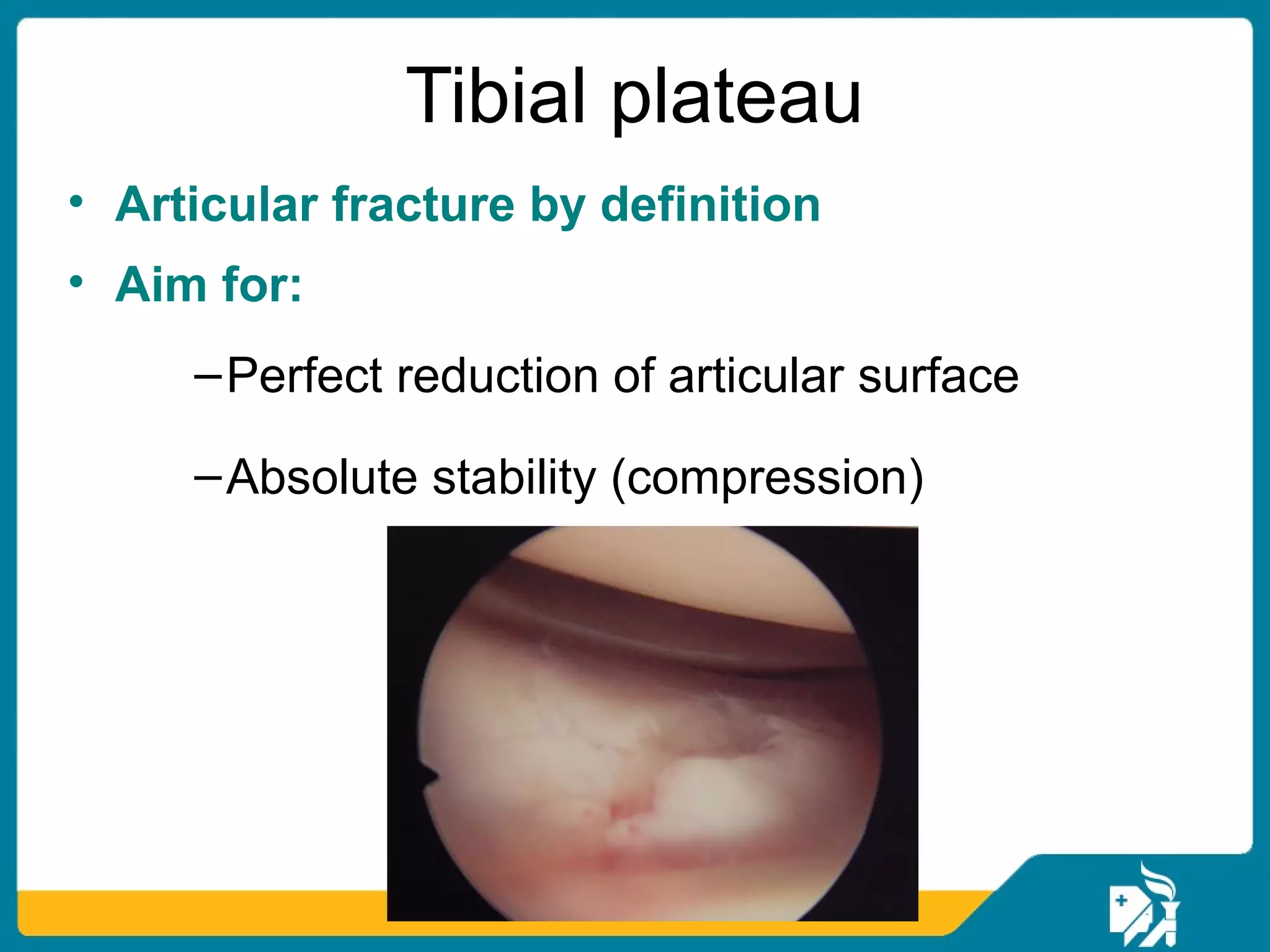
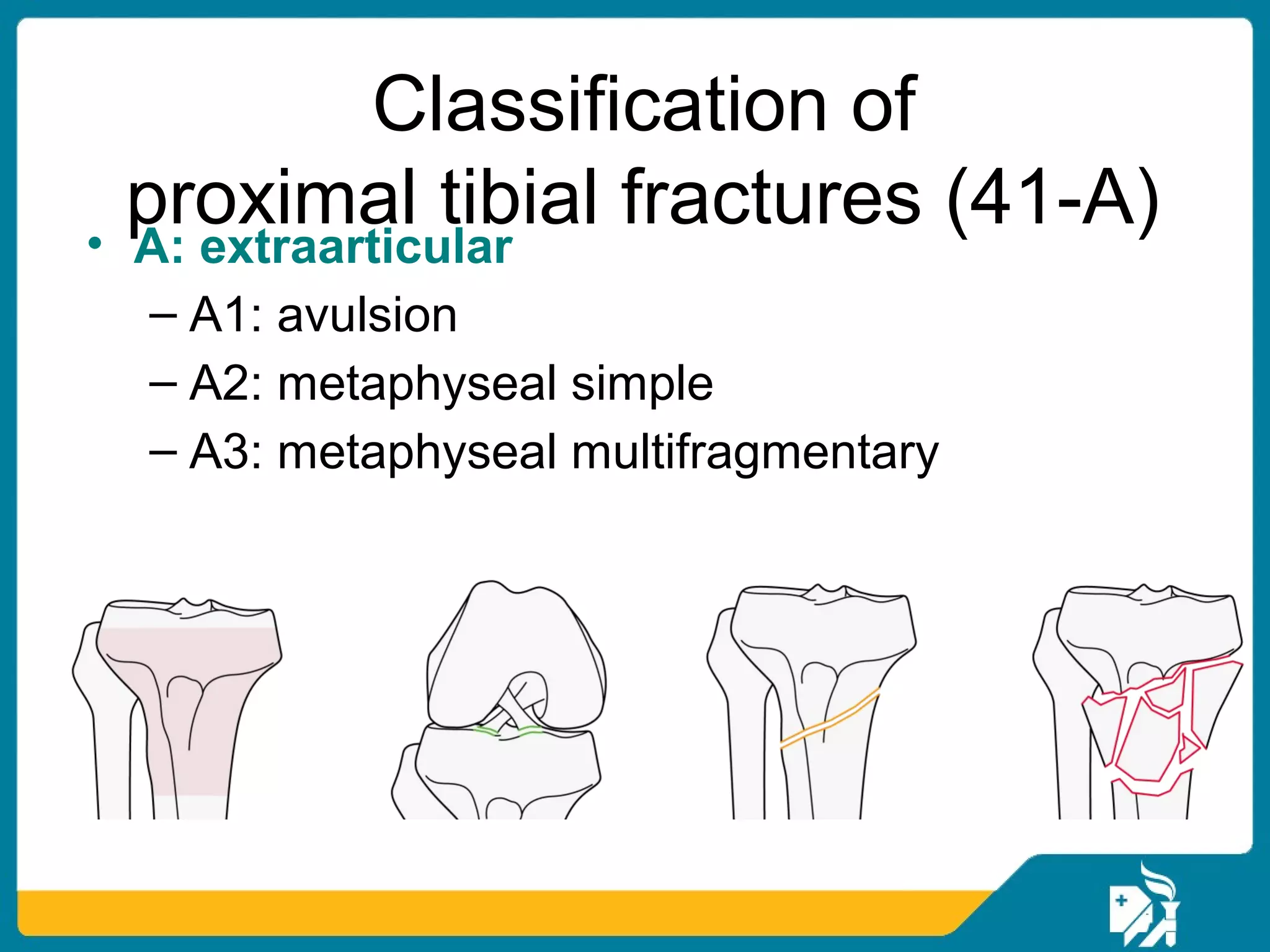
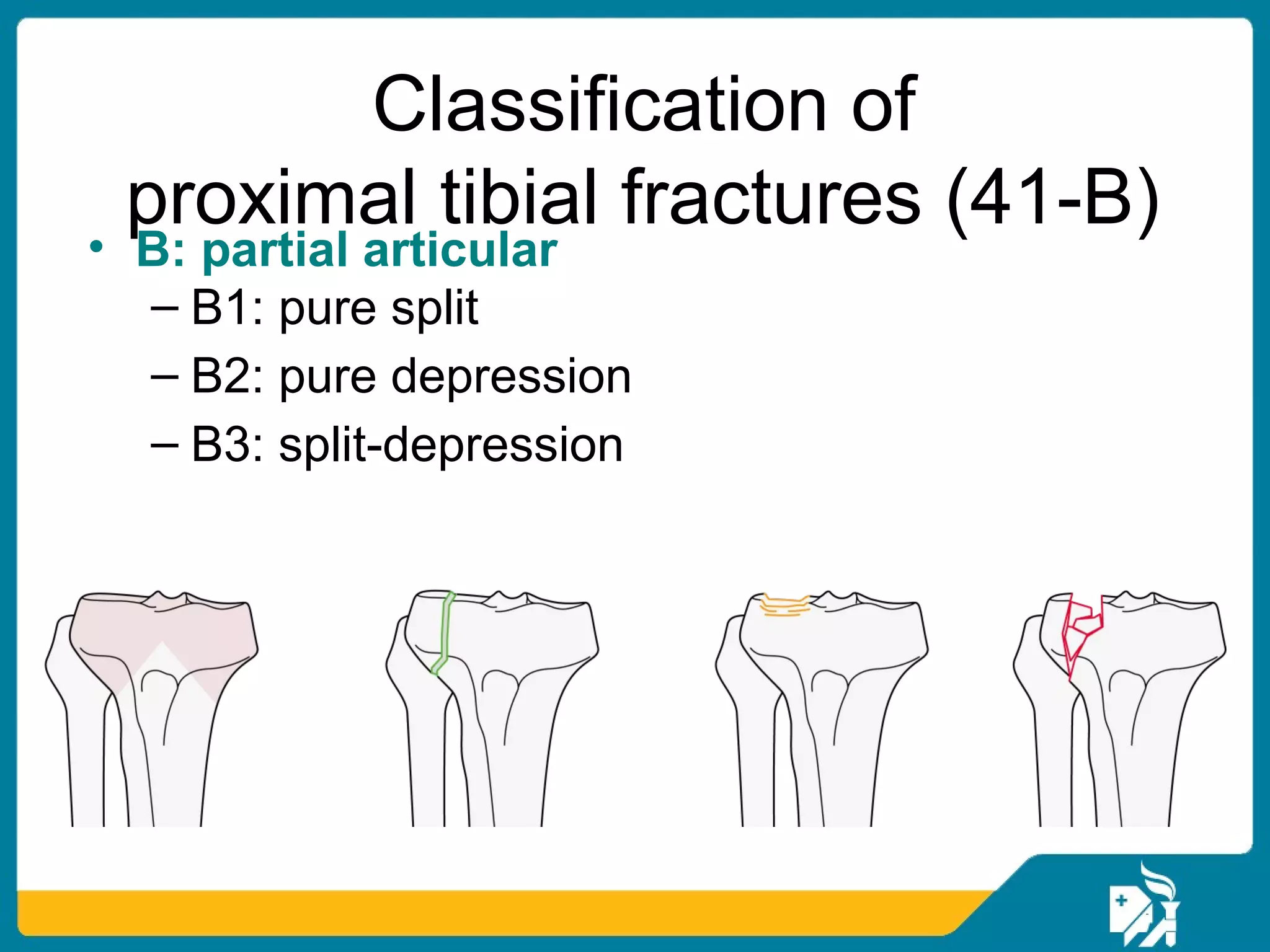
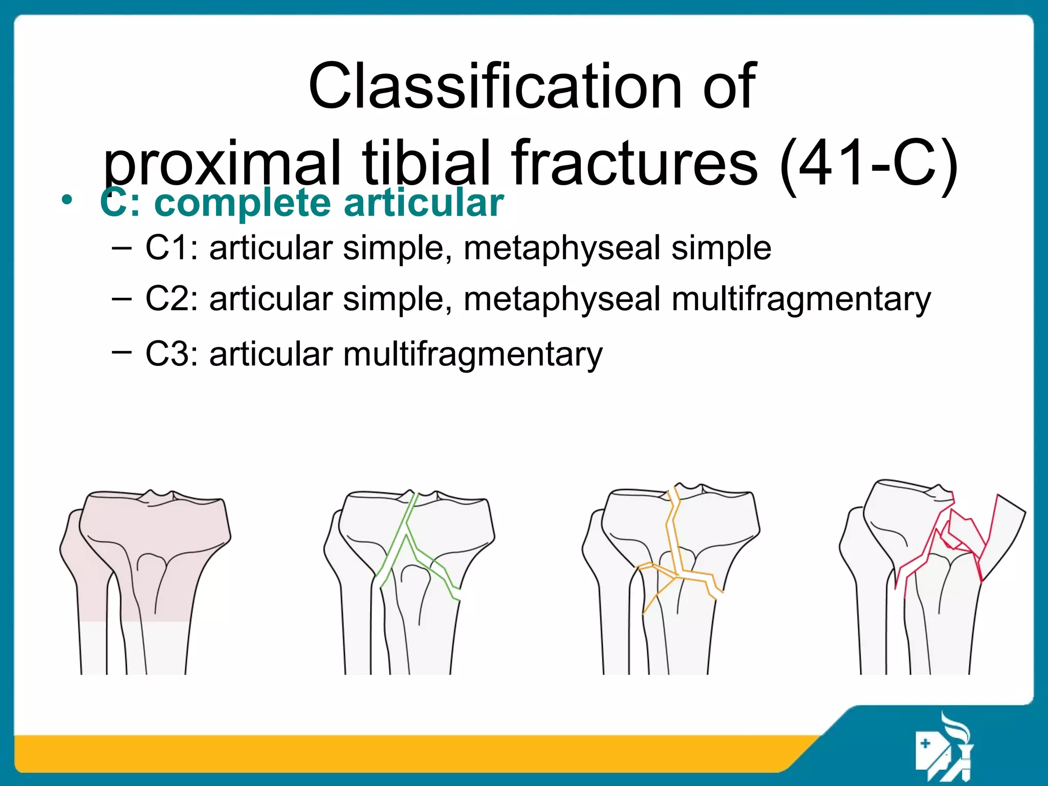
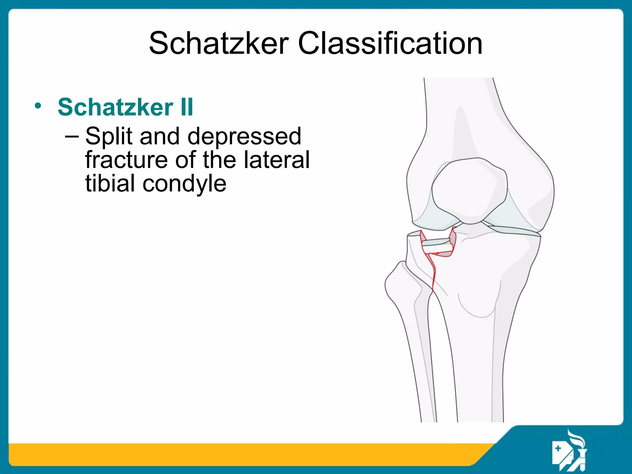
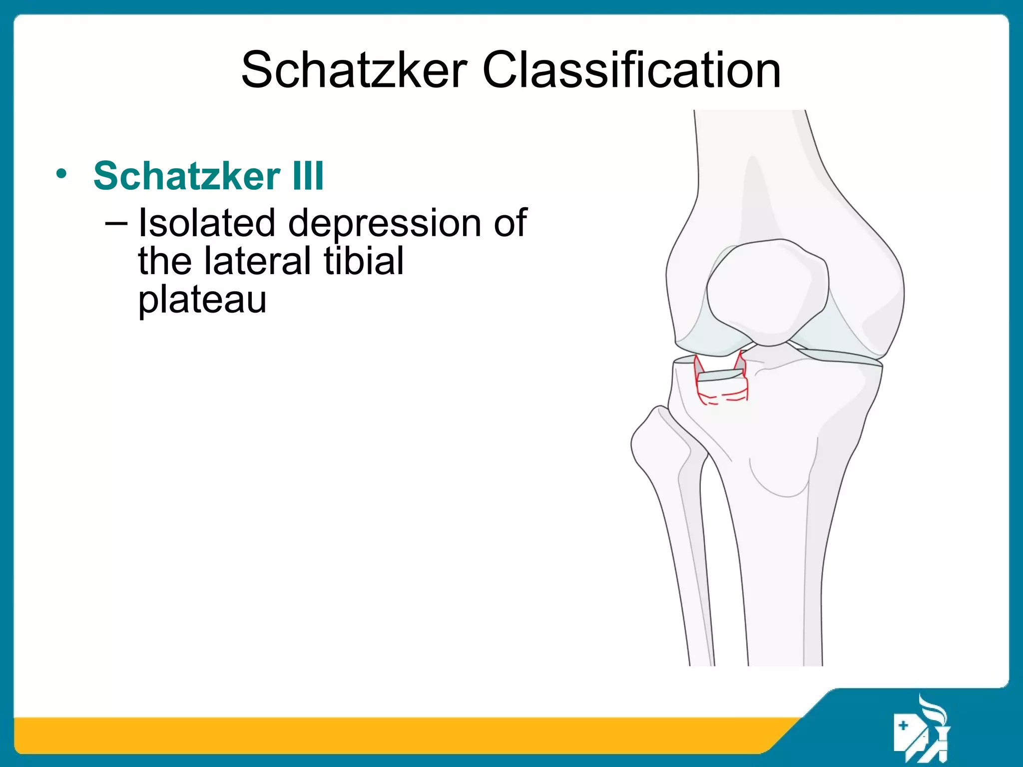
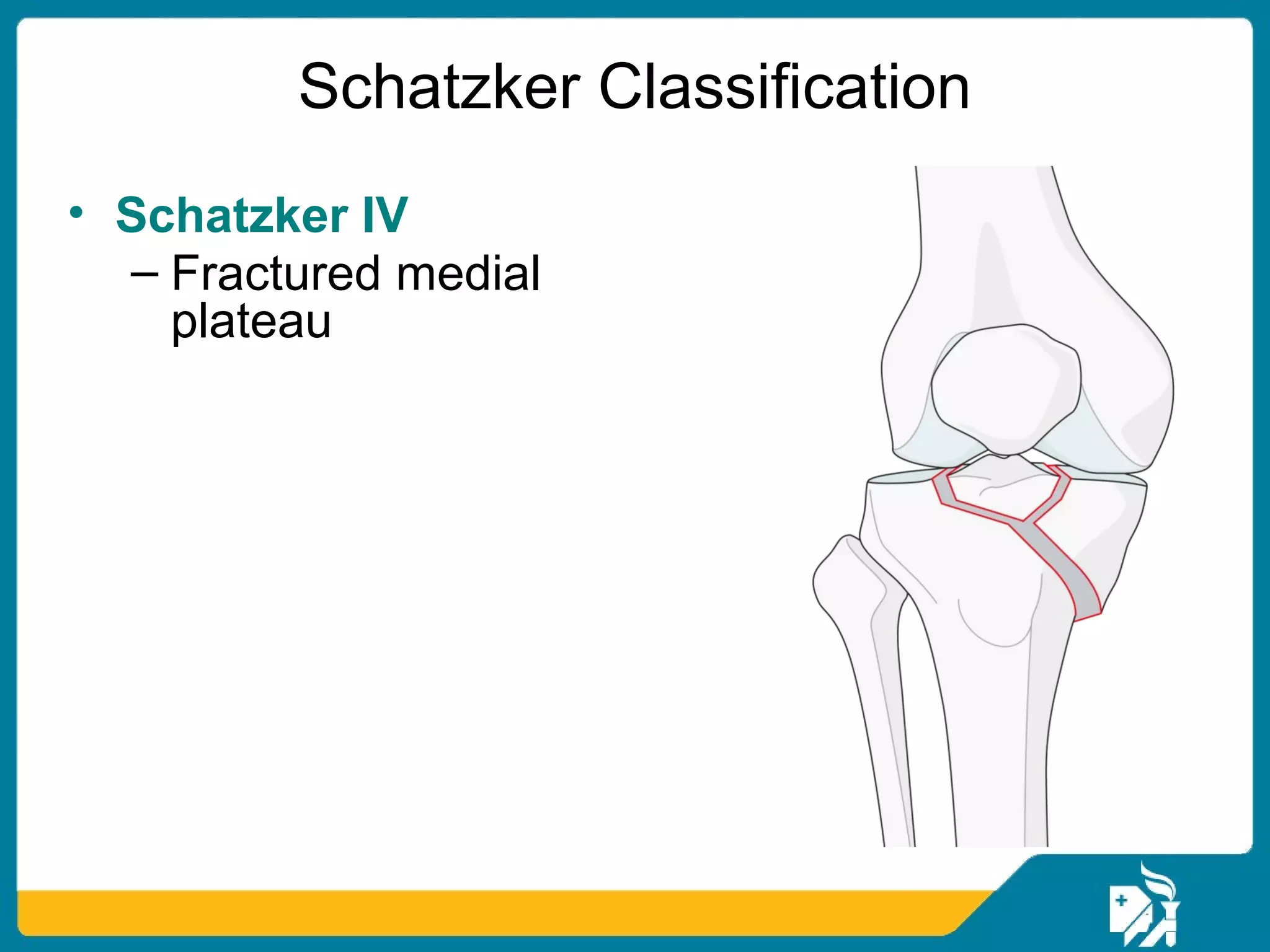
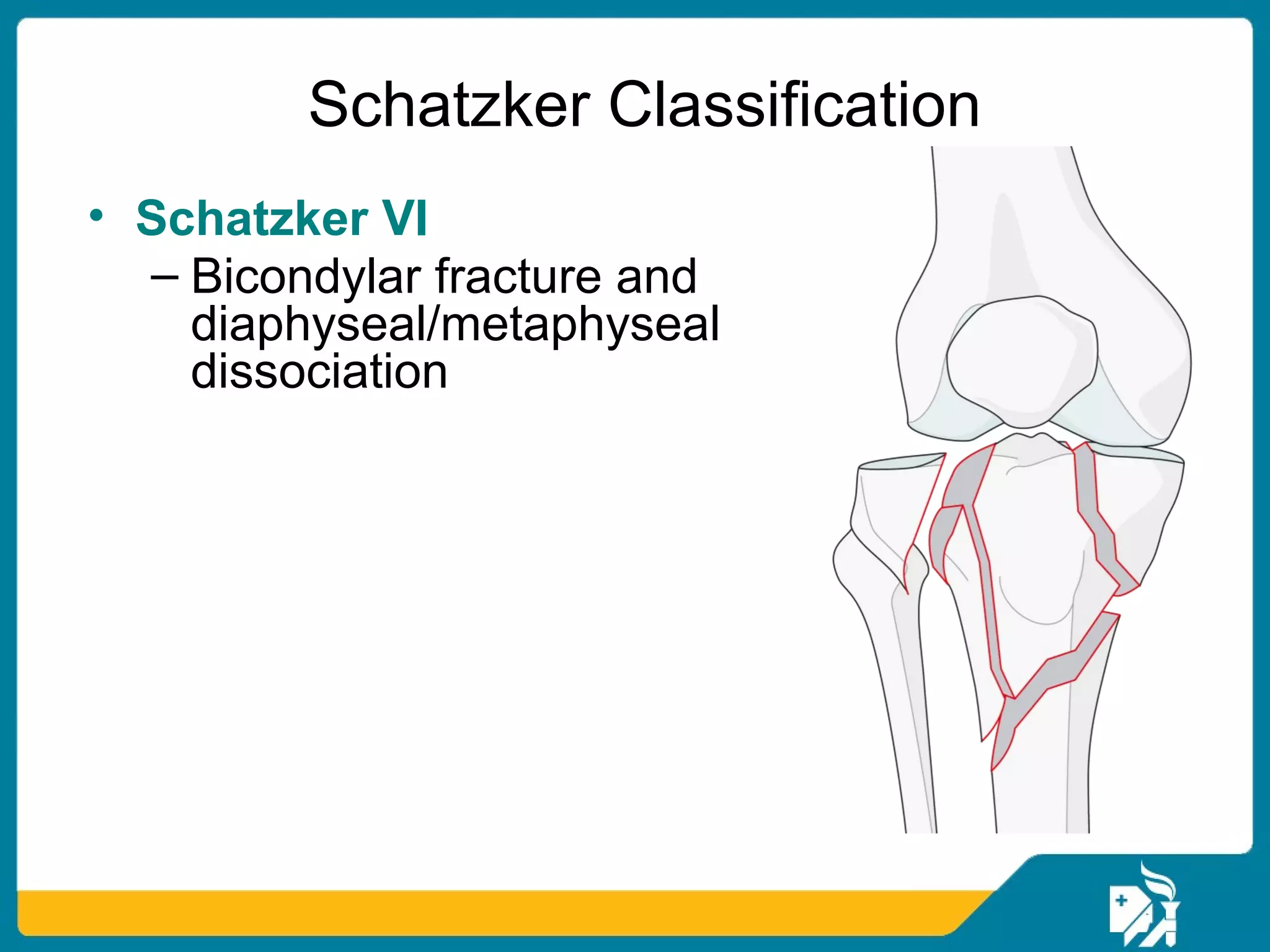
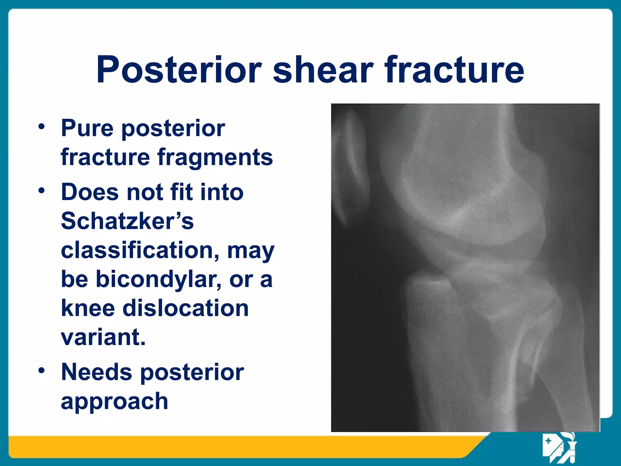
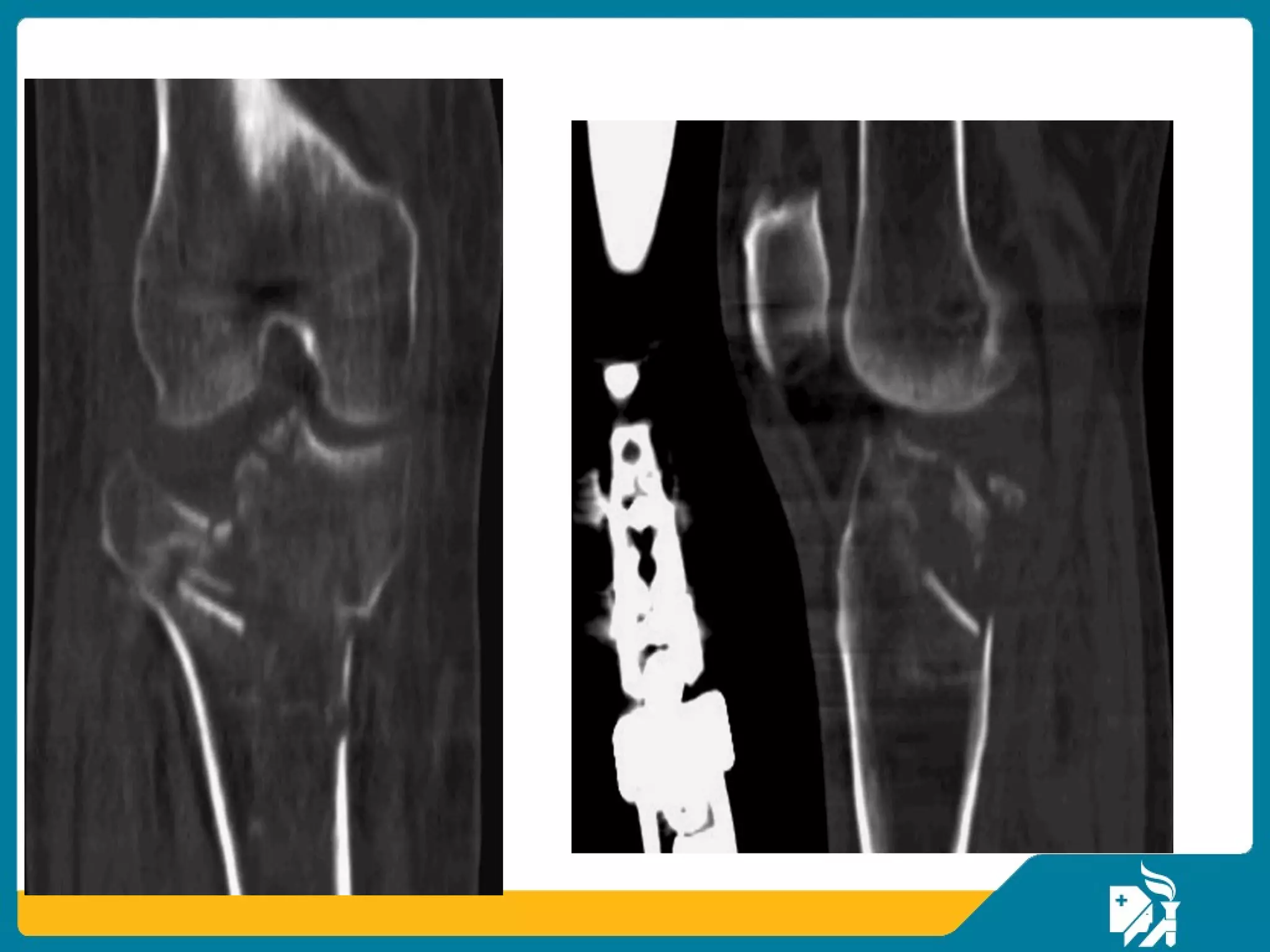
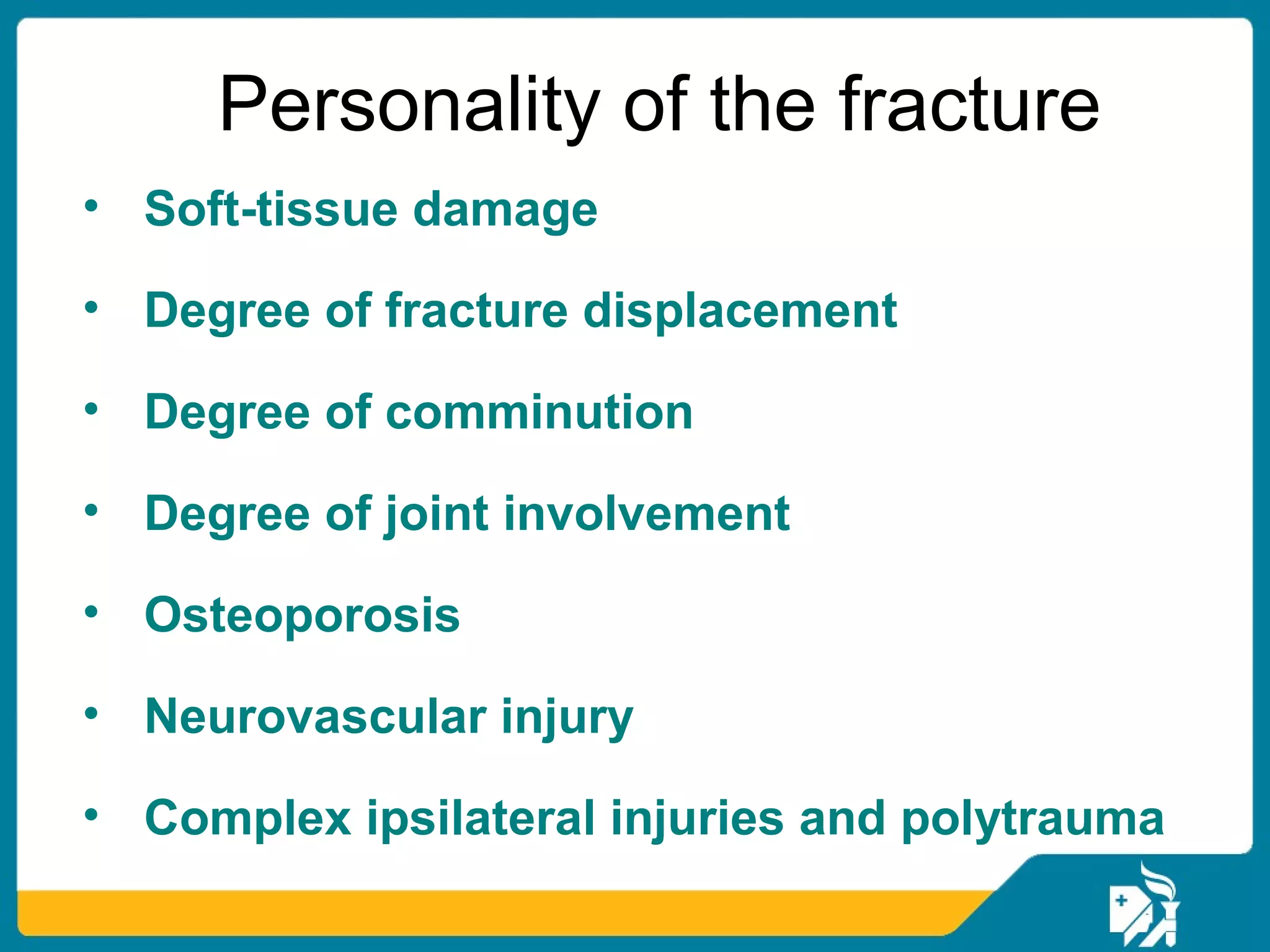
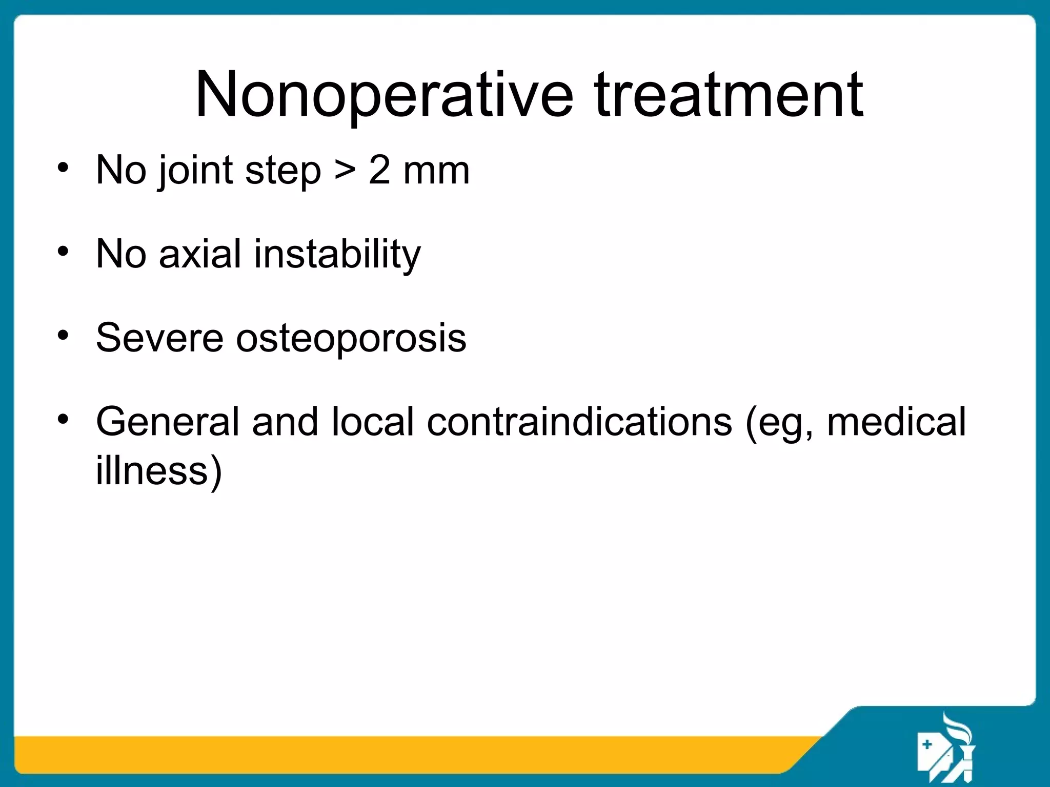
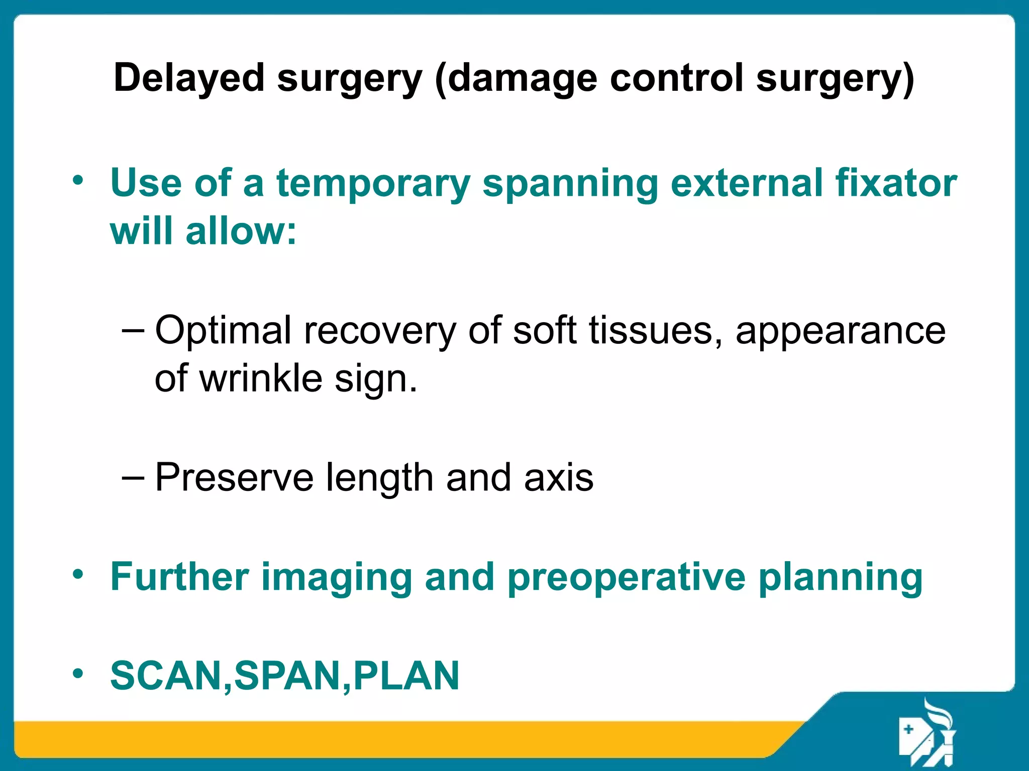
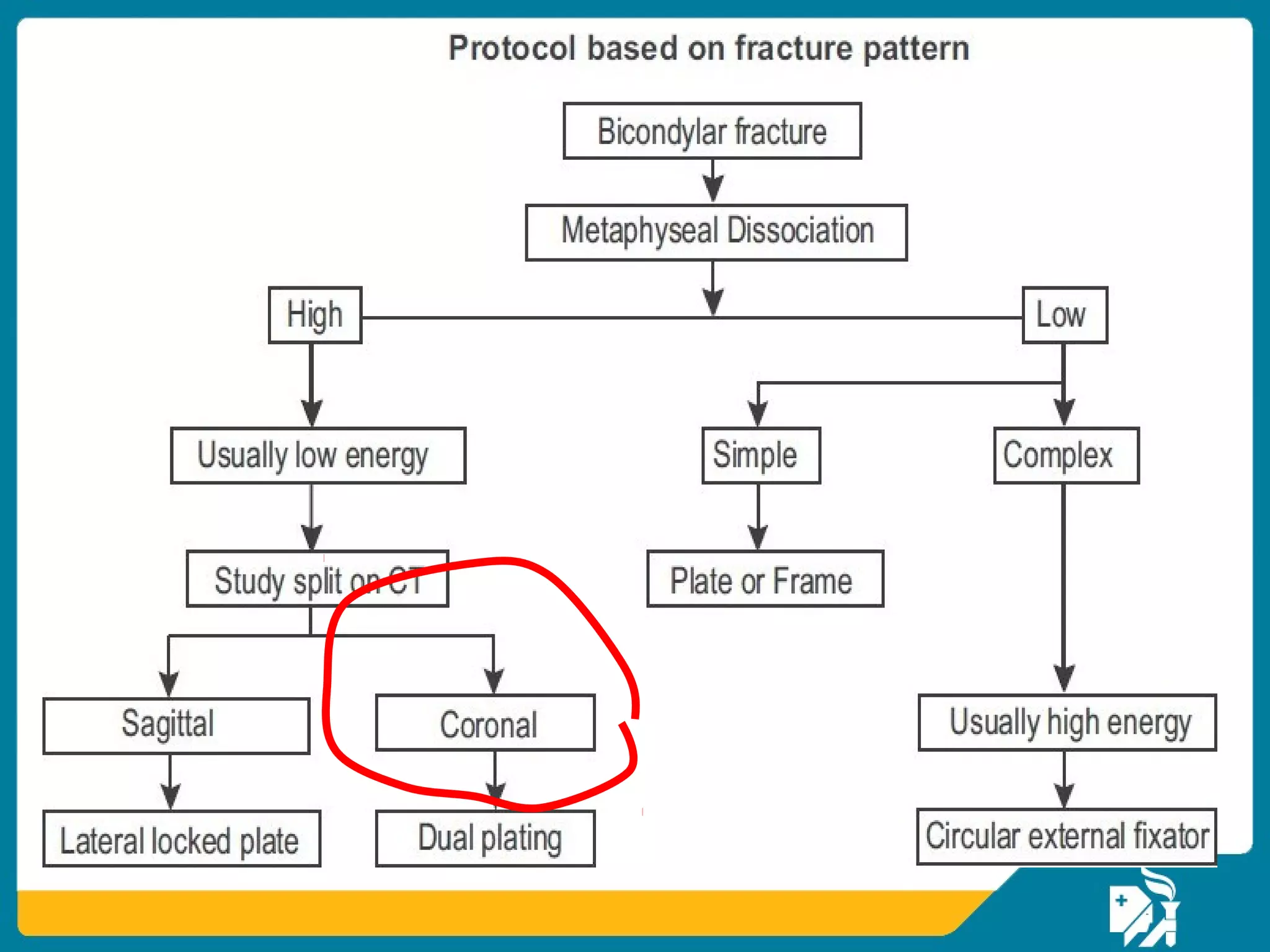
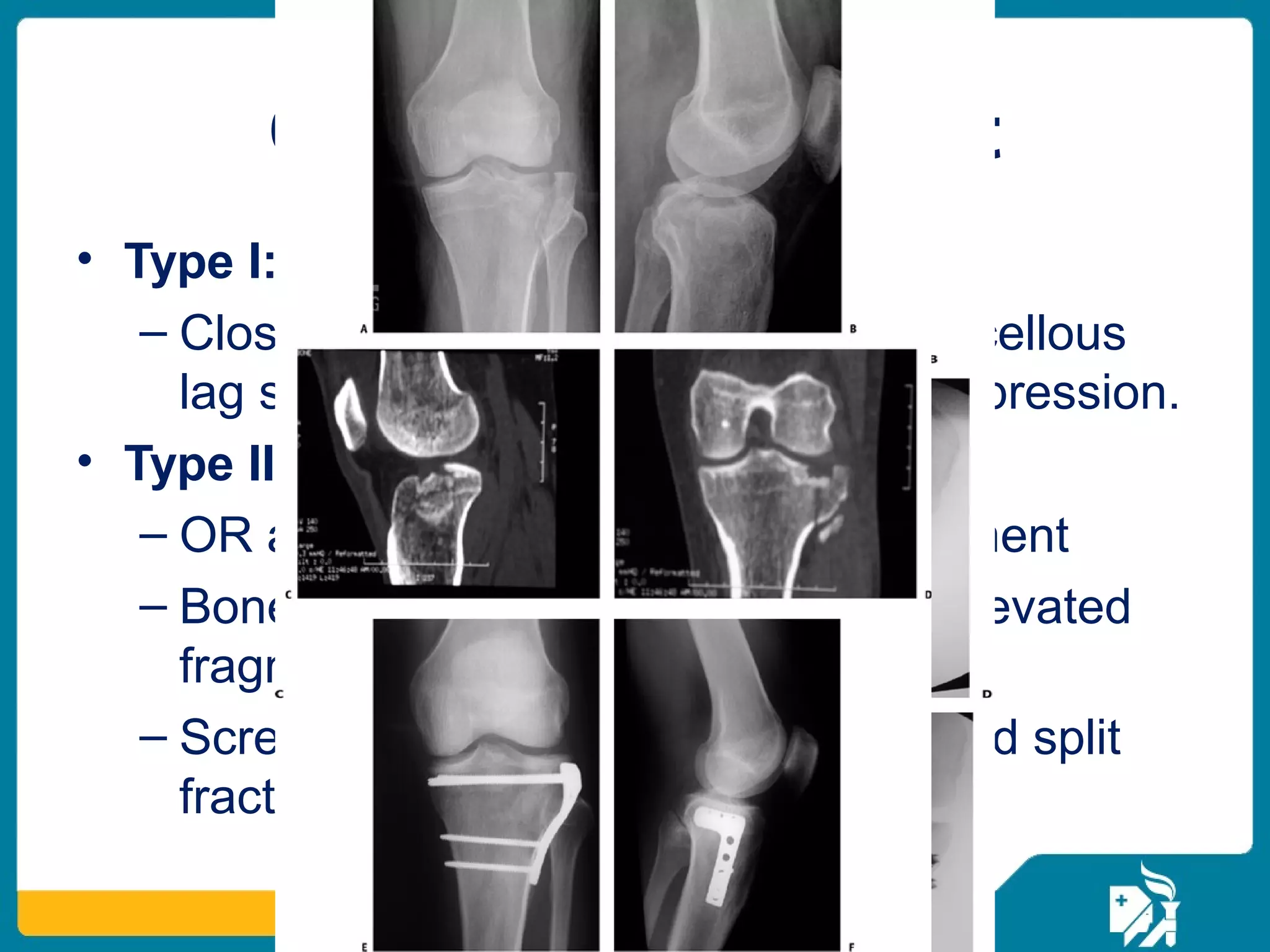
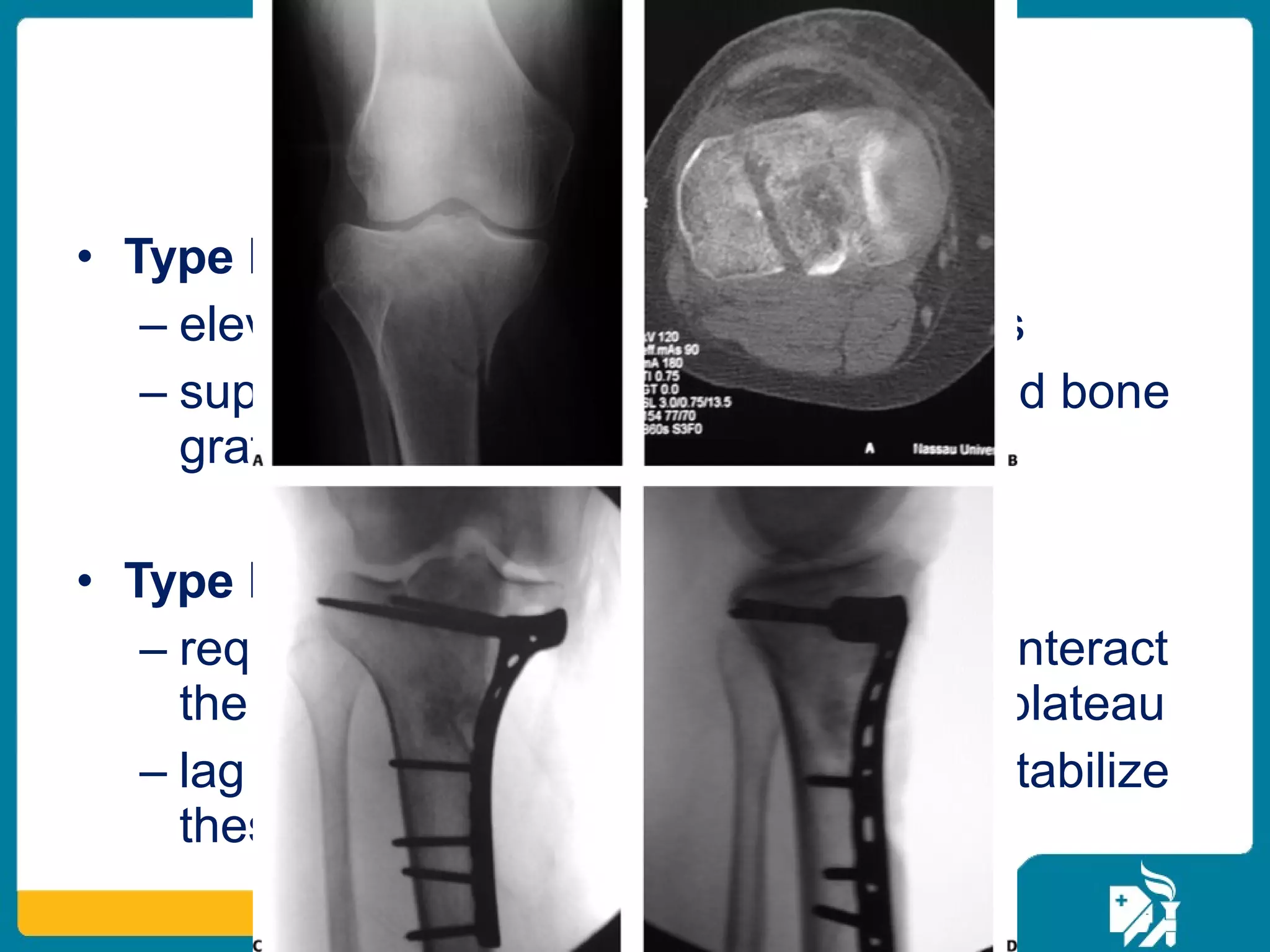
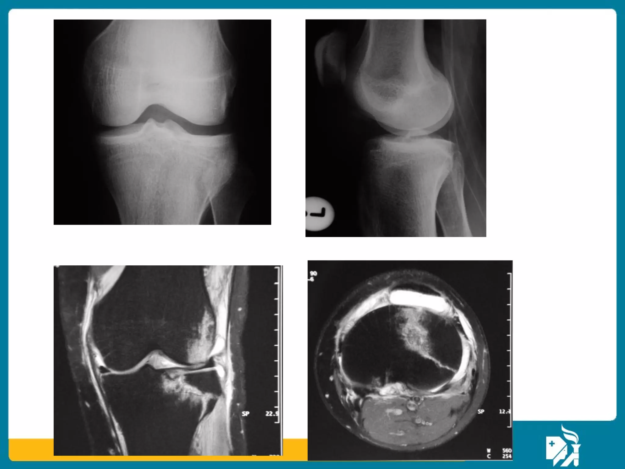
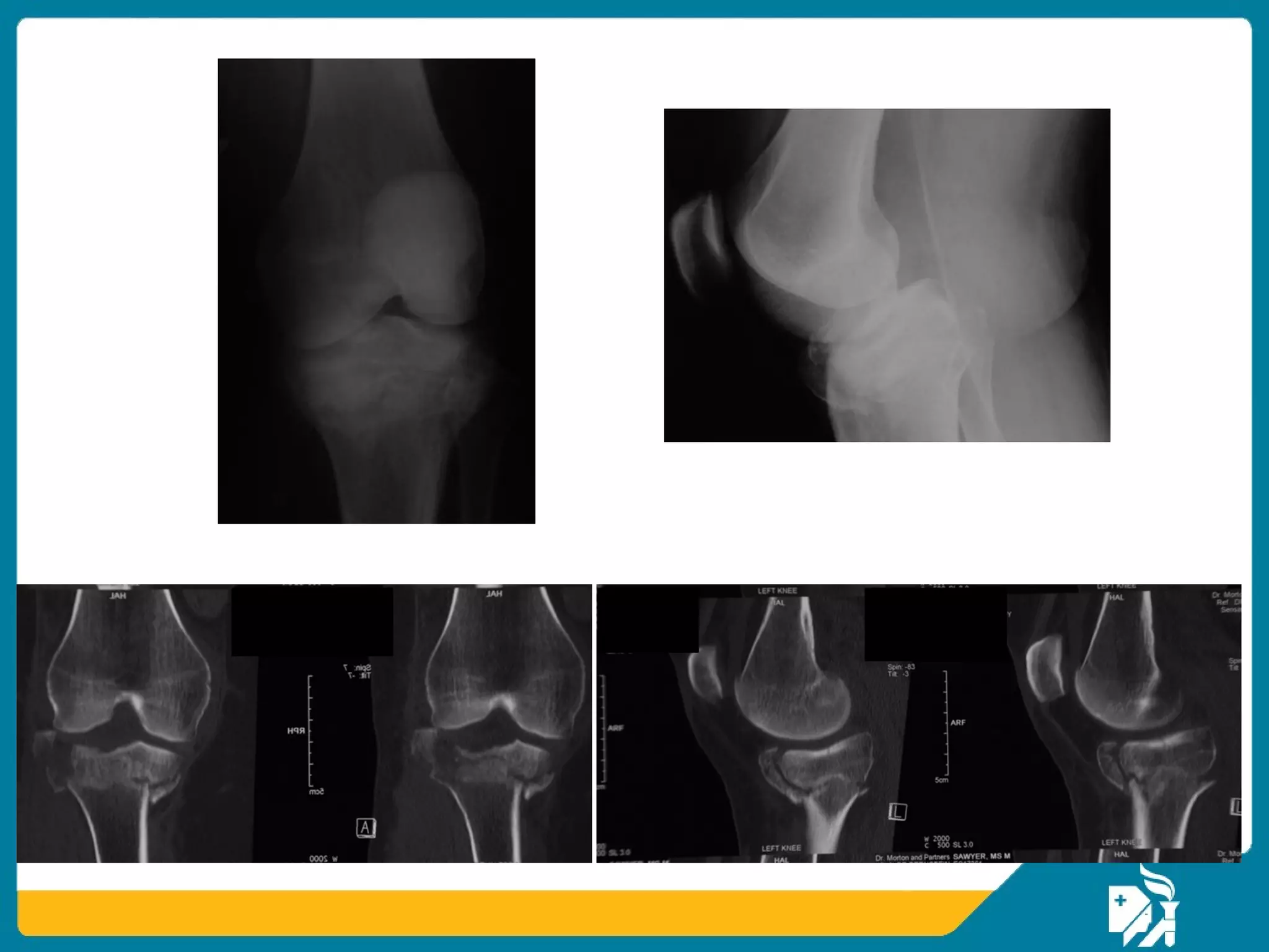
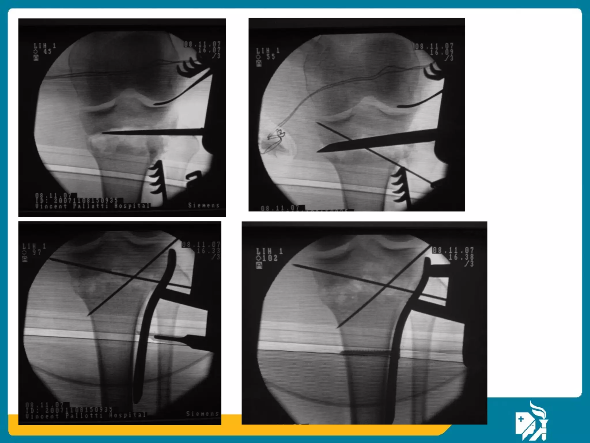
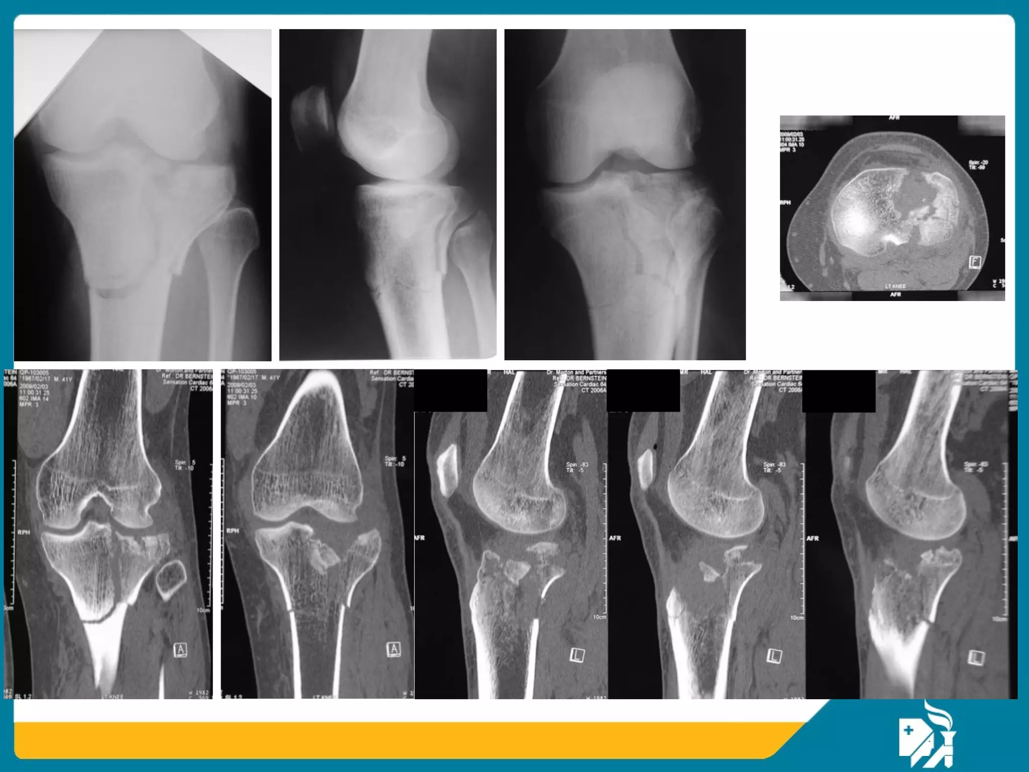
In the elderly osteoporotic fractures although the principles are the same but some special considerations in management of the soft tissues and the bony injuries are considered.
All Time Service Available Call Girls Marine Drive đł 9820252231 For 18+ VIP C...
Â
Proximal tibial fracture
1. Dr Smarajit Patnaik DNB
Senior Consultant Orthopaedic Surgeon
Apollo Hospitals
Bhubaneswar
Proximal Tibial Fractures in
the elderly: Surgical
considerations
3. Tibial plateau
⢠Articular fracture by definition
⢠Aim for:
âPerfect reduction of articular surface
âAbsolute stability (compression)
4. Caused by a variety of forces:
â˘Valgus/varus deformation
â˘Torsional forces due to slip and fall
â˘Axial compression
â˘Flexion/extension
â˘Direct trauma
Fracture mechanisms
5. Fracture mechanisms
⢠1% of all fractures:
â Lateral plateau: 60%
â Medial plateau: 25%
â Bi-condylar: 15%
⢠Two subgroups exist
⢠Young patients with good bone stockâhigh-
energy
⢠Elderly patients with osteoporosisâlow-
energy
15. Posterior shear fracture
⢠Pure posterior
fracture fragments
⢠Does not fit into
Schatzkerâs
classification, may
be bicondylar, or a
knee dislocation
variant.
⢠Needs posterior
approach
17. Etiology: high-energy trauma
Extensive damage to the soft tissues:
⢠Contusions
⢠Open injuries
⢠Compartment syndrome
⢠Peroneal and tibial nerve injury
⢠Popliteal artery injury
18. Etiology: low-energy trauma
⢠Axial trauma
⢠No contusions
⢠Closed injuries
⢠Less soft-tissue problems
⢠Axis deviation
⢠Fixation problem (osteoporosis)
19. Etiology
⢠In low-energy trauma the problem is:
â Mechanicalâfixation in osteoporotic bone
⢠In high-energy trauma the problem is:
â Biological and associated with damage to the
soft tissues
20. Investigations
⢠X-ray
â AP and lateral views
â 45°oblique views
⢠Computed tomography (CT)
⢠Magnetic resonance imaging (MRI)
⢠Angiography
21. ⢠Plain X-Ray:
⢠Supine AP and lateral view for all patients
⢠Internal and external oblique view
⢠Obtain contralateral AP and Lateral (compare)
⢠Tibial plateau view: AP with knee extended and beam
directed 15 degrees caudally
⢠CT scan:
⢠increases the diagnostic accuracy
⢠indicated in cases of articular depression
⢠shown to increase the interobserver and intraobserver
agreement on classification in tibial plateau fractures
⢠excellent adjuncts in the preoperative planning
Radiology
22. ⢠MRI:
⢠alternative to CT scan or arthroscopy
⢠osseous as well as the soft tissue
components of the injury
⢠cost prohibitive for use in standard situations
⢠Duplex US and Arteriography:
â To evaluate associated arterial injury.
Radiology
25. Personality of the fracture
⢠Soft-tissue damage
⢠Degree of fracture displacement
⢠Degree of comminution
⢠Degree of joint involvement
⢠Osteoporosis
⢠Neurovascular injury
⢠Complex ipsilateral injuries and polytrauma
26. Goals of treatment
⢠Decompression and preservation of soft-tissues
⢠Reconstruction of joint surfaces
⢠Reconstruction of normal mechanical axis
⢠Early motion
27. Nonoperative treatment
⢠No joint step > 2 mm
⢠No axial instability
⢠Severe osteoporosis
⢠General and local contraindications (eg, medical
illness)
28. ⢠Non-operative management:
â Indicated for non-displaced or minimally
displaced fractures
⢠Method:
â Protected weight bearing and early range-of-knee
motion in a hinged fracture brace.
â Isometric quadriceps exercises and progressive
passive, active-assisted, and active range-of-
knee motion exercises.
â Partial-weight bearing (30-40 Ib) for 8 to 12
weeks with progression to full weight bearing.
Management
29. Schatzkerâs principles of treatment
⢠Immobilization > 4 weeks: residual stiffness
⢠ORIF and immobilization: even more residual
stiffness
⢠Regardless of treatment: mobilize early
⢠As long as mobility is preserved a secondary
reconstructive procedure is possible
30. ⢠Impacted fractures cannot be dislodged by
traction or manipulation
⢠Depressed articular surfaces remain permanent
defects
Schatzkerâs principles of treatment
33. What if a proximal tibia comes on
day 2 of injury ?
⢠1) Take up immediately for
surgery
⢠2) Wait for investigations and
operate on second
⢠3) Wait for 8-10 days and
operate later
⢠4) Would operate after 1
month
34. Delayed surgery (damage control surgery)
⢠Use of a temporary spanning external fixator
will allow:
â Optimal recovery of soft tissues, appearance
of wrinkle sign.
â Preserve length and axis
⢠Further imaging and preoperative planning
⢠SCAN,SPAN,PLAN
35.
36. Surgical approach
⢠Minimally invasive versus ORIF
â ORIF: anterior, antero lateral, (postero)medial,
(postero)lateral
⢠Prepare for bone grafting
⢠Knee flexed position, floating position and
combined approach
⢠Tourniquet
⢠Fluoroscopy
38. Intraoperative procedure
⢠Expose ligamentous and meniscal structures
⢠Reconstruct the joint surface!
⢠Support the joint surface with bone or substitute
⢠Buttress with a plate (conventional)
⢠Repair the ligaments and menisci to achieve joint
stability
39. ⢠Type I:
â Closed reduction then stabilized cancellous
lag screws with washers to gain compression.
⢠Type II:
â OR and elevation of depressed fragment
â Bone graft is placed to support the elevated
fragments
â Screws are placed across the reduced split
fracture fragments in lag mode
Operative treatment
40. ⢠Type III:
â elevation through cortical fenestrations
â supported with subchondral screws and bone
graft
⢠Type IV:
â requires a medial buttress plate to counteract
the shear forces acting on the medial plateau
â lag screws alone are not sufficient to stabilize
these fractures
Operative treatment
45. Locked internal fixators
⢠Tibial locked internal fixators are available
⢠Locking head screws provide better
support than conventional screws in a
short metaphyseal fragment
⢠Percutaneous insertion preserves soft
tissues
56. Fine wire fixator for severe soft-tissue injuries
⢠Reconstruction of the
joint surface
⢠Reconstruction of
stable axes
⢠Early motion
⢠Excellent results
- (Schatzker IV, V, and
VI)
57.
58. Fine wire/Hybrid
⢠Exoskeleton allows:
â Attention to soft tissues
â Application of relative stability to
the metaphyseal/diaphyseal
component
⢠Problems:
â Fine wire irritation
â Intracapsular portals
â Must get articular reduction first!
â Patients unhappy!
59. Results of ORIF on tibial plateau in general
⢠Depends on the fracture type
⢠Depends on soft-tissue management
⢠Depends on realization of goals
⢠Can be excellent even in high-energy
trauma:
â Average range of motion 0â120°(87%)
â No deterioration in the 2nd 5 years
â Good prognosis
60.
61. Take-home messages
⢠Anatomical reduction and rigid fixation of
joint surfaceâabsolute stability
⢠Functional reduction and stable fixation of
metaphysisârelative stability
⢠Restoration of joint stability by appropriate
soft-tissue reconstruction
⢠Early active movement
⢠Non operative treatment has a role in
severe osteoporosis in elderly
⢠Respect the soft tissues!!!
Editor's Notes
Published: August 2013
References:
Kennedy JC, Bailey WH. Experimental tibial-plateau fractures. Studies of the mechanism and a classification. J Bone Joint Surg Am. 1968 Dec;50(8):1522-34.
Images courtesy of AO Surgery Reference.
The screws indicated should probably be omitted in line with current bridging osteosynthesis practice. Lecturer can choose to challange participants on this point prior to showing this slide.