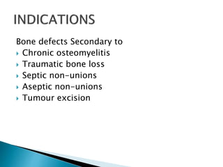
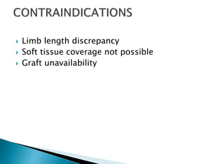
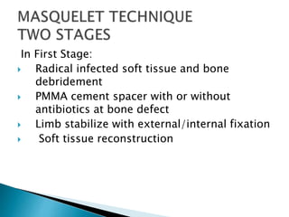
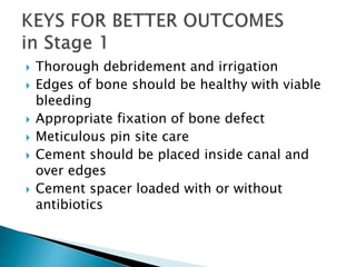
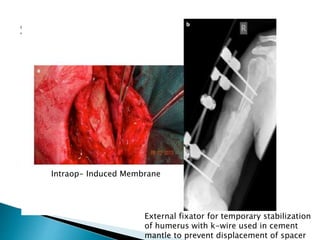
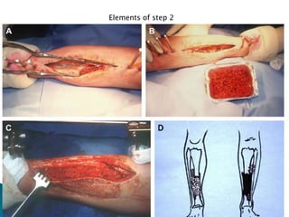
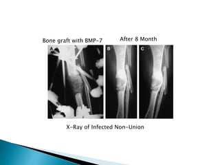
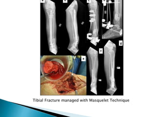
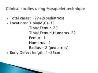
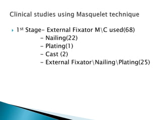
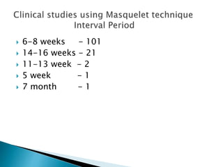
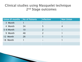
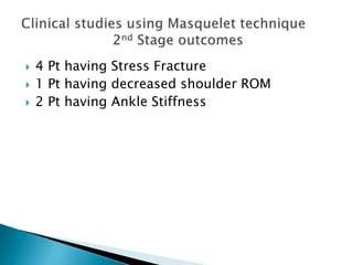
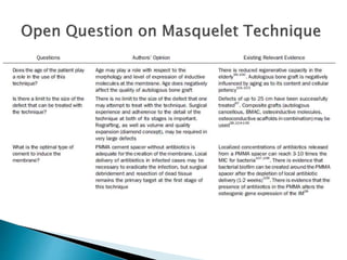
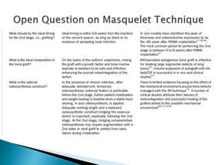
Dr. Shubhanshu
Orthopedics Surgeon
 The Concept of Induced Membrane for
Reconstruction of Long Bone Defects by Alain
C.Masquelet,Thierry Begue in Elsevier journal
2010
 Masquelet technique for the treatment of
bone defects:Tips-tricks and future directions
by Peter V. Giannoudis et al in Elsevier 2011
 Current Concepts Review - Bone Repair Using
the Masquelet Technique by Alain Masquelet
et al in JBJS journal 2019
 Masquelet technique for infected distal radius
fractures with gaps in paediatric age group by
John Mukhopadhaya, Janki Sharan Bhadani in
Elsevier 2021
 A case report of extensive segmental defect
of the humerus due to thermal necrosis
treated with Masquelet technique by John
Mukhopadhaya, Kumar Gautam, Janki Sharan
Bhadani in Elsevier 2021
 INTRODUCTION
 EFFECT OF THE INDUCED MEMBRANE
 INDICATIONS
 CONTRAINDICATIONS
 KEYS FOR BETTER OUTCOMES
 DRAWBACKS
 VARIOUS REPORT OUTCOMES
 Masquelet first described in 1986
 2 Steps procedure
for bone defect (upto25cm) and
non-union by induced membrane
osteogenesis
Induced membrane :
 Pseudosynovial membrane formed around
cement spacer due to foreign body
reaction(stage-1)
 Acts chamber around the bony defect to
contain the bone graft and stimulate bone
regeneration(stage-2)
A. Mechanical effects
◦ Cement spacer maintains the length of bone and
prevent soft tissue interposition
◦ Creates contained space for the placement of bone
graft
◦ Prevent the reabsortion of the bone graft in the
defects and remains non-adherent to cement
spacer
◦ Cement spacer may be used as a carrier for
antibiotics to achieve high local concentration of
antibiotics without systemic toxicity
B. Biological effects
◦ Membrane rich in type 1 collagen, fibroblasts and
myofibroblasts
◦ Highly vascular due to high concentration of VEGF
which promotes angiogenesis
◦ Increased levels of TGF-beta, BMP-2 which
promotes osteogenesis
Bone defects Secondary to
 Chronic osteomyelitis
 Traumatic bone loss
 Septic non-unions
 Aseptic non-unions
 Tumour excision
 Limb length discrepancy
 Soft tissue coverage not possible
 Graft unavailability
In First Stage:
 Radical infected soft tissue and bone
debridement
 PMMA cement spacer with or without
antibiotics at bone defect
 Limb stabilize with external/internal fixation
 Soft tissue reconstruction
 Thorough debridement and irrigation
 Edges of bone should be healthy with viable
bleeding
 Appropriate fixation of bone defect
 Meticulous pin site care
 Cement should be placed inside canal and
over edges
 Cement spacer loaded with or without
antibiotics
 Soft tissue envelop should have adequate
blood supply
 Good soft tissue coverage
 Wound closure must not be under tension
After Debridement
Punctate Bleeding at bony edges
Cement over the edges of bone
Maintain space of reconstruction
 Usually 6-8 weeks
 No evidences showing any relation in interval
duration between using external
fixatornailingplating in 1st stage.
 Clinical studies shows that using antibiotics
loaded cement spacer decrease infection
 In clinical study by Biau shown that Femur
(c/o Ewing Sarcoma) takes longer Interval
period than rest locations i.e 7 months
Mechanical stability using temporary external fixator
In Second stage:
 6-8 weeks later
 Cement spacer is carefully removed
 Formed “INDUCED MEMBRANE” is minimally
disturbed
 Defect is filled with morcellised cancellous
autologous bone graft(with additional bone
graft substitutes, 1:3)
 Fixation is mandatory for bone stabilization.
 Culture prior to administration of antibiotics
intraoperatively
 Membrane must be incised with caution
 Cement spacer removed with saw or
osteotome
 IM canal is prepared with hand reamers and
curette
 All non-vitals tissue must be removed
 Depending on size adequate volume of graft
material should be available
 Autologous bone graft from Iliac crest or
intramedullary canal by
Reamer/Irrigator/Aspirator(RIA)
 For large defect can be augmented with
allograft or bone substitute
 Bone graft material can be enhanced with
osteoprogenitor cells or osteoinductive
growth factors
 Graft material must be enclosed within
membrane
 Adequate mechanical stability
 Adequate soft tissue coverage
 Wound closure must not be under tension
Intraop- Induced Membrane
External fixator for temporary stabilization
of humerus with k-wire used in cement
mantle to prevent displacement of spacer
Elements of step 2
Bone graft with BMP-7 After 8 Month
X-Ray of Infected Non-Union
Tibial Fracture managed with Masquelet Technique
 Total cases: 127+2(pediatrics)
 Locations: Tibia(MC)-35
TibiaFemur-25
TibiaFemurHumerus-22
Femur- 1
Humerus- 2
Radius – 2 (pediatrics)
 Bony Defect length: 1-25cm
 1st Stage- External Fixator MC used(68)
- Nailing(22)
- Plating(1)
- Cast (2)
- External FixatorNailingPlating(25)
 6-8 weeks - 101
 14-16 weeks - 21
 11-13 week - 2
 5 week - 1
 7 month - 1
Union @ months No of Patients Infection Non Union
3 Month 1 - -
4 Month 34 5 4
5-6 Month 10 2 -
9 Month 48 2 1
12 Month 26 1 1
18 Month 1 - 1
 4 Pt having Stress Fracture
 1 Pt having decreased shoulder ROM
 2 Pt having Ankle Stiffness
 Two different interventions
 Availability of graft
- Limited
- Donor site morbidity
 Supplementary procedures for soft tissue
transfer
 Masquelet technique provides good
functional outcome in patients with severe
bone defect and non-union
 Meticulous debridement in stage one and
preservation of Induced Membrane in stage
two are key to this techniques
 Using antibiotics cement spacer decreases
further infection
 By using this technique can achieve functional
range of motion
 Autogenous bone graft helped in improving
the stability of fixation and reduced risk of
fixation failure

Masquelet Technique

  • 1.
  • 2.
     The Conceptof Induced Membrane for Reconstruction of Long Bone Defects by Alain C.Masquelet,Thierry Begue in Elsevier journal 2010  Masquelet technique for the treatment of bone defects:Tips-tricks and future directions by Peter V. Giannoudis et al in Elsevier 2011  Current Concepts Review - Bone Repair Using the Masquelet Technique by Alain Masquelet et al in JBJS journal 2019
  • 3.
     Masquelet techniquefor infected distal radius fractures with gaps in paediatric age group by John Mukhopadhaya, Janki Sharan Bhadani in Elsevier 2021  A case report of extensive segmental defect of the humerus due to thermal necrosis treated with Masquelet technique by John Mukhopadhaya, Kumar Gautam, Janki Sharan Bhadani in Elsevier 2021
  • 5.
     INTRODUCTION  EFFECTOF THE INDUCED MEMBRANE  INDICATIONS  CONTRAINDICATIONS  KEYS FOR BETTER OUTCOMES  DRAWBACKS  VARIOUS REPORT OUTCOMES
  • 6.
     Masquelet firstdescribed in 1986  2 Steps procedure for bone defect (upto25cm) and non-union by induced membrane osteogenesis
  • 7.
    Induced membrane : Pseudosynovial membrane formed around cement spacer due to foreign body reaction(stage-1)  Acts chamber around the bony defect to contain the bone graft and stimulate bone regeneration(stage-2)
  • 8.
    A. Mechanical effects ◦Cement spacer maintains the length of bone and prevent soft tissue interposition ◦ Creates contained space for the placement of bone graft ◦ Prevent the reabsortion of the bone graft in the defects and remains non-adherent to cement spacer ◦ Cement spacer may be used as a carrier for antibiotics to achieve high local concentration of antibiotics without systemic toxicity
  • 9.
    B. Biological effects ◦Membrane rich in type 1 collagen, fibroblasts and myofibroblasts ◦ Highly vascular due to high concentration of VEGF which promotes angiogenesis ◦ Increased levels of TGF-beta, BMP-2 which promotes osteogenesis
  • 10.
    Bone defects Secondaryto  Chronic osteomyelitis  Traumatic bone loss  Septic non-unions  Aseptic non-unions  Tumour excision
  • 11.
     Limb lengthdiscrepancy  Soft tissue coverage not possible  Graft unavailability
  • 12.
    In First Stage: Radical infected soft tissue and bone debridement  PMMA cement spacer with or without antibiotics at bone defect  Limb stabilize with external/internal fixation  Soft tissue reconstruction
  • 13.
     Thorough debridementand irrigation  Edges of bone should be healthy with viable bleeding  Appropriate fixation of bone defect  Meticulous pin site care  Cement should be placed inside canal and over edges  Cement spacer loaded with or without antibiotics
  • 14.
     Soft tissueenvelop should have adequate blood supply  Good soft tissue coverage  Wound closure must not be under tension
  • 15.
    After Debridement Punctate Bleedingat bony edges Cement over the edges of bone Maintain space of reconstruction
  • 16.
  • 17.
     No evidencesshowing any relation in interval duration between using external fixatornailingplating in 1st stage.  Clinical studies shows that using antibiotics loaded cement spacer decrease infection  In clinical study by Biau shown that Femur (c/o Ewing Sarcoma) takes longer Interval period than rest locations i.e 7 months
  • 18.
    Mechanical stability usingtemporary external fixator
  • 19.
    In Second stage: 6-8 weeks later  Cement spacer is carefully removed  Formed “INDUCED MEMBRANE” is minimally disturbed  Defect is filled with morcellised cancellous autologous bone graft(with additional bone graft substitutes, 1:3)  Fixation is mandatory for bone stabilization.
  • 20.
     Culture priorto administration of antibiotics intraoperatively  Membrane must be incised with caution  Cement spacer removed with saw or osteotome  IM canal is prepared with hand reamers and curette  All non-vitals tissue must be removed
  • 21.
     Depending onsize adequate volume of graft material should be available  Autologous bone graft from Iliac crest or intramedullary canal by Reamer/Irrigator/Aspirator(RIA)  For large defect can be augmented with allograft or bone substitute  Bone graft material can be enhanced with osteoprogenitor cells or osteoinductive growth factors
  • 22.
     Graft materialmust be enclosed within membrane  Adequate mechanical stability  Adequate soft tissue coverage  Wound closure must not be under tension
  • 23.
    Intraop- Induced Membrane Externalfixator for temporary stabilization of humerus with k-wire used in cement mantle to prevent displacement of spacer
  • 24.
  • 25.
    Bone graft withBMP-7 After 8 Month X-Ray of Infected Non-Union
  • 26.
    Tibial Fracture managedwith Masquelet Technique
  • 27.
     Total cases:127+2(pediatrics)  Locations: Tibia(MC)-35 TibiaFemur-25 TibiaFemurHumerus-22 Femur- 1 Humerus- 2 Radius – 2 (pediatrics)  Bony Defect length: 1-25cm
  • 28.
     1st Stage-External Fixator MC used(68) - Nailing(22) - Plating(1) - Cast (2) - External FixatorNailingPlating(25)
  • 29.
     6-8 weeks- 101  14-16 weeks - 21  11-13 week - 2  5 week - 1  7 month - 1
  • 30.
    Union @ monthsNo of Patients Infection Non Union 3 Month 1 - - 4 Month 34 5 4 5-6 Month 10 2 - 9 Month 48 2 1 12 Month 26 1 1 18 Month 1 - 1
  • 31.
     4 Pthaving Stress Fracture  1 Pt having decreased shoulder ROM  2 Pt having Ankle Stiffness
  • 34.
     Two differentinterventions  Availability of graft - Limited - Donor site morbidity  Supplementary procedures for soft tissue transfer
  • 35.
     Masquelet techniqueprovides good functional outcome in patients with severe bone defect and non-union  Meticulous debridement in stage one and preservation of Induced Membrane in stage two are key to this techniques
  • 36.
     Using antibioticscement spacer decreases further infection  By using this technique can achieve functional range of motion  Autogenous bone graft helped in improving the stability of fixation and reduced risk of fixation failure

Editor's Notes

  • #33 BMAC bone marrow aspirate concentration Diamond concept- Mechanical/osteoconductive\osteogenic\growth factor
  • #34 Hydroxyapatite tricalciumphospahte