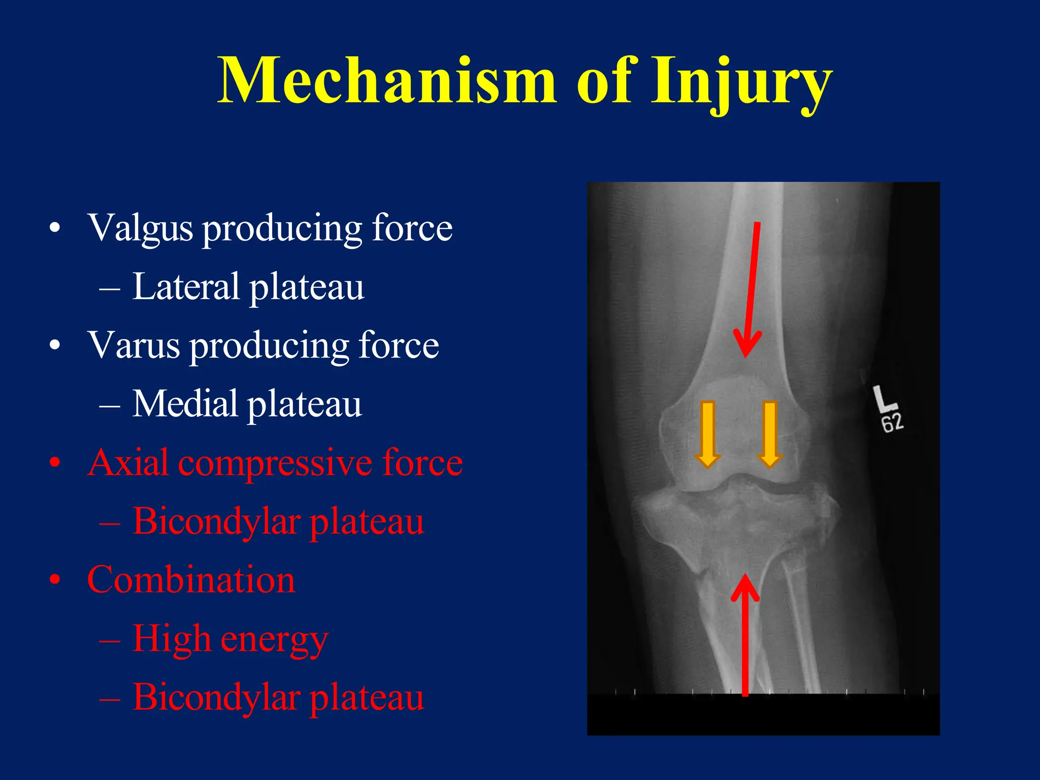
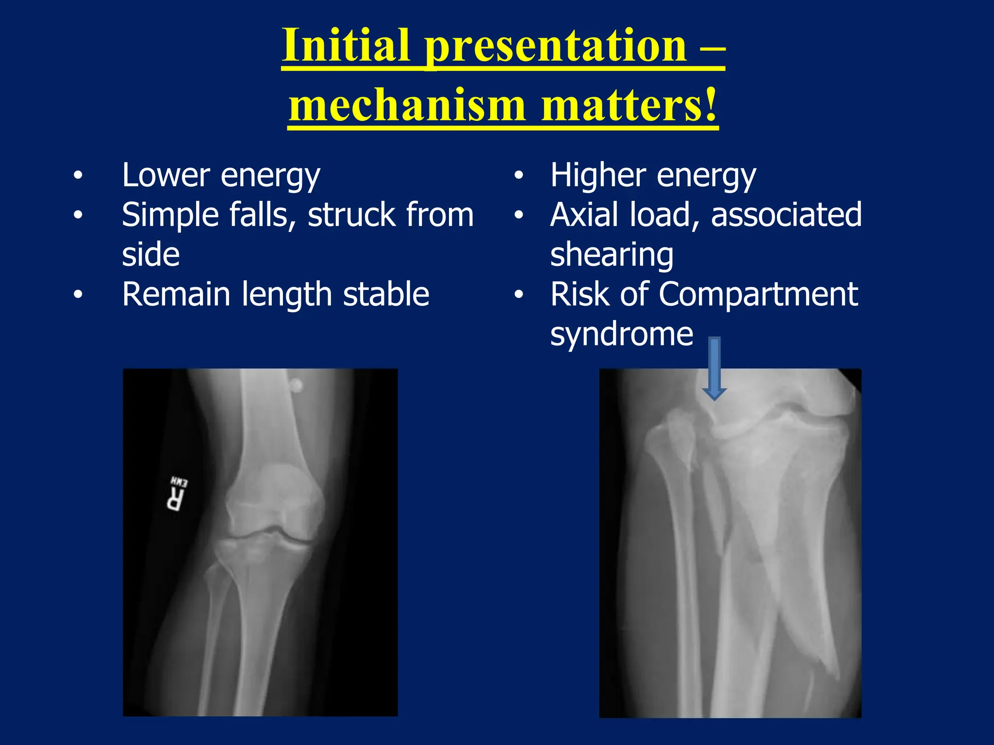
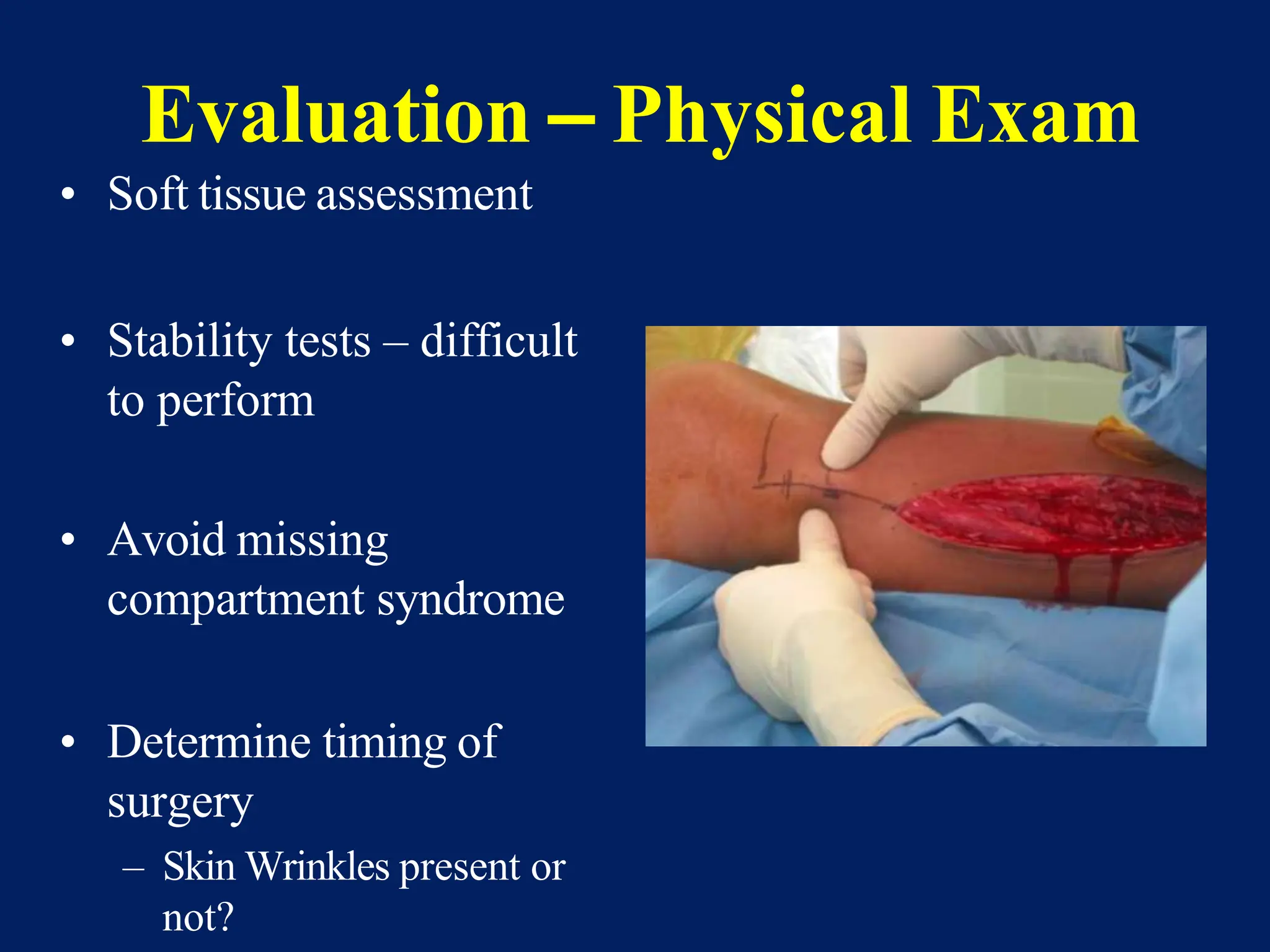
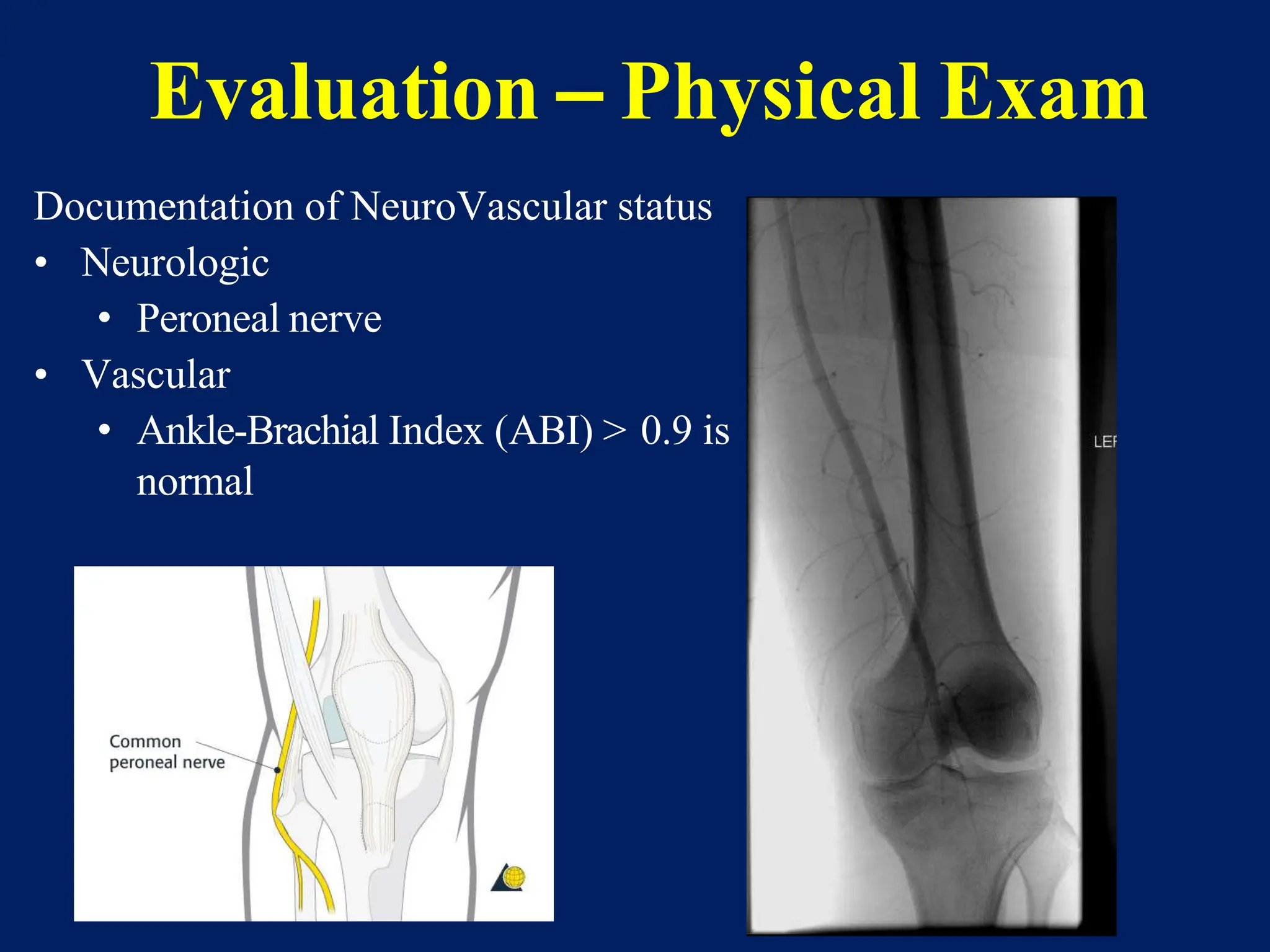
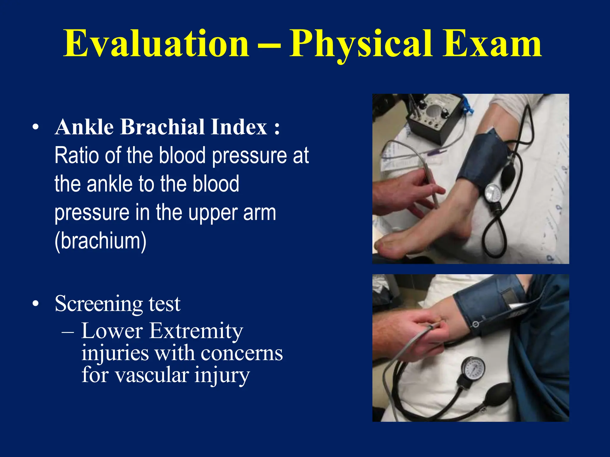
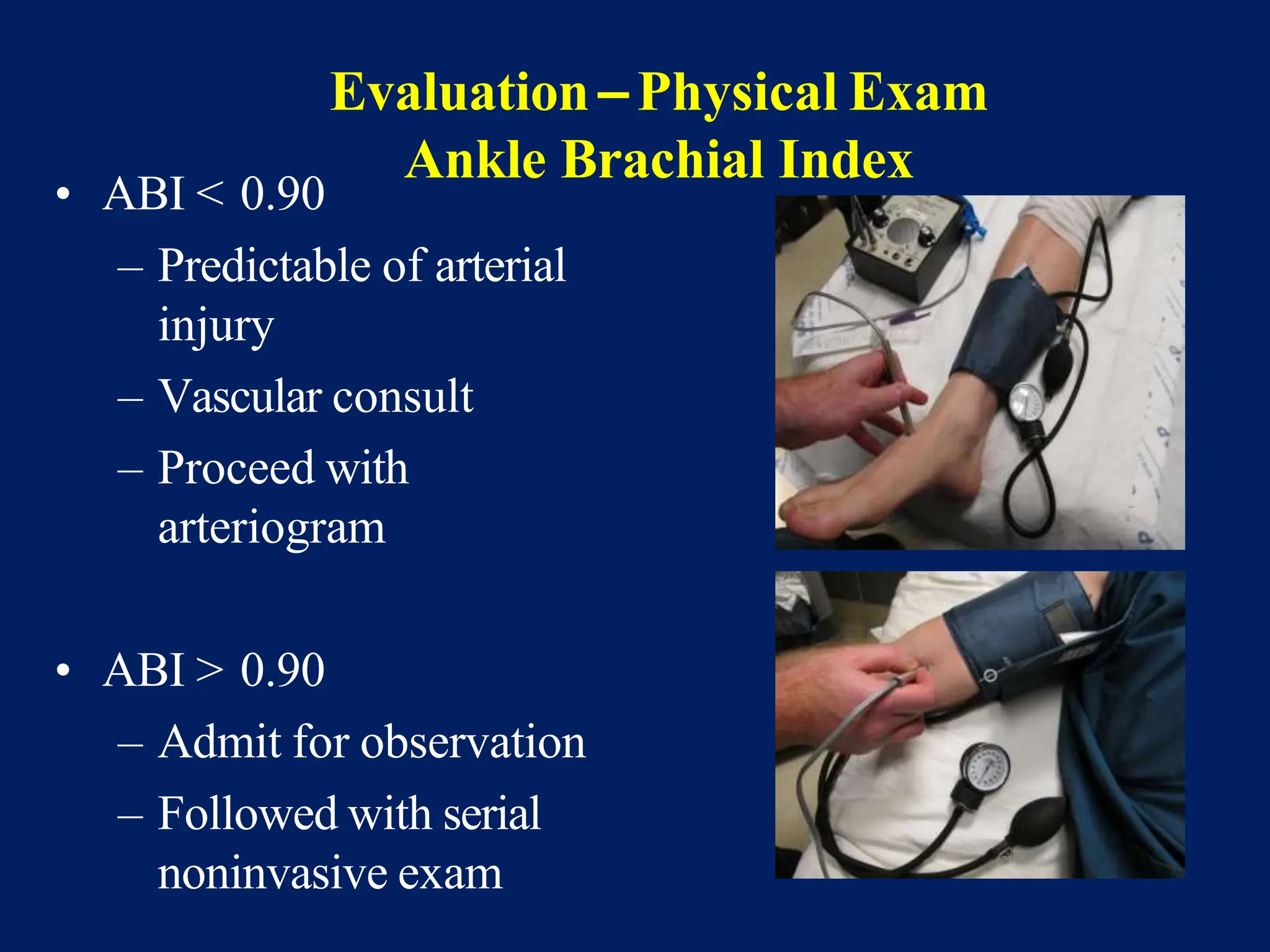
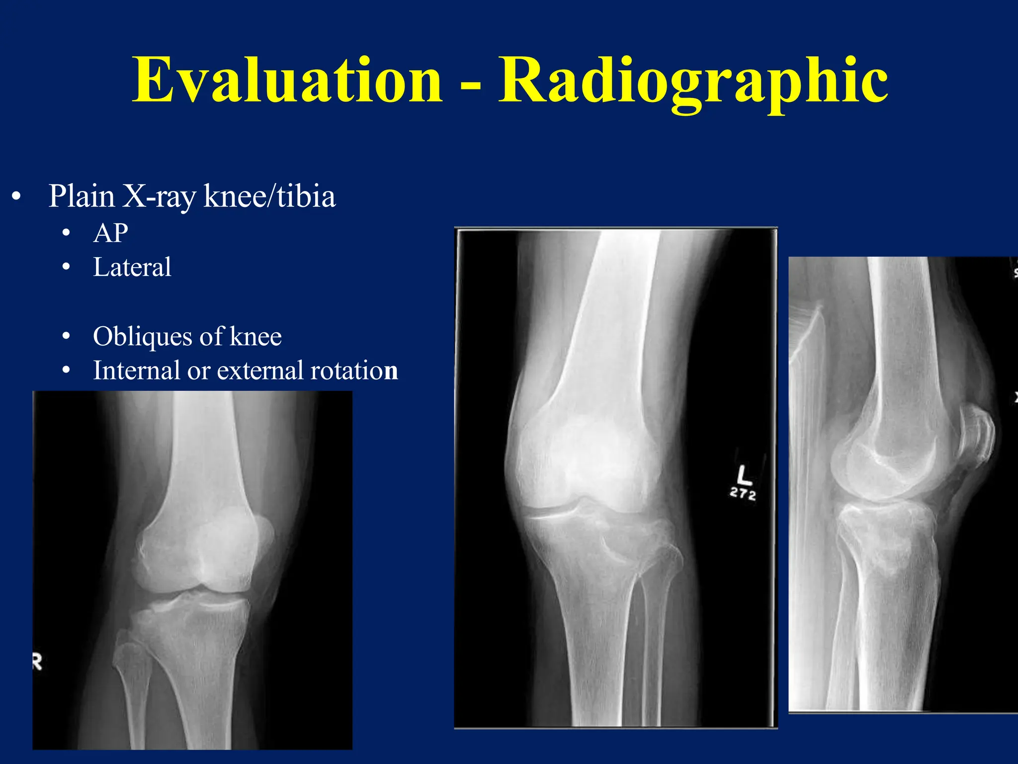
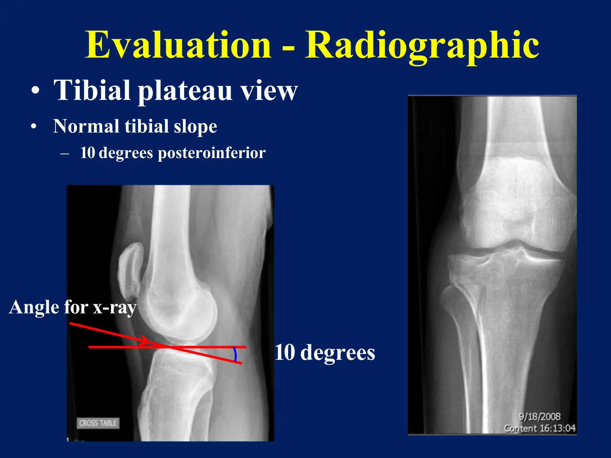
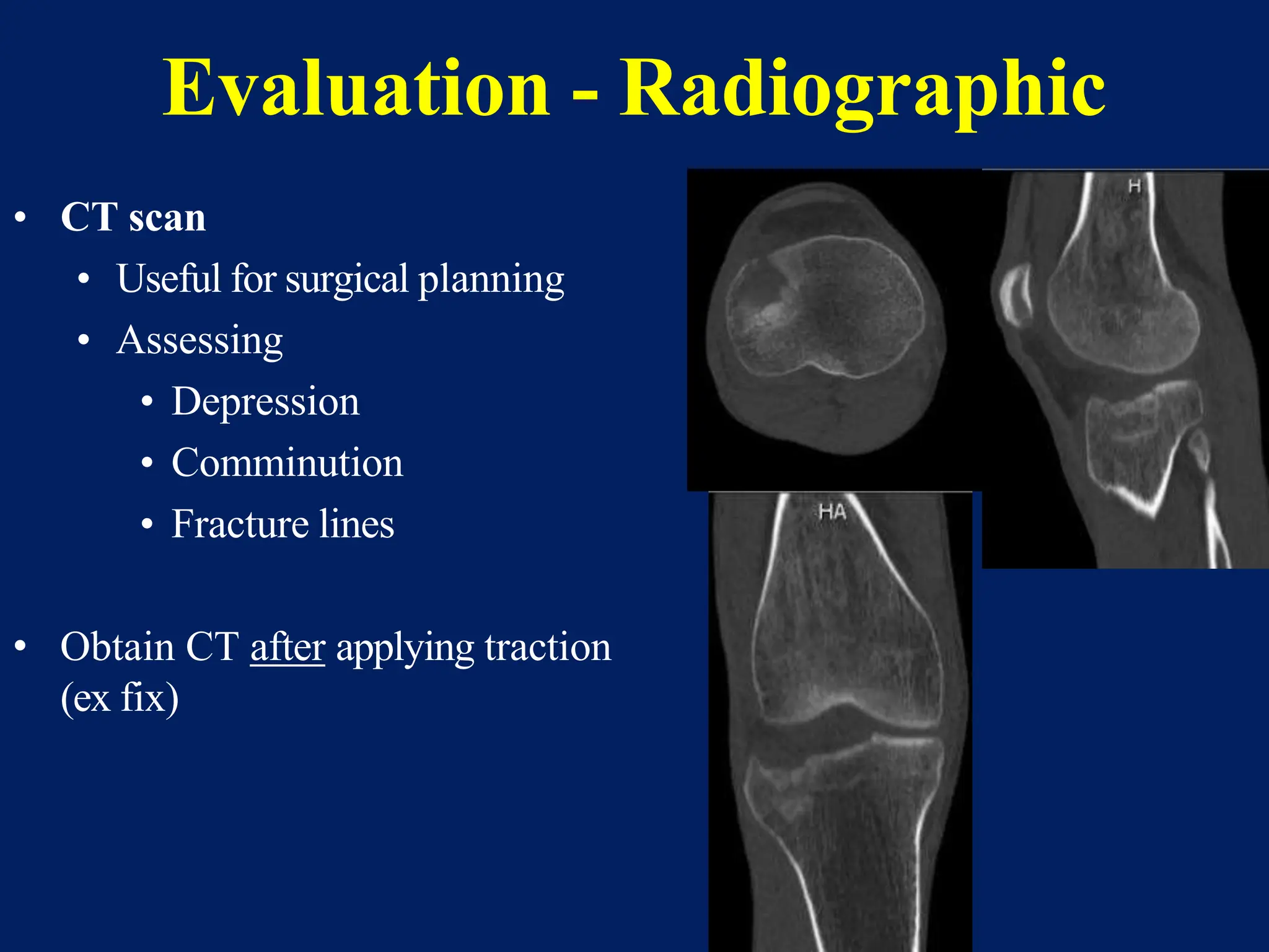
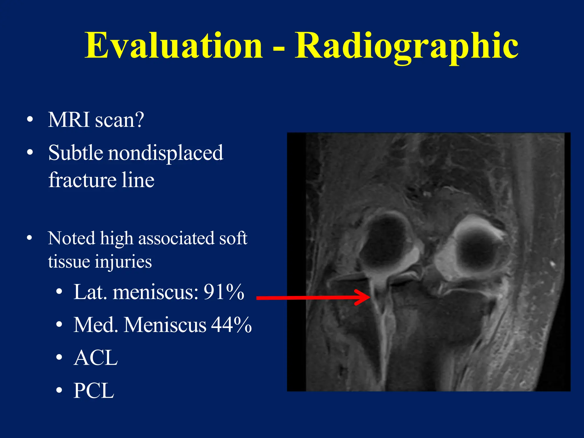
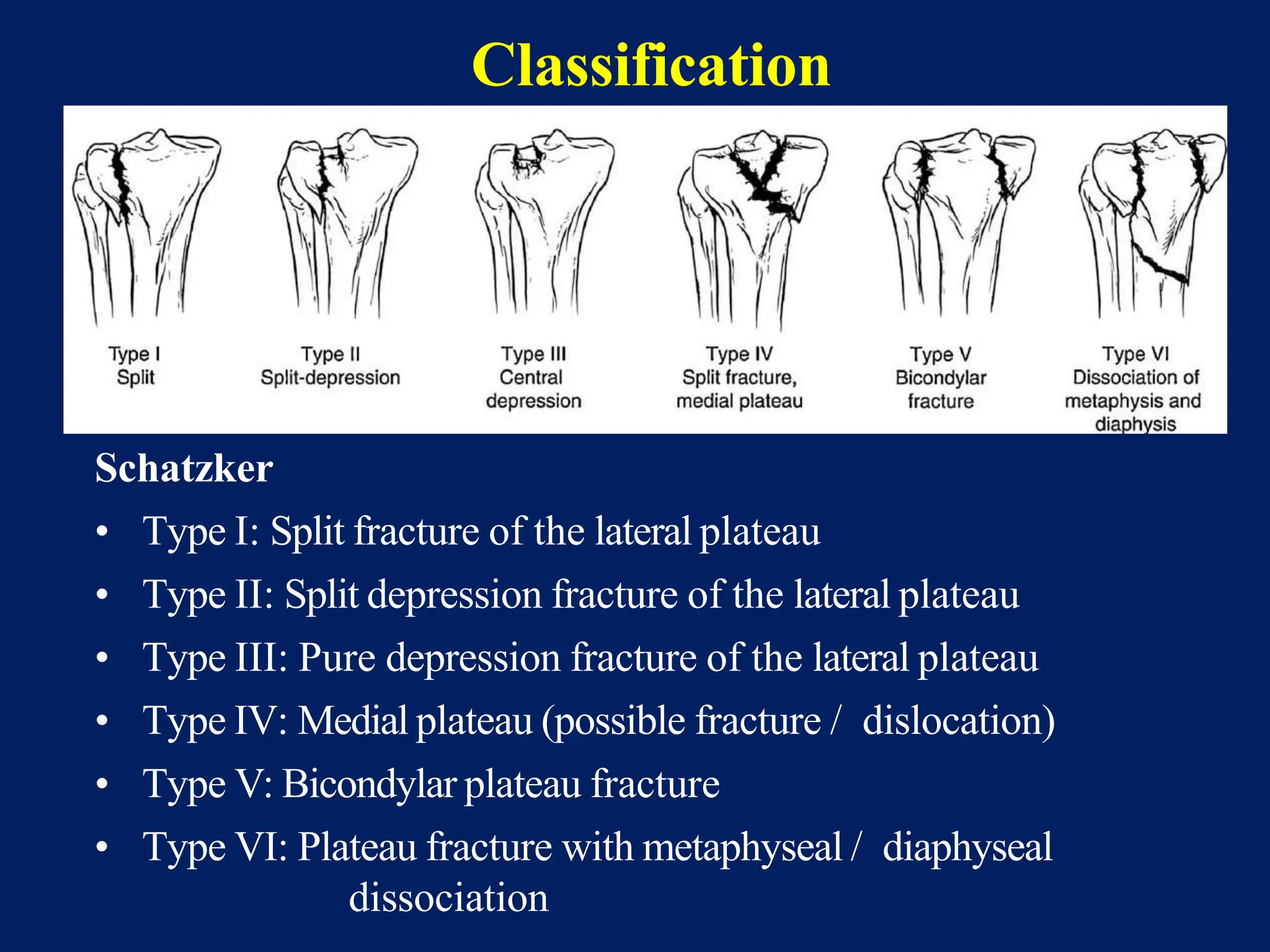
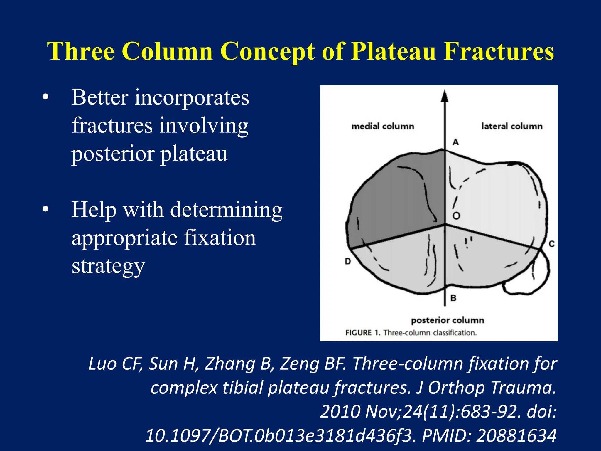
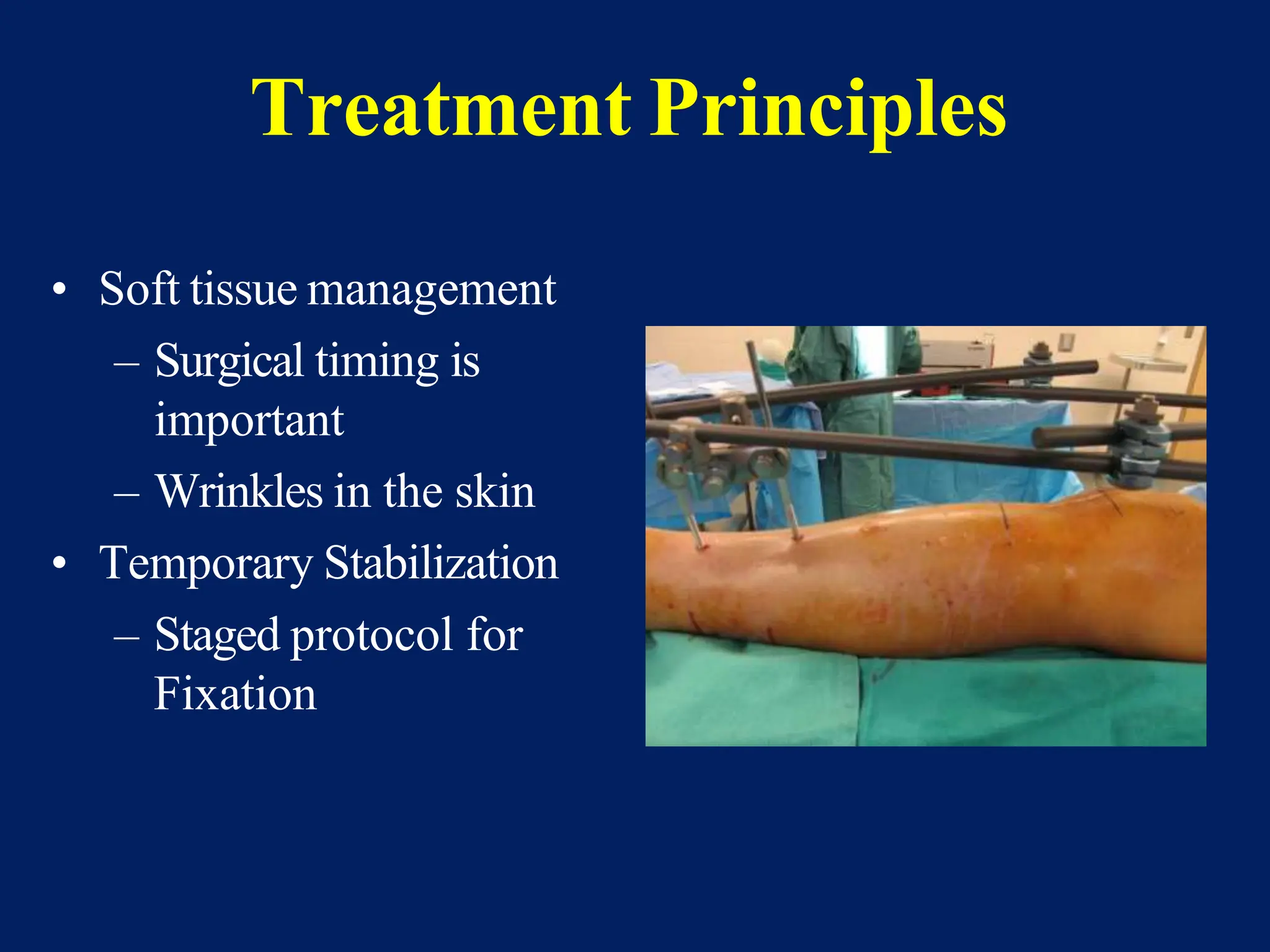
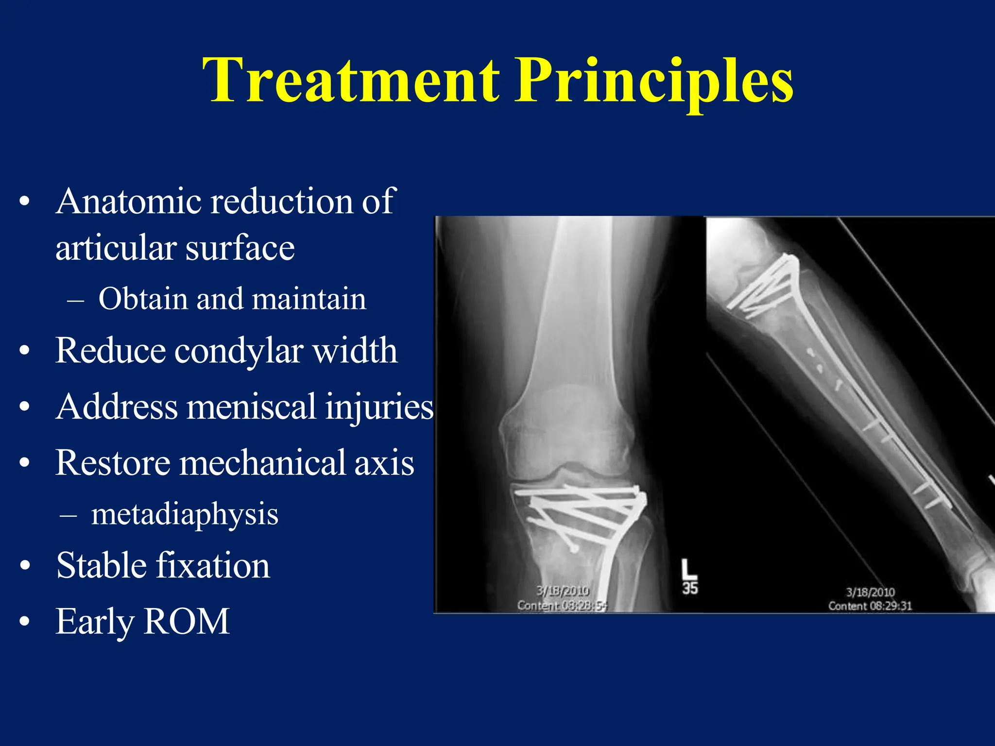
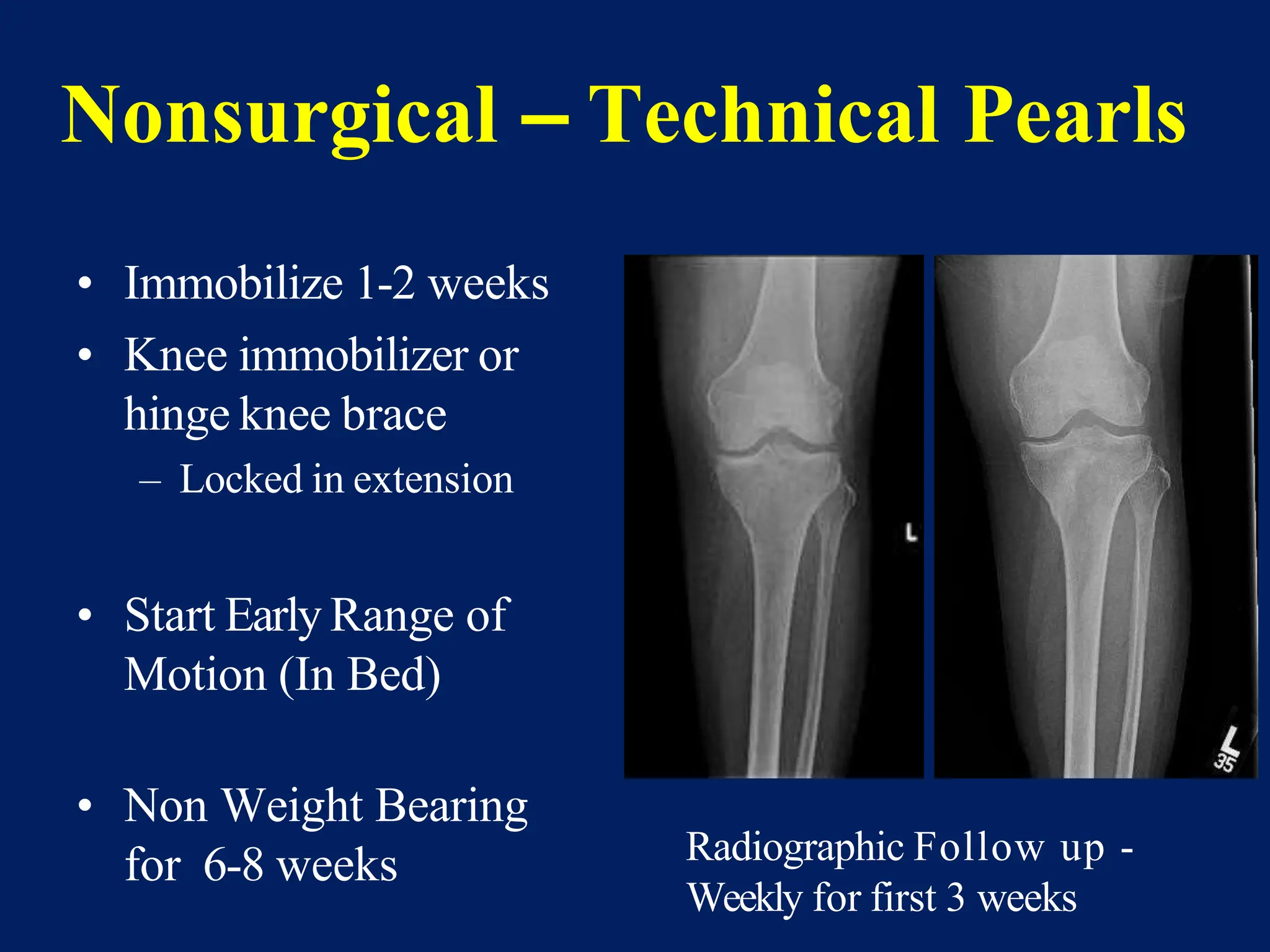
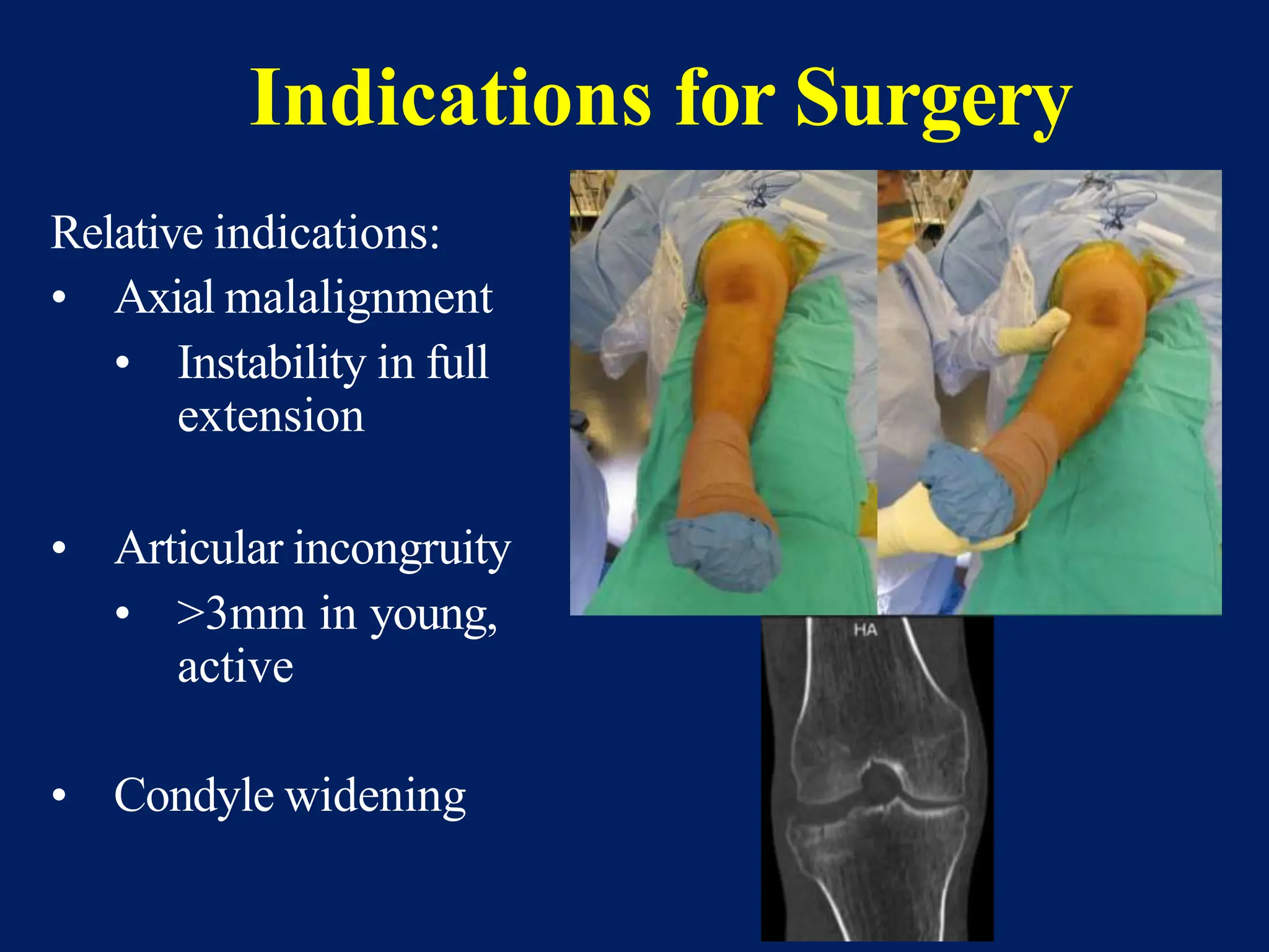
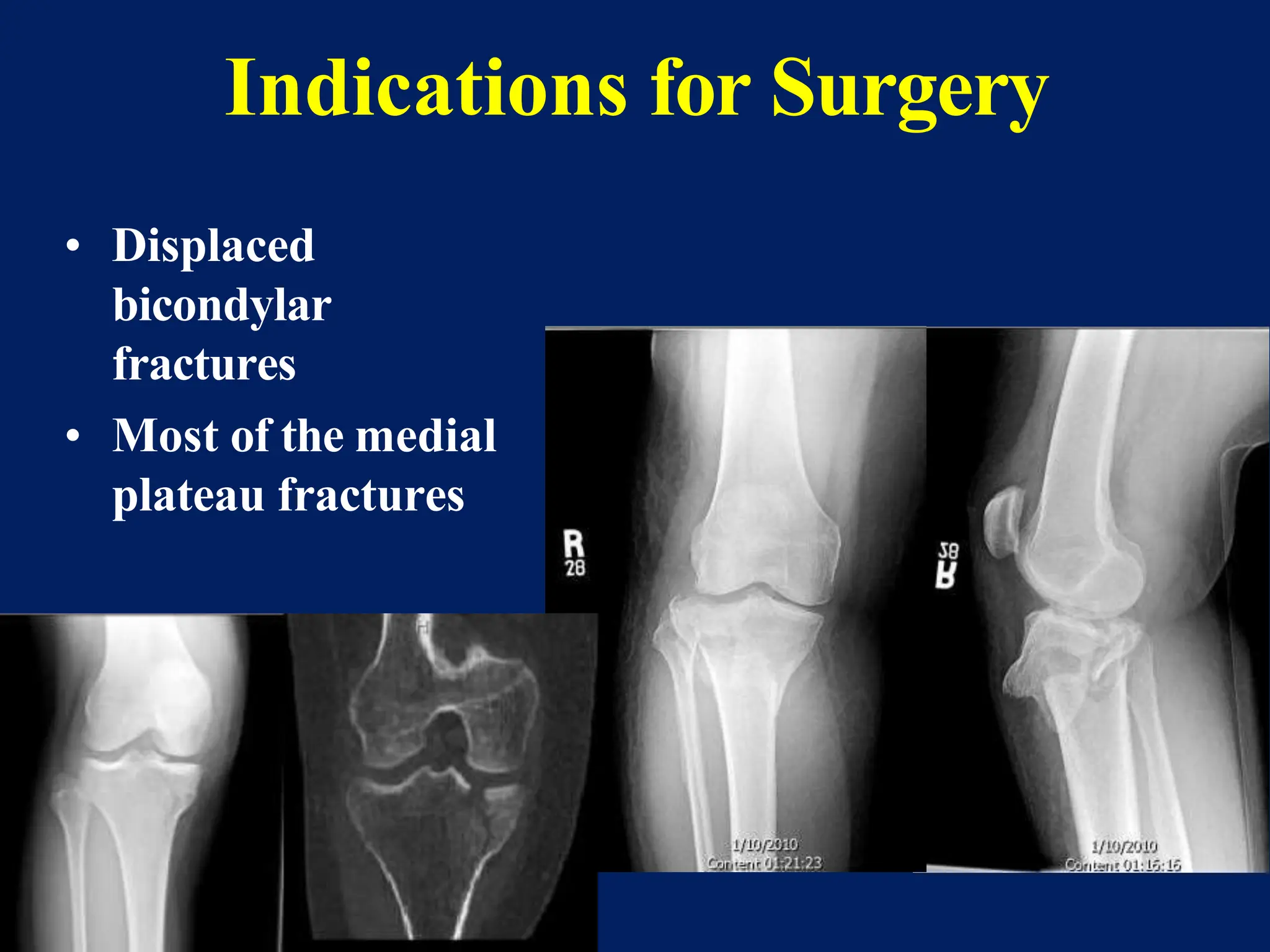
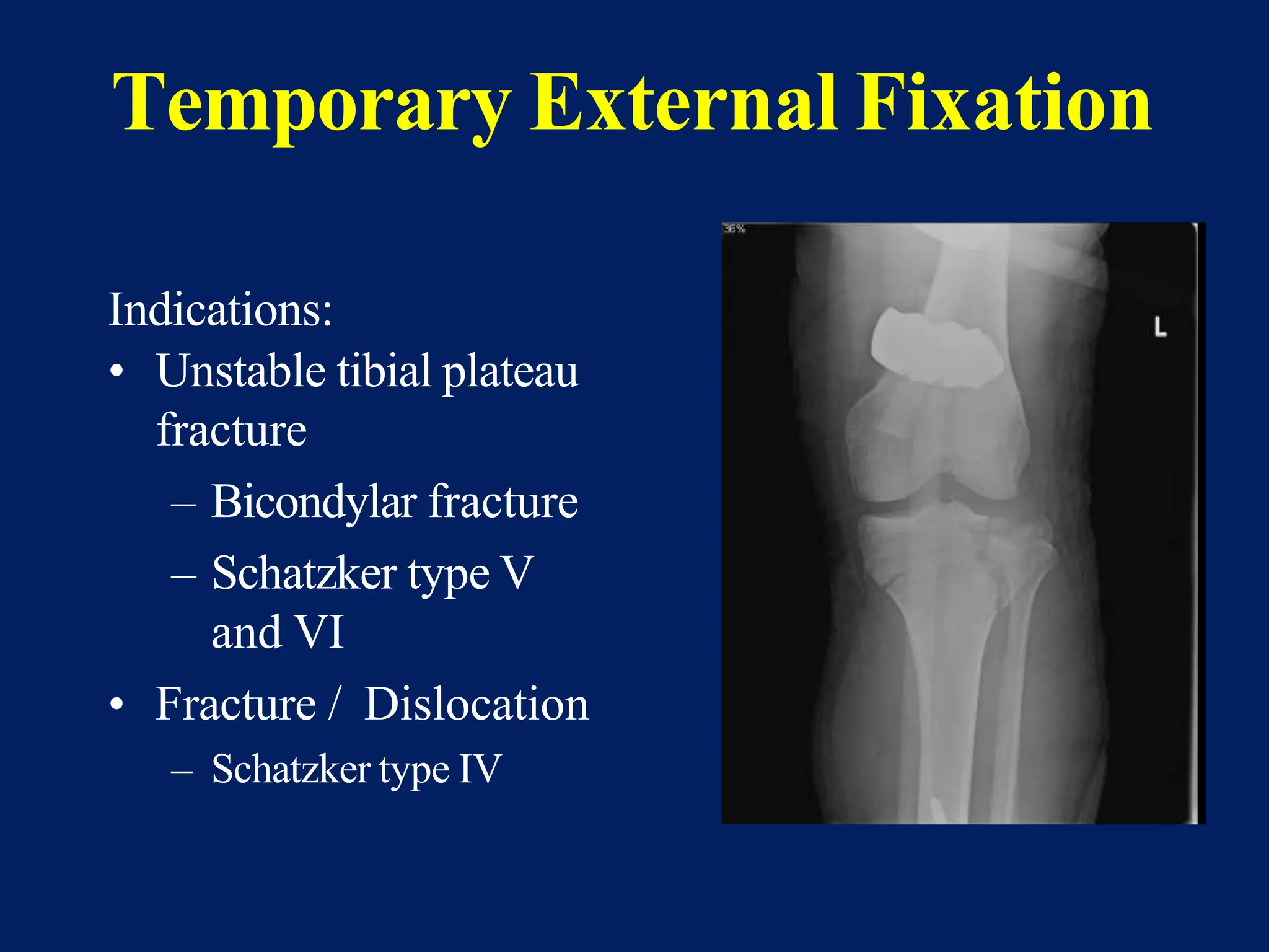
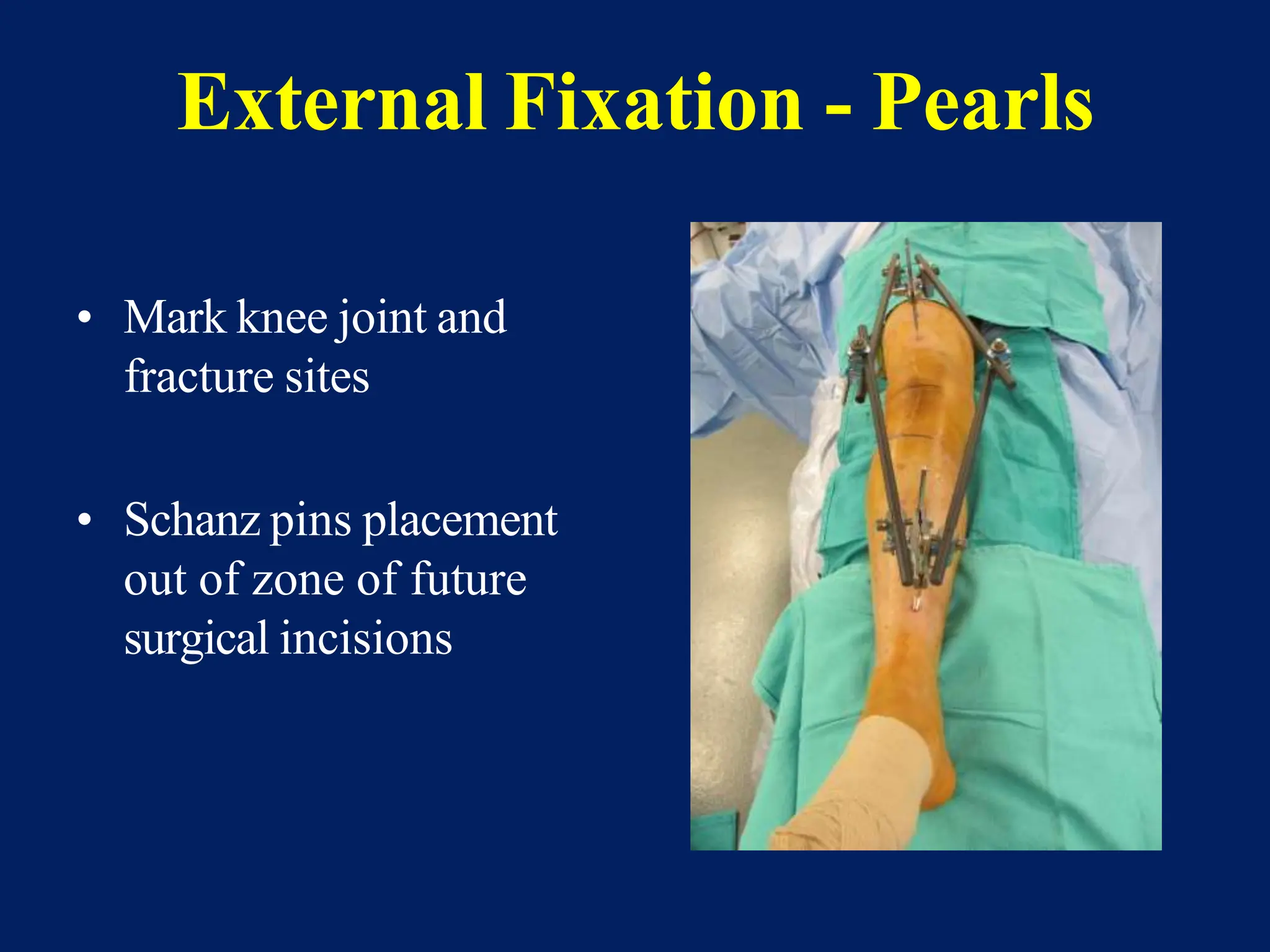
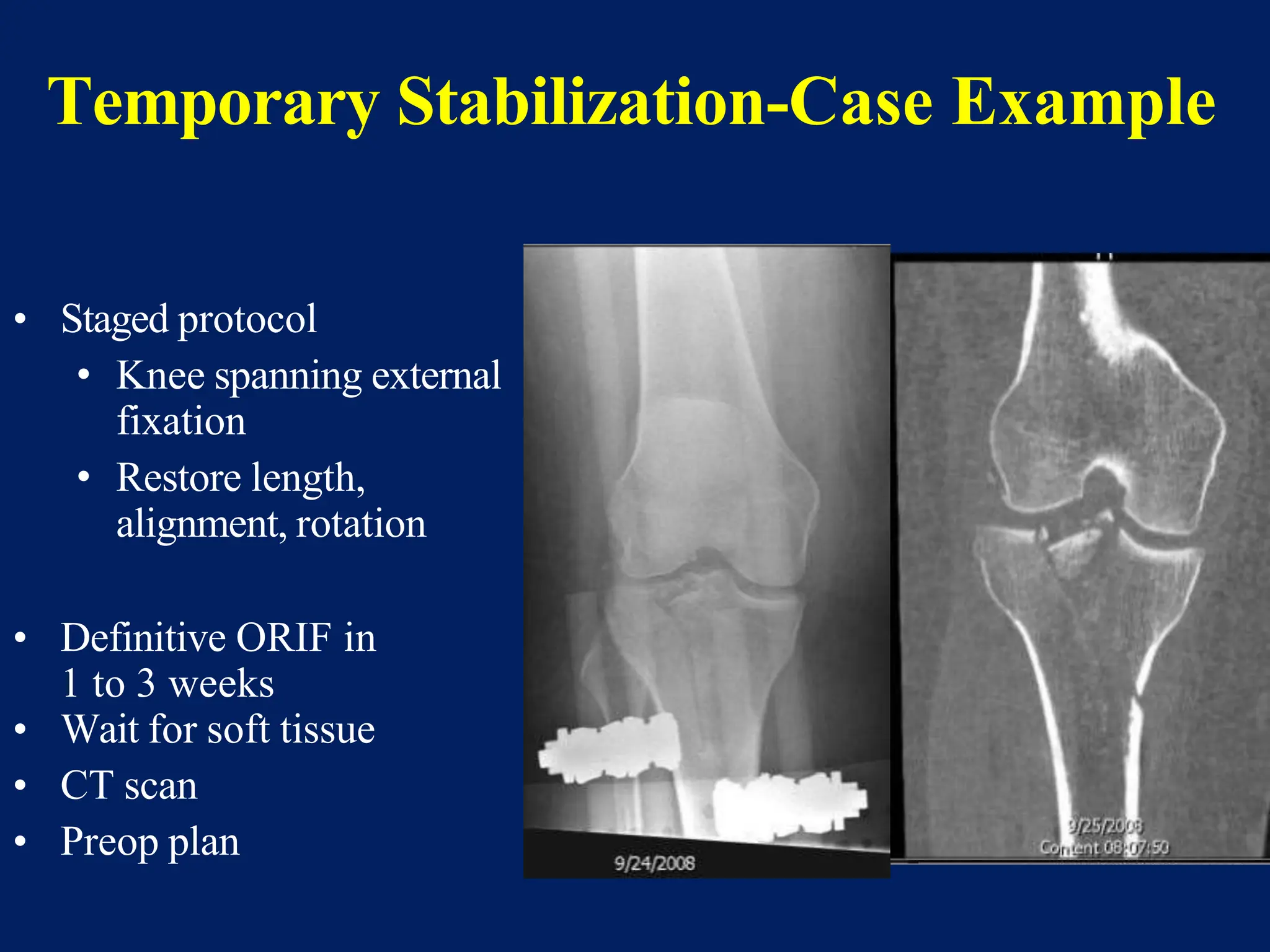
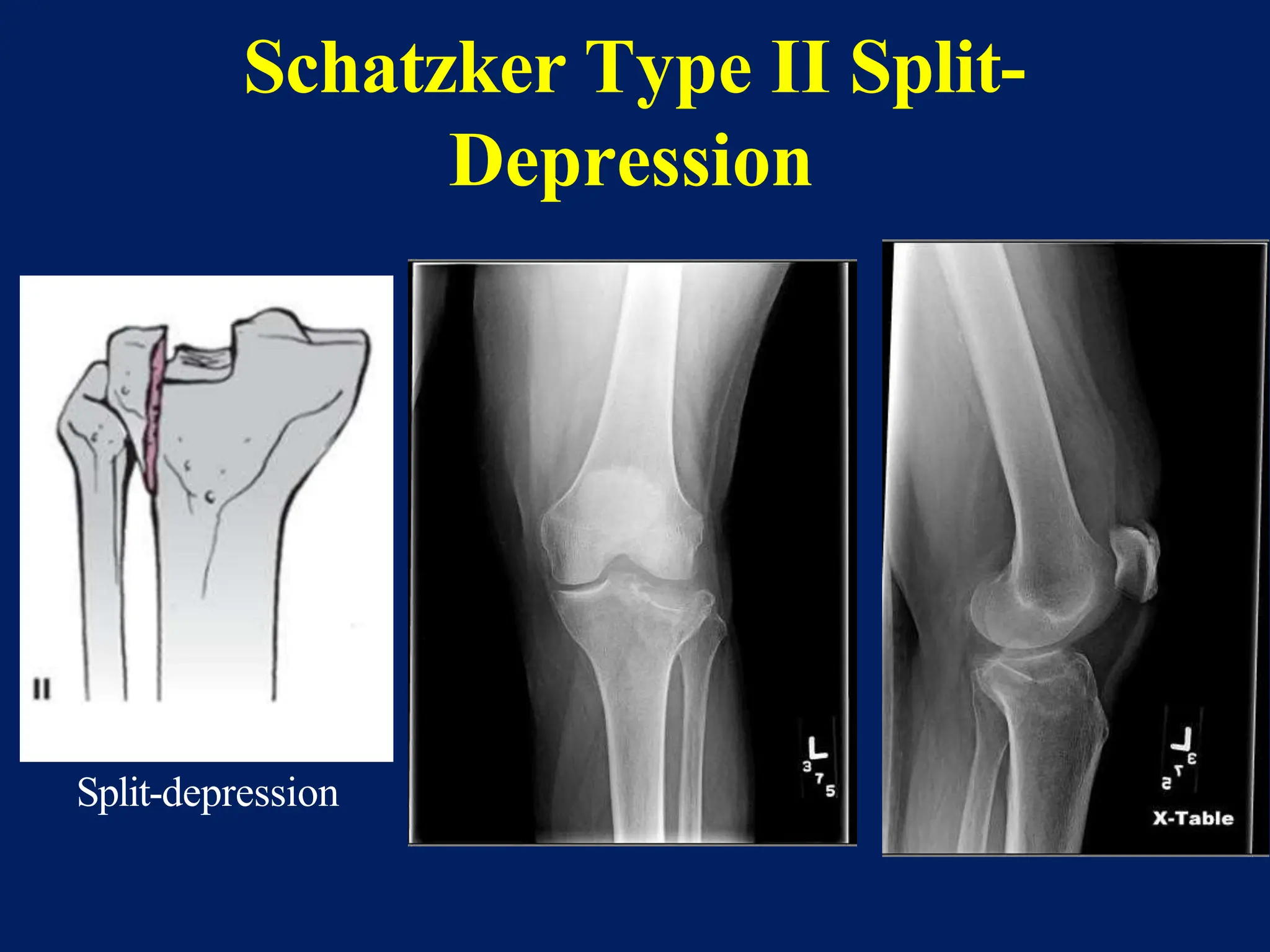
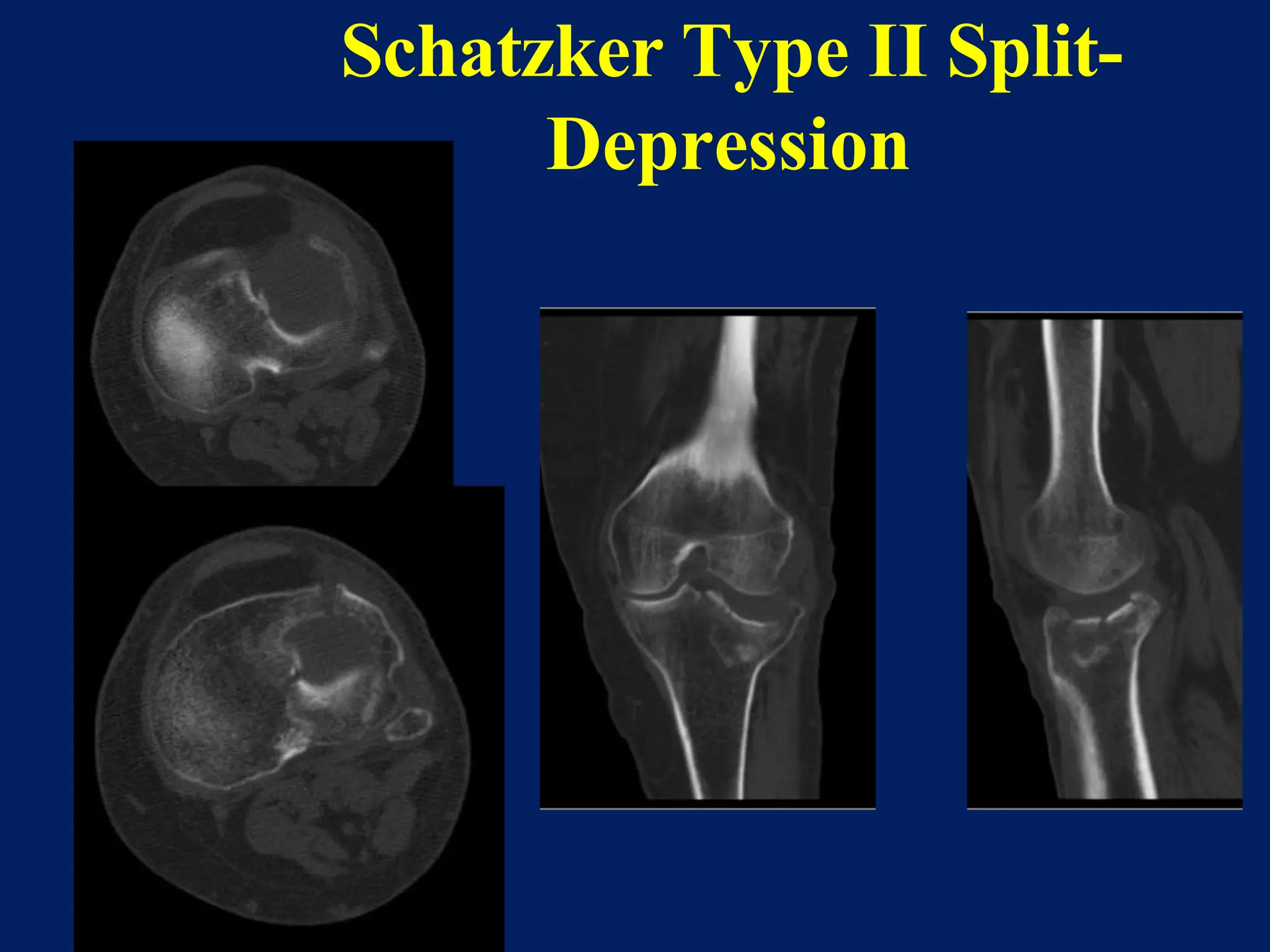
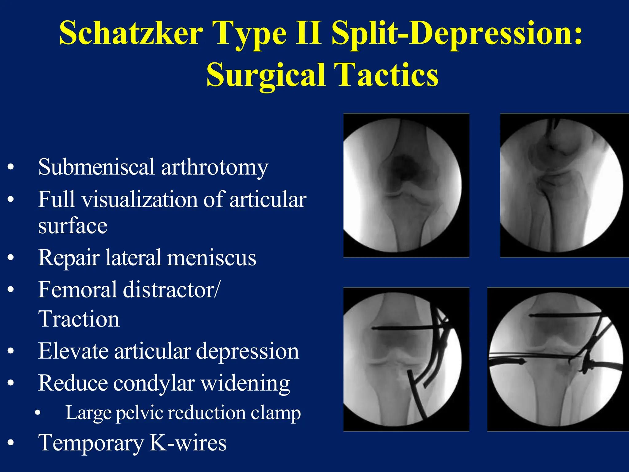
Objectives:
-Recognize the anatomy of the proximal tibia
-Describe initial evaluation and management
-Identify common fracture patterns
-Apply treatment principles and strategies for Partial articular fractures and Complete articular fractures
-Discuss rehabilitation and complications
-Learn Management in selected tibial plateau case scenarios
High tibial osteotomy (HTO) is a common and widely accepted procedure in orthopaedic surgery. In the literature, we find descriptions of the technique dating back to the 50s, with Jackson (Jackson, 1958). However, it was not until the 70s, with the publications of Conventry (Coventry, 1969 and 1973) and Insall (Insall, 1975), that proximal tibial osteotomy became common practice as a treatment option for medial compartment osteoarthritis of the knee usually associated to varus deformity. At that time, closing wedge osteotomies were performed, despite the greater technical difficulty and risks involved, as there were no fixation materials available that could enable opening wedge osteotomy. Only after the development of medial wedge plate fixation that opening wedge osteotomy became applicable (Puddu, 2004).
The goals of HTO are:
1. To reduce knee pain by transferring weight-bearing loads to the relatively unaffected compartment;
2. To increase the life span of the knee joint, by slowing or stopping the destruction of the medial joint compartment. This could delay the need of a joint replacement.
Can read freely here
https://sethiortho.blogspot.com/
Challenges and Solutions in
Management of Distal Humerus Fractures
Epidemiology
Anatomy
Classification
Controversies and Recent studies
Approach
Implants selection
Plate configuration
Ulnar nerve transposition
Role of total elbow arthroplasty in DHF
Role of hemiarthroplasty in DHF
Metaphyseal comminution –
Anatomic complexity of the distal humerus
Positioning of the plates
TBW –
Skin closure
Osteoporotic nature of the bone –
Less BMD/Thin metaphysis
Screw Pullout strength is low
DHF account for 2% of all adult fractures
The common pattern of fracture
Intraarticular and involves both columns
Bimodal distribution
Peak incidence in young male and in older female patients
Young male – High-velocity injury
Older female - Osteoporosis
The distal humerus is flattened and expanded bony structure
It is composed of lateral and medial columns with the trochlea situated between these columns.
The location of the trochlea is central rather than medial
Formed by Medial SCR + M/Epicondyle
The distal end has 450 angulation with humeral shaft
M/ Epicondyle gives attachment for MCL & Common Flexor Origin
The MCL originates from the undersurface of the medial epicondyle where it is vulnerable to excessive dissection
Ulnar nerve
Formed by Lateral SCR and L/Epicondyle and Capitulum
Distal end has 200 with humeral shaft
L/ epicondyle gives attachment for LCL & common extensor origin
Its posterior surface is non articular and can be used as a site for a plate fixation
The lateral column curves anteriorly
Placement of a straight plate on the posterolateral surface of the humerus risks straightening of distal humerus.
The medial column including the medial epicondyle is in line with the humeral shaft.
It forms the center of the triangle
It has 30 - 80 – external rotation & 250 anterior divergent with the shaft
It forms a 40 - 80 degree valgus direction
X-ray -
Anterior-posterior view
lateral View
Traction View – This can help to define articular fragments and aid in pre-operative classification of the fracture.
NCCT – Elbow
Articular surfaces
Position of the fracture fragments
useful for identifying impacted fracture fragments that make reduction challenging
Olecranon Osteotomy Approach – 52-57%
Triceps sparing VS Olecranon osteotomy approach
The lateral column was often the first to fail as a result of excessive varus forces acting on the elbow during normal activities of daily living. Small anterior-posterior diameter
Smaller diameter of the humerus, permitting only one or two short screws for fixation.
Interruption of blood supply to the lateral column
blood supply to the lateral column is also derived from posterior segmental vessels. Sagittal plane plating has less risk of injuring these structures, which may improve the chances of union
Distal femur fractures what makes it complex ,dr mohamed ashraf,hod orthopae...drashraf369
distal femur fractures are notorious for post operative complications due to malreduction and improper fixation.unless plan and execute a sound and stable fixation,this injury will lead to undesirable results.dr mohamed ashraf HOD orthopaedics govt TD medical college is presenting how to avoid complications in surgical management of these fractures..
Please find the power point on Fracture of Talus with well diagrammatic explanation from very reliable sources. If you need such a power point on different topics related with MBBS then please write it on comment section. Thank you
Posterior Pelvic Injury need correct squeal procedure reduction and fixation.Here we hare our experience in China Medical University Hospital , Taichung,Taiwan. This topic also presented in the meeting in TOA.
In the elderly osteoporotic fractures although the principles are the same but some special considerations in management of the soft tissues and the bony injuries are considered.
Telehealth Psychology Building Trust with Clients.pptxThe Harvest Clinic
Telehealth psychology is a digital approach that offers psychological services and mental health care to clients remotely, using technologies like video conferencing, phone calls, text messaging, and mobile apps for communication.
Navigating Challenges: Mental Health, Legislation, and the Prison System in B...Guillermo Rivera
This conference will delve into the intricate intersections between mental health, legal frameworks, and the prison system in Bolivia. It aims to provide a comprehensive overview of the current challenges faced by mental health professionals working within the legislative and correctional landscapes. Topics of discussion will include the prevalence and impact of mental health issues among the incarcerated population, the effectiveness of existing mental health policies and legislation, and potential reforms to enhance the mental health support system within prisons.
Health Education on prevention of hypertensionRadhika kulvi
Hypertension is a chronic condition of concern due to its role in the causation of coronary heart diseases. Hypertension is a worldwide epidemic and important risk factor for coronary artery disease, stroke and renal diseases. Blood pressure is the force exerted by the blood against the walls of the blood vessels and is sufficient to maintain tissue perfusion during activity and rest. Hypertension is sustained elevation of BP. In adults, HTN exists when systolic blood pressure is equal to or greater than 140mmHg or diastolic BP is equal to or greater than 90mmHg. The
Empowering ACOs: Leveraging Quality Management Tools for MIPS and BeyondHealth Catalyst
Join us as we delve into the crucial realm of quality reporting for MSSP (Medicare Shared Savings Program) Accountable Care Organizations (ACOs).
In this session, we will explore how a robust quality management solution can empower your organization to meet regulatory requirements and improve processes for MIPS reporting and internal quality programs. Learn how our MeasureAble application enables compliance and fosters continuous improvement.
The Importance of Community Nursing Care.pdfAD Healthcare
NDIS and Community 24/7 Nursing Care is a specific type of support that may be provided under the NDIS for individuals with complex medical needs who require ongoing nursing care in a community setting, such as their home or a supported accommodation facility.
One of the most developed cities of India, the city of Chennai is the capital of Tamilnadu and many people from different parts of India come here to earn their bread and butter. Being a metropolitan, the city is filled with towering building and beaches but the sad part as with almost every Indian city
Medical Technology Tackles New Health Care Demand - Research Report - March 2...pchutichetpong
M Capital Group (“MCG”) predicts that with, against, despite, and even without the global pandemic, the medical technology (MedTech) industry shows signs of continuous healthy growth, driven by smaller, faster, and cheaper devices, growing demand for home-based applications, technological innovation, strategic acquisitions, investments, and SPAC listings. MCG predicts that this should reflects itself in annual growth of over 6%, well beyond 2028.
According to Chris Mouchabhani, Managing Partner at M Capital Group, “Despite all economic scenarios that one may consider, beyond overall economic shocks, medical technology should remain one of the most promising and robust sectors over the short to medium term and well beyond 2028.”
There is a movement towards home-based care for the elderly, next generation scanning and MRI devices, wearable technology, artificial intelligence incorporation, and online connectivity. Experts also see a focus on predictive, preventive, personalized, participatory, and precision medicine, with rising levels of integration of home care and technological innovation.
The average cost of treatment has been rising across the board, creating additional financial burdens to governments, healthcare providers and insurance companies. According to MCG, cost-per-inpatient-stay in the United States alone rose on average annually by over 13% between 2014 to 2021, leading MedTech to focus research efforts on optimized medical equipment at lower price points, whilst emphasizing portability and ease of use. Namely, 46% of the 1,008 medical technology companies in the 2021 MedTech Innovator (“MTI”) database are focusing on prevention, wellness, detection, or diagnosis, signaling a clear push for preventive care to also tackle costs.
In addition, there has also been a lasting impact on consumer and medical demand for home care, supported by the pandemic. Lockdowns, closure of care facilities, and healthcare systems subjected to capacity pressure, accelerated demand away from traditional inpatient care. Now, outpatient care solutions are driving industry production, with nearly 70% of recent diagnostics start-up companies producing products in areas such as ambulatory clinics, at-home care, and self-administered diagnostics.
1. Proximal Tibia
Fractures Dr. Kota Aditya
MS Orthopaedics
Fellow in Ilizarov Surgery
Fellow in Sports Medicine
Fellow in Joint Replacement
Member - AO Trauma
& Young Surgeon Committee – SICOT
Professor
Department of Orthopaedics
Curriculum Committee Member
Medical Education Unit
ASRAM Medical College
2. • Recognize the anatomy of the proximal tibia
• Describe initial evaluation and management
• Identify common fracture patterns
• Apply treatment principles and strategies
• Partial articular fractures
• Complete articular fractures
• Discuss rehabilitation and complications
• Learn Management in selected tibial plateau case
scenarios
Learning Objectives
3. Epidemiology
• 1-2% of all fractures
• Bimodal distribution
• Young adults: high energy
mechanism
• Highest in 5th decade
• Male > Female
• Elderly: low energy mechanism
• Osteoporotic bone
• Female > Male
• Lateral Plateau: 70% of fractures
• Medial Plateau: 10% of fractures
• Bicondylar Plateau: 20% of fractures
4. Anatomy
Consist of Medial
and Lateral Plateau
– Medial larger &
harder (thus less
likely to fracture)
– Medial lower &
concave
– Lateral higher &
convex
– Lateral cartilage
thicker (3 vs 4
mm)
Medial
Lateral Medial
Strong
Lateral
Weak
Medial
concave
Lateral
convex
6. Mechanism of Injury
• Valgus producing force
– Lateral plateau
• Varus producing force
– Medial plateau
• Axial compressive force
– Bicondylar plateau
• Combination
– High energy
– Bicondylar plateau
– Soft tissue injury
7. Mechanism of Injury
• Valgus producing force
– Lateral plateau
• Varus producing force
– Medial plateau
• Axial compressive force
– Bicondylar plateau
• Combination
– High energy
– Bicondylar plateau
– Soft tissue injury
8. Mechanism of Injury
• Valgus producing force
– Lateral plateau
• Varus producing force
– Medial plateau
• Axial compressive force
– Bicondylar plateau
• Combination
– High energy
– Bicondylar plateau
9. Initial presentation –
mechanism matters!
• Lower energy
• Simple falls, struck from
side
• Remain length stable
• Higher energy
• Axial load, associated
shearing
• Risk of Compartment
syndrome
13. Evaluation – Physical Exam
• Soft tissue assessment
• Stability tests – difficult
to perform
• Avoid missing
compartment syndrome
• Determine timing of
surgery
– Skin Wrinkles present or
not?
14. Evaluation – Physical Exam
Documentation of NeuroVascular status
• Neurologic
• Peroneal nerve
• Vascular
• Ankle-Brachial Index (ABI) > 0.9 is
normal
15. Evaluation – Physical Exam
• Ankle Brachial Index :
Ratio of the blood pressure at
the ankle to the blood
pressure in the upper arm
(brachium)
• Screening test
– Lower Extremity
injuries with concerns
for vascular injury
16. Evaluation–Physical Exam
Ankle Brachial Index
• ABI < 0.90
– Predictable of arterial
injury
– Vascular consult
– Proceed with
arteriogram
• ABI > 0.90
– Admit for observation
– Followed with serial
noninvasive exam
17. Evaluation - Radiographic
• Plain X-ray knee/tibia
• AP
• Lateral
• Obliques of knee
• Internal or external rotation
21. Classification
Schatzker
• Type I: Split fracture of the lateral plateau
• Type II: Split depression fracture of the lateral plateau
• Type III: Pure depression fracture of the lateral plateau
• Type IV: Medial plateau (possible fracture / dislocation)
• Type V: Bicondylar plateau fracture
• Type VI: Plateau fracture with metaphyseal / diaphyseal
dissociation
22. Three Column Concept of Plateau Fractures
• Better incorporates
fractures involving
posterior plateau
• Help with determining
appropriate fixation
strategy
Luo CF, Sun H, Zhang B, Zeng BF. Three-column fixation for
complex tibial plateau fractures. J Orthop Trauma.
2010 Nov;24(11):683-92. doi:
10.1097/BOT.0b013e3181d436f3. PMID: 20881634
23. Treatment Principles
• Soft tissue management
– Surgical timing is
important
– Wrinkles in the skin
• Temporary Stabilization
– Staged protocol for
Fixation
24. Treatment Principles
• Anatomic reduction of
articular surface
– Obtain and maintain
• Reduce condylar width
• Address meniscal injuries
• Restore mechanical axis
– metadiaphysis
• Stable fixation
• Early ROM
26. Nonsurgical – Technical Pearls
• Immobilize 1-2 weeks
• Knee immobilizer or
hinge knee brace
– Locked in extension
• Start Early Range of
Motion (In Bed)
• Non Weight Bearing
for 6-8 weeks Radiographic Follow up -
Weekly for first 3 weeks
27. Indications for Surgery
Absolute indications:
• Open tibial plateau
• Associated
compartment syndrome
• Associated vascular
injury
28. Indications for Surgery
Relative indications:
• Axial malalignment
• Instability in full
extension
• Articular incongruity
• >3mm in young,
active
• Condyle widening
30. Timing of Surgery
Low Energy:
Fixed electively and early
High Energy:
Don’t Hurry, Have patience
31. Temporary External Fixation
• Knee spanning external
fixation
• Ligamentotaxis
• Improve fracture fragment
gross alignment
– Length and alignment
• Minimize further damage
to articular surface
• Soft tissue assessment and
wound care
32. Temporary External Fixation
Indications:
• Unstable tibial plateau
fracture
– Bicondylar fracture
– Schatzker type V
and VI
• Fracture / Dislocation
– Schatzker type IV
33. External Fixation
• 2 pins in femur
– Anterior or lateral
• 2 pins in tibia
– Antero-medial
34. External Fixation - Pearls
• Mark knee joint and
fracture sites
• Schanz pins placement
out of zone of future
surgical incisions
35. Temporary Stabilization-Case Example
• Staged protocol
• Knee spanning external
fixation
• Restore length,
alignment, rotation
• Definitive ORIF in
1 to 3 weeks
• Wait for soft tissue
• CT scan
• Preop plan
36. Calcaneum Traction if Ex Fix is
Not Applied in Complex cases
Using Ilizarov Wire and Half Ring
39. ORIF- Implant Options
• Angular stable (Locking)
implants
• Precontoured for
proximal tibia
• Bicondylar tibia plateau
with metadiaphyseal
involvement
• Spanning or bridging
across fracture zone
• Selected fracture, allows
stabilization of medial
plateau
40. External Fixation
• Limited internal fixation
– Small incisions or
percutaneous
• Thin-wire ring fixators
– Connect to the shaft
– Fixation distally with 5mm
half-pins
• Advantages
– Minimize soft tissue injury
• Still need to reduce articular
surface!!! May need a
screw!
41. Surgical Approaches
• Anterolateral
– Lateral plateau involvement
– Combination with medial
for complex plateau
• Posteromedial
– Medial plateau
– Coronal split
• Posterior
• Dual approaches
– Anterolateral
– Posteromedial
Copyright by AO Foundation, Switzerland
42. Surgical Approaches
• Other surgical approaches
• Direct medial or midline
parapatellar anterior
– Isolated medial tibia
fractures
• Direct posterior approach
– Posterior shear fractures
– Prone
– Inability to treat
anterolateral fracture
48. • Submeniscal arthrotomy
• Full visualization of articular
surface
• Repair lateral meniscus
• Femoral distractor/
Traction
• Elevate articular depression
• Reduce condylar widening
• Large pelvic reduction clamp
• Temporary K-wires
Schatzker Type II Split-Depression:
Surgical Tactics
49. Schatzker Type II Split-Depression
• Fill defect
• Allograft
• Autograft
• Bone substitutes
• Buttress plate
• Nonlocking: Most
• Locked: osteoporotic
bone
• Subchondral raft screws
51. • Surgical technique
• Open approach
• Submeniscal
• Arthroscopic
• Elevate depressed fragment
• Fill defect
• Stabilization
• Subchondral raft screws
Schatzker Type III Pure Depression
52. Schatzker Type III Pure Depression
• Surgical technique
• Submeniscal arthrotomy
• Arthroscopic
• Elevate depressed fragment
• Fill defect
• Stabilization
• Subchondral screws
54. Schatzker Type IV Medial plateau
CT post ext. fix
Split fracture,
Medial plateau
55. Schatzker Type IV Medial plateau
• Surgical approach
• Posteromedial
• Interval between
• Pes anserine tendons and Medial head gastrocnemius
Copyright by AO Foundation, Switzerland
56. Schatzker Type IV Medial
plateau
• Don’t forget about
possible lateral
plateau depression
• May need
anterolateral
incision to reduce
depression
57. Schatzker Type IV Medial plateau
• Fixation
• Straight medial
plating
• Posteromedial
plating
• Combination
58. Schatzker Type V, VI Bicondylar
Bicondylar fracture Metadiaphyseal
dissociation
64. Schatzker V, VI Bicondylar
• Single lateral fixed angle
implant
• Ability to capture medial
condyle with laterally
based implant
• Medial apex cortical
contact with minimal
comminution
65. Rehabilitation
Postoperative Care
• Knee brace
• Elevate leg
• Non Weight Bearing for
10-12 weeks
Physical therapy
• Early ROM
• Quadriceps Strengthening
• Gait training
– Crutches for non
weight bearing in
young patients who
are compliant
74. Selected Cases – Case 3
Bicondylar with tibial tuberosity fracture
• Must address
tuberosity
– Allow early ROM
• Options for
tuberosity fixation
– Lag screws
– Plates/screws
82. Case 6
• Minimally displaced
Bicondylar Fracture
• Fixed with
Posteromedial
Buttress using Semi
tubular Plate and
percutaneous CC
screws for Lateral
Condyle split
fracture
83. Case 7
• Type 6 Scahtzker
with Poor Soft
Tissue Condition
(skin Bleps and
Blisters)
• Ilizarov Externeral
Fixator after
Interfragmentary
screw to maintain
intra articular
congruity
84. Case 8
• Type 6 Scahtzker
with Poor Soft
Tissue Condition
(skin Bleps and
Blisters)
• Ilizarov Externeral
Fixator after
Interfragmentary
screw to maintain
intra articular
congruity
85. Summary
• Understand the fracture pattern
• Respect the soft tissues
• Partial articular (Schatzker 1-3)
• Buttress: Plates +/- interfragmentary screws
• Beware of Medial plateau (Schatzker 4)
• Posteromedial approach
• Complete articular (Schatzker 5,6)
• External fixation is beneficial
• Plan, ORIF Dual Approach may be necessary
86. References
• OTA PPT Education Resources
• Rockwood and Green’s Fractures in Adults Philadelphia: Lippincott
Williams & Wilkins, 2014.
• Katsenis D. JOrthop Trauma 2009;23(7):493-501.
• Lansinger O. JBone Joint Surg Am 1986;68(1):13-19.
• Rademakers M.V. JOrthop Trauma 2007;21(1):5-10).
• Schatzker J. Clin Orthop Relat Res 1979;138:94-104.
• Stevens DG. JOrthop Trauma 2001;15(5):312-320.
• Weigel DP. JBone Joint Surg Am 2002;84(9):1541-1551.
• The Schatzker Classification Figure 55-9. Court-Brown C, Heckman JD,
McKee M, et al. Rockwood and Green’s Fractures in Adults Philadelphia:
Lippincott Williams & Wilkins, 2014.
• Hansen, Matthias; Pesantez, Rodrigo. AO Surgery Reference.