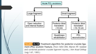
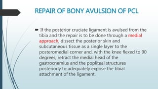
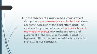
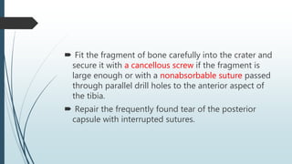
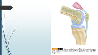
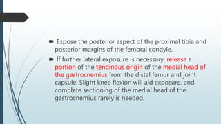
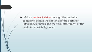
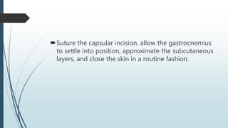
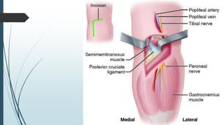
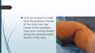
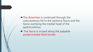
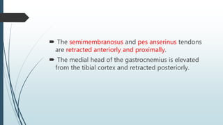
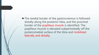
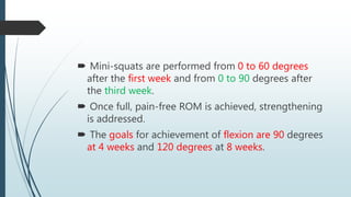
PCL Avulsion
CAMPBELL’S OPRATIVE ORTHOPAEDICS 2013
By: Dr Hamid Hejrati
Resident of Orthopedic Surgery
Iran, Mashhad university of medical science
 If the isolated posterior cruciate ligament disruption
is characterized by avulsion of a large piece of bone
from the posterior aspect of the tibia and a posterior
approach is planned, the knee must be examined
arthroscopically to rule out other orthopaedic
disorders before making the approach. This approach
does not permit exploration of the knee or correction
of any other disorder.
REPAIR OF BONY AVULSION OF PCL
 If the posterior cruciate ligament is avulsed from the
tibia and the repair is to be done through a medial
approach, dissect the posterior skin and
subcutaneous tissue as a single layer to the
posteromedial corner and, with the knee flexed to 90
degrees, retract the medial head of the
gastrocnemius and the popliteal structures
posteriorly to adequately expose the tibial
attachment of the ligament.
 In the absence of a major medial compartment
disruption, a posteromedial capsular incision allows
adequate exposure of the tibial attachment. The
most medial portion of an intact posterior horn of
the medial meniscus may make exposure and
placement of the suture in the distal end of the
ligament difficult, but excision of the intact medial
meniscus is not necessary.
 Fit the fragment of bone carefully into the crater and
secure it with a cancellous screw if the fragment is
large enough or with a nonabsorbable suture passed
through parallel drill holes to the anterior aspect of
the tibia.
 Repair the frequently found tear of the posterior
capsule with interrupted sutures.
BURKS AND SCHAFFER
 With the patient prone, make a gently curved
incision, with a horizontal limb near the flexion crease
of the knee and a vertical limb overlying the medial
aspect of the gastrocnemius muscle.
 Carry the dissection to the deep fascial layer and
incise it vertically over the medial head of the
gastrocnemius.
 Protect the medial sural cutaneous nerve (posterior
cutaneous nerve of the calf), which usually perforates
the deep fascia distal to the horizontal limb of the
incision.
 Identify the medial border of the medial gastrocnemius
and bluntly develop the interval between it and the
semimembranosus tendon, exposing the posterior joint
capsule. The middle geniculate artery may be
encountered near the midposterior capsule and can be
ligated if necessary. By lateral retraction on the medial
head of the gastrocnemius, no tension is directly applied
to the motor branch to the medial head of the
gastrocnemius, the only motor branch from the tibial
nerve in the popliteal fossa that traverses medially.
 The thick muscle belly protects the neurovascular
structures as the capsule is exposed. Dissection on
this protected medial side of the popliteal fossa is
therefore relatively safe.
 Expose the posterior aspect of the proximal tibia and
posterior margins of the femoral condyle.
 If further lateral exposure is necessary, release a
portion of the tendinous origin of the medial head of
the gastrocnemius from the distal femur and joint
capsule. Slight knee flexion will aid exposure, and
complete sectioning of the medial head of the
gastrocnemius rarely is needed.
 Make a vertical incision through the posterior
capsule to expose the contents of the posterior
intercondylar notch and the tibial attachment of the
posterior cruciate ligament.
Suture the capsular incision, allow the gastrocnemius
to settle into position, approximate the subcutaneous
layers, and close the skin in a routine fashion.
Tibial Avulsion of PCL
 The PCl tibial avulsion is approached similarly to
tibial inlay reconstruction.
 The patient is positioned supine, to facilitate
arthroscopic examination.
 The leg is brought into a figure 4 position, with the
knee flexed to 90 degrees and the bump
repositioned under the lateral ankle.
 A 6-cm incision is made
over the posterior border
of the tibia from the
crease of the popliteal
fossa and curving distally
along the posteromedial
border of the tibia.
The dissection is continued through the
subcutaneous fat to the sartorius fascia and the
fascia overlying the medial head of the
gastrocnemius.
 The fascia is incised along the palpable
posteromedial tibial border
 The semimembranosus and pes anserinus tendons
are retracted anteriorly and proximally.
 The medial head of the gastrocnemius is elevated
from the tibial cortex and retracted posteriorly.
The medial border of the gastrocnemius is followed
distally along the posterior tibia, and the proximal
border of the popliteus muscle is identified. The
popliteus muscle is elevated subperiosteally off the
posteromedial surface of the tibia and mobilized
laterally and distally.
 A vertical arthrotomy is made, and the avulsed
fragment of the tibia with the attached PCl is
identified.
 The bone fragment and PCl are reduced and secured
with a 4.0-mm cortical or a 6.5-mm cancellous screw
and spiked washer, depending on the size of the
fragment.
 The reduction is confirmed with fluoroscopy or a
radiograph.
POSTOPERATIVE CARE
 A hinged knee brace is applied and locked in
extension. The patient is awakened and taken to the
recovery room, where pain and neurovascular status
are reevaluated.
 Patients may be kept overnight for pain
management and to monitor their neurovascular
status.
Patients are given instructions for exercises (quadriceps
sets, straight-leg raises, and calf pumps) and crutch
use.
 All dressing changes are performed while an anterior
tibial force is applied.
 Patients are instructed to maintain touch-down weight
bearing for 1 week.
 Partial weight bearing is initiated after the first
postoperative visit.
 The brace is unlocked after 4 to 6 weeks, and usually
is discontinued after 8 weeks.
 Symmetric full hyperextension is achieved, and
passive prone knee flexion, quadriceps sets, and
patellar mobilization exercises are performed with
the assistance of a physical therapist for the first
month.
 Mini-squats are performed from 0 to 60 degrees
after the first week and from 0 to 90 degrees after
the third week.
 Once full, pain-free ROM is achieved, strengthening
is addressed.
 The goals for achievement of flexion are 90 degrees
at 4 weeks and 120 degrees at 8 weeks.

Pcl avulsion

  • 1.
    PCL Avulsion CAMPBELL’S OPRATIVEORTHOPAEDICS 2013 By: Dr Hamid Hejrati Resident of Orthopedic Surgery Iran, Mashhad university of medical science
  • 3.
     If theisolated posterior cruciate ligament disruption is characterized by avulsion of a large piece of bone from the posterior aspect of the tibia and a posterior approach is planned, the knee must be examined arthroscopically to rule out other orthopaedic disorders before making the approach. This approach does not permit exploration of the knee or correction of any other disorder.
  • 4.
    REPAIR OF BONYAVULSION OF PCL  If the posterior cruciate ligament is avulsed from the tibia and the repair is to be done through a medial approach, dissect the posterior skin and subcutaneous tissue as a single layer to the posteromedial corner and, with the knee flexed to 90 degrees, retract the medial head of the gastrocnemius and the popliteal structures posteriorly to adequately expose the tibial attachment of the ligament.
  • 5.
     In theabsence of a major medial compartment disruption, a posteromedial capsular incision allows adequate exposure of the tibial attachment. The most medial portion of an intact posterior horn of the medial meniscus may make exposure and placement of the suture in the distal end of the ligament difficult, but excision of the intact medial meniscus is not necessary.
  • 6.
     Fit thefragment of bone carefully into the crater and secure it with a cancellous screw if the fragment is large enough or with a nonabsorbable suture passed through parallel drill holes to the anterior aspect of the tibia.  Repair the frequently found tear of the posterior capsule with interrupted sutures.
  • 8.
    BURKS AND SCHAFFER With the patient prone, make a gently curved incision, with a horizontal limb near the flexion crease of the knee and a vertical limb overlying the medial aspect of the gastrocnemius muscle.  Carry the dissection to the deep fascial layer and incise it vertically over the medial head of the gastrocnemius.
  • 9.
     Protect themedial sural cutaneous nerve (posterior cutaneous nerve of the calf), which usually perforates the deep fascia distal to the horizontal limb of the incision.
  • 10.
     Identify themedial border of the medial gastrocnemius and bluntly develop the interval between it and the semimembranosus tendon, exposing the posterior joint capsule. The middle geniculate artery may be encountered near the midposterior capsule and can be ligated if necessary. By lateral retraction on the medial head of the gastrocnemius, no tension is directly applied to the motor branch to the medial head of the gastrocnemius, the only motor branch from the tibial nerve in the popliteal fossa that traverses medially.
  • 11.
     The thickmuscle belly protects the neurovascular structures as the capsule is exposed. Dissection on this protected medial side of the popliteal fossa is therefore relatively safe.
  • 12.
     Expose theposterior aspect of the proximal tibia and posterior margins of the femoral condyle.  If further lateral exposure is necessary, release a portion of the tendinous origin of the medial head of the gastrocnemius from the distal femur and joint capsule. Slight knee flexion will aid exposure, and complete sectioning of the medial head of the gastrocnemius rarely is needed.
  • 13.
     Make avertical incision through the posterior capsule to expose the contents of the posterior intercondylar notch and the tibial attachment of the posterior cruciate ligament.
  • 14.
    Suture the capsularincision, allow the gastrocnemius to settle into position, approximate the subcutaneous layers, and close the skin in a routine fashion.
  • 16.
    Tibial Avulsion ofPCL  The PCl tibial avulsion is approached similarly to tibial inlay reconstruction.  The patient is positioned supine, to facilitate arthroscopic examination.  The leg is brought into a figure 4 position, with the knee flexed to 90 degrees and the bump repositioned under the lateral ankle.
  • 17.
     A 6-cmincision is made over the posterior border of the tibia from the crease of the popliteal fossa and curving distally along the posteromedial border of the tibia.
  • 18.
    The dissection iscontinued through the subcutaneous fat to the sartorius fascia and the fascia overlying the medial head of the gastrocnemius.  The fascia is incised along the palpable posteromedial tibial border
  • 19.
     The semimembranosusand pes anserinus tendons are retracted anteriorly and proximally.  The medial head of the gastrocnemius is elevated from the tibial cortex and retracted posteriorly.
  • 20.
    The medial borderof the gastrocnemius is followed distally along the posterior tibia, and the proximal border of the popliteus muscle is identified. The popliteus muscle is elevated subperiosteally off the posteromedial surface of the tibia and mobilized laterally and distally.
  • 21.
     A verticalarthrotomy is made, and the avulsed fragment of the tibia with the attached PCl is identified.  The bone fragment and PCl are reduced and secured with a 4.0-mm cortical or a 6.5-mm cancellous screw and spiked washer, depending on the size of the fragment.  The reduction is confirmed with fluoroscopy or a radiograph.
  • 23.
    POSTOPERATIVE CARE  Ahinged knee brace is applied and locked in extension. The patient is awakened and taken to the recovery room, where pain and neurovascular status are reevaluated.  Patients may be kept overnight for pain management and to monitor their neurovascular status.
  • 24.
    Patients are giveninstructions for exercises (quadriceps sets, straight-leg raises, and calf pumps) and crutch use.  All dressing changes are performed while an anterior tibial force is applied.  Patients are instructed to maintain touch-down weight bearing for 1 week.  Partial weight bearing is initiated after the first postoperative visit.
  • 25.
     The braceis unlocked after 4 to 6 weeks, and usually is discontinued after 8 weeks.  Symmetric full hyperextension is achieved, and passive prone knee flexion, quadriceps sets, and patellar mobilization exercises are performed with the assistance of a physical therapist for the first month.
  • 26.
     Mini-squats areperformed from 0 to 60 degrees after the first week and from 0 to 90 degrees after the third week.  Once full, pain-free ROM is achieved, strengthening is addressed.  The goals for achievement of flexion are 90 degrees at 4 weeks and 120 degrees at 8 weeks.