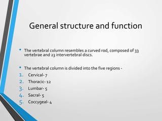
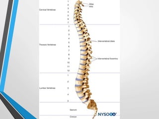
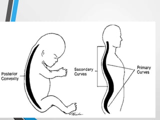
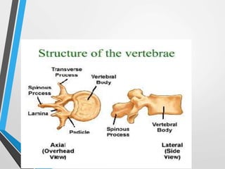
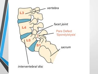
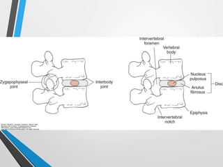
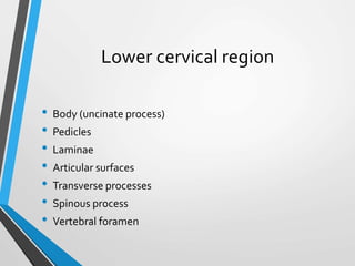
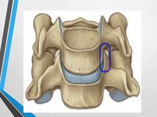
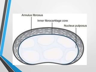
SlideShare a Scribd company logo
Biomechanics of
Vertebral Column
Dr. Rushi Gajjar
General structure and function
• The vertebral column resembles a curved rod, composed of 33
vertebrae and 23 intervertebral discs.
• The vertebral column is divided into the five regions -
1. Cervical- 7
2. Thoracic- 12
3. Lumbar- 5
4. Sacral- 5
5. Coccygeal- 4
• The vertebrae increase in size from the cervical to the lumbar
regions and then decrease in size from the Sacral to coccygeal
region.
• 7 vertebrae are located in cervical region, 12 in the thoracic
region, 5 in the lumbar region.
• 5 vertebrae are fused to form the sacrum and remaining 4
constitute a coccygeal region.
Curves of spine
• The curve of the vertebral column of a fetus exhibits one
long curve that is convex Posteriorly, secondary curves
develop in infancy.
• The two curves (thoracic and Sacral) that retain the
original posterior convexity through life are called
primary curves and are referred as kyphotic curves.
• The two curves (cervical and lumbar) that show a reversal
of original posterior convexity are called secondary
curves and are called lordotic curves.
• The secondary curves develop as a result of the
accommodation of the skeleton to the upright posture.
Typical vertebrae
• The structure of typical vertebra consist of two parts -
1. Anterior, cylindrical shaped vertebral body
2. Posteriorly, irregularly shaped neural arch
• The vertebral body is designed to be the weight bearing
structure of spinal column.
• The neural arch can be further divided into the pedicles
and the posterior elements.
• The pedicles are the portion of the neural arch that lie
anterior to the articular processes on either side and
serve as a connection between the posterior elements
and the vertebral body.
• The function of the pedicles is to transmit the load and
bending forces from the posterior elements to the
vertebral bodies. આ
• The pedicles increase in size from the cervical to lumbar
regions, which makes sense because greater forces are
transmitted through the pedicles in the lumbar region.
• The remaining posterior elements are the laminae, articular
processes, spinous process, and the transverse process.
• The laminae are thin, vertically oriented pieces of bone that
serve as a 'roof' to the neural arch, which protects the spinal
cord.
• It transmits weight from the posterior elements to pedicles
and through them to the vertebral bodies.
• This force transfer occurs through a region of laminae called
pars interarticularis.
• It is a portion of laminae that is between superior and
inferior articular process.
• It is subjected to bending forces and there for it is
susceptible to stress fracture.
• The spinous process and two transverse processes are
sites for muscle attachments and serve to increase lever
arm for the muscles.
• The articular processes consist of two superior and two
inferior facets for articulation with facets from cranial and
caudal vertebra respectively
The Intervertebral Disc
• The intervertebral disc has two main functions-
1. To separate vertebral bodies
2. To transmit load from one vertebral body to next.
• The intervertebral discs, which make up about 20%-33%
of the Length of theVertebral column, increase in size
from cervical to the lumbar region.
• The disc thickness varies approximately 3 mm in the
cervical region, where the weight bearing loads are the
lowest, to about 9 mm in the lumbar region, where the
weight bearing loads are highest.
• The ratio between disc thickness and vertebral body
height determines the available motion.
• Greater the ratio, greater the mobility.
• Ratio is grateest in the cervical region, followed by
lumbar region, and ratio is smallest in thoracic region.
• Discs are composed of 3 parts:
1. The nucleus pulposus
2. The anulus fibrosus
3. The vertebral end plates
• The nucleus pulposus is the gelatinous mass found in the
center, the anulus fibrosus is a fibrous outer ring, and the
vertebral end plates is the cartilagenous layer covering
the superior and inferior aspect of disc.
NUCLEUS PULPOSUS :
• The nucleus pulposus is 70%-90% water, depending on
age and time of day.
• Proteogycans make up approximately 65% of the dry
weight and have ability to attract water molecules.
• Collagen fibers contribute 15%-20% of dry weight.
• It has both type 1 and type 2 collagen, however type 2
predominates because of its ability to resist compressive
loads.
• The nucleus pulposus has been frequently likened to a
water balloon.
• When compressed, it deforms, and the increased
pressure stretches the s of the nucleus pulposus in all
direction.
ANULUS FIBROSUS :
• It has 60%-70% of water.
• Collagen fibres make up 50%-60% of dry weight, with
Proteogycans contributing only 20% of dry weight.
• Remaining 10% is of elastin fibres and cells such as fibroblasts and
chondrocytes.
• Type 1 and type 2 collagen are present, however type 1
collagen predominates in the anulus fibrosus, particularly
in the outer portions.
• The anulus fibres are attached by Sharpe fibres to the
cartilagenous end plates on the inferior and superior
vertebral plateaus of adjacent vertebrae.
VERTEBRAL END PLATES :
• It is layer of cartilage 0.6 to 1 mm thick that cover the region of
the vertebral bodies.
• They cover the entire nucleus pulposus but not the entire anulus
fibrosus.
• It consist of Proteogycans, collagen, and water.
• The cartilage ofVertebral end plates is both hyaline
cartilage and fibro cartilage.
• Hyaline cartilage is present closest to the vertebral body
and fibro cartilage is present closest to the nucleus
pulposus.
d
INNER ACTION AND NUTRITION:
• The intervertebral discs are innervated in the outer 1/3 to
1/2 of the fibers of anulus fibrosus.
• In the cervical and lumbar regions, the intervertebral
discs are innervated by branches from the vertebra and
sinuvertebral nerves.
• Outer surface of anulus fibrosus receives blood supply
from the small branches of metaphyseal arteries.
• The remainder of the disc receives its nutrition via
diffusion.
Articulations
• Mainly two joints are found in vertebral column.
1. Interbody joints
2. Zygapophyseal joints (Facet joint)
1. INTERBODY JOINTS-
• Joints between the vertebral bodies.
• Available movements at this joint include sliding, distraction and
compression, and rotation (tilt).
• Sliding can occur in following direction: anterior to posterior,
medial to lateral and torsional.
• Tilt can occur in anterior to posterior and in lateral
direction.
• The amount of each of these motions are small and vary
by region according to structural differences in discs and
vertebral bodies, as well as in the ligament pus support.
2. ZYGAPOPHYSEAL JOINTS-
• That is a articulation between the right and left superior
articulating facets of a vertebra and the right and left
inferior facets of adjacent cranial vertebra.
• These joints are diarthrodial joints and have regional
variation in structure.
Ligaments and Joint Capsules
Six ligaments-
1. Anterior longitudinal ligament
2. Posterior longitudinal ligament
3. Ligamentum flavum
4. Interspinous ligament
5. Supra spinous ligament
6. Intertransverse ligament
1. ANTERIOR LONGITUDINAL LIGAMENT -
• It runs along the anterior and lateral surfaces of the vertebral
bodies from the sacrum to C2.
• Extension of ligament from C2 to occipht are called anterior
atlanto occipital and anterior atlanto axial ligaments.
• It is well developed in the lordotic sections but has little substance
in kyphotic region.
• The tensile strength of ligament is greatest in high
cervical, lower thoracic and lumbar region.
• It is reported to be twice as strong as the posterior
longitudinal ligament.
• It is compressed in flexion and stretched in extension.
2. POSTERIOR LONGITUDINAL LIGAMENT -
• It runs over on the posterior aspects ofVertebral bodies from C2
to the sacrum.
• Superiorly, the ligament becomes the tectorial membrane from
C2 to the occiput.
• It is stretched in flexion and slack in extension.
3. LIGAMENTUM FLAVUM-
• It is a thick, elastic ligament that connects lamina to lamina from C2
to the sacrum.
• From C2 to the occiput, this ligament continues as the posterior
atlanto occipital and posterior atlanto axial membranes.
• It is strongest in lower thoracic region and weakest in mid cervical
region.
• Highest strain occurs during flexion when this ligament is
stretched, although this ligament is under constant
tension even when the spine is in neutral position,
because of its elastic nature.
4. INTERSPINOUS LIGAMENT -
• It connects spinous processes of adjacent vertebra.
• It is the first to be damaged with excessive flexion.
• It has been found to contribute to lumbar spine mobility
and to degenerate with aging.
5. SUPRA SPINOUS LIGAMENT -
• It is a strong cord like structure that connects the tips of spinous
processes from C7 to L3 or L4.
• In the cervical region, the ligament becomes Ligamentum
nuchae.
• It is stretched in flexion and its fibres resist separation of the
spinous processes during flexion.
6. INTERTRANSVERSE LIGAMENT -
• The ligament pass between the transverse processes and
attach to the deep muscles of the back.
• The ligaments are alternatively stretched and
compressed during lateral bending.
ZYGAPOPHYSEAL JOINT CAPSULES-
• It assist the ligaments in providing limitation to motion
and stability for vertebral column.
• The capsules are strongest in theThoraco lumbar region
and at cervicothoracic junction, where the spinal
configuration changes.
Kinematics
• The motions available to the column as a whole are flexion and
extension, lateral flexion and rotation.
• Coupling is defined as the consistent association of one motion
about an axis with another motion around a different axis.
• The most predominant coupled motion is lateral flexion and
rotation.
FLEXION-
• In flexion, anterior tilting and gliding of the superior
vertebra occur and cause widening of the intervertebral
foramen and separation of the spinous process.
• With flexion, the anterior portion of anulus fibrosus is
compressed and bulges Anteriorly, while the posterior
portion is stretched.
EXTENSION -
• In extension, posterior tilting and gliding of the superior
vertebra occur and cause narrowing of the intervertebral
foramen, and the spinous processes move closer together.
• It is limited by bony contact of the spinous process and
tension in Zygapophyseal joint capsule and ALL.
LATERAL FLEXION -
• In lateral flexion, the superior vertebra laterally tilts,
rotates and translates over adjacent vertebra below.
• The intervertebral foramen is widened on the side to
contralateral to the lateral flexion and narrowed on the
ipsilateral side.
• The anulus fibrosus is compressed on the con cavity of
the curve and stretched on the convexity of the curve.
ROTATION-
• Rotation is available in each spinal region, but due to the
drastically different shapes of Zygapophyseal joints, the
kinematics vary by region.
Kinetics
• The vertebral column is subjected to following loads-
1. Axial compression
2. Bending
3. Torsion
4. Shear
AXIAL COMPRESSION -
• It is a force acting through the long axis of the spine at right
angles to the discs.
• It occurs as a result of the force of gravity, groung reaction
forces and forces produced by the ligaments and muscular
contraction.
• The discs and vertebral bodies resist most of the
compressive force, but neural arches and Zygapophyseal
joints share some of the load.
• The compressive load is transmitted from superior end
plate to the inferior end plate.
• The nucleus pulposus acts as a ball of fluid that can be
deformed by a compressive force.
• When a weight is applied to the nucleus pulposus from
the above, the nucleus pulposus exhibits a swelling
pressure and tries to expand outward toward the anulus
fibrosus.
• The end plates are able to undergo the least amount of
deformation and therefore will be the first to fail
(fracture) under high compressive loading.
• The discs will be the last to fail or rupture.
• Under sustained compressive loading, the rise in the
swelling pressure causes fluid to be expressed from the
nucleus pulposus and anulus fibrosus.
• The expressed fluid is absorbed through microspic pores
in the cartilagenous end plates.
• When the compressive loads are decreased, the disc
absorb fluid back from the vertebral body.
• The recovery of fluid that returns the disc to its original
state explains why a person getting up from bed is taller
in the morning than in the evening.
BENDING-
• Bending causes both compression and tension on the
structures of spine.
• In flexion, the anterior structures are subjected to
compression and the posterior structures are subjected to
tension.
• The resistance offered to the tensile forces by collagen
fibres in the posterior outer anulus fibrosus,
Zygapophyseal joint capsule, and posterior ligaments
provide stability in flexion.
• Creep occurs in extreme flexion or extreme extension.
• In extension, the posterior structures generally are either
unloaded or subjected to compression while anterior
structures are subjected to tension.
• Resistance to extension is provided by anterior outer
fibers of anulus fibrosus, Zygapophyseal joint capsule,
passive tension in ALL and contact of spinous process.
• In lateral bending, ipsilateral side of disc is compressed
and contralateral side is stretched.
• Therefore, the contralateral fibres of outer anulus
fibrosus and the contralateral Intertransverse ligament
help to provide stability.
TORSION-
• Torsional forces are created during axial rotation that occurs
as a part of the coupled motions that takes place in spine.
• When the disc is subjected toTorsion, half of the anulus
fibrosus fibres resist clockwise rotations while fibres
oriented in the opposite direction resist counterclockwise
rotation.
SHEAR-
• Shear forces act on the midplane of the disc and tend to
cause each vertebra to undergo translation (move
Anteriorly Posteriorly, or from side to side in relation to
the inferior vertebra).
Structure of cervical
spine
• The cervical vertebral column consists of seven vertebrae.
• Morphologically and functionally, the cervical column is
divided into two distinct regions: the
1. upper cervical spine, or craniovertebral region, and the
2. lower cervical spine
• The craniovertebral region includes the occipital condyles
and the first two cervical vertebrae, C1 and C2, or,
respectively, the atlas and axis.
• The lower cervical spine includes the vertebrae of C3 to
C7.
• The vertebrae from C3 to C6 display similar
characteristics and are therefore considered to be the
typical cervical vertebrae.
• The atlas, axis, and C7 exhibit unique characteristics and
are considered the atypical cervical vertebrae
Upper cervical region
ATLAS-
• The atlas (C1) is frequently described to be like a washer
sitting between the occipital condyles and the axis.
• The functions of the atlas are to cradle the occiput and to
transmit forces from the occiput to the lower cervical
vertebrae.
• The atlas is different from other vertebrae in that it has
no vertebral body or spinous process and is shaped like a
ring.
• There are two large lateral masses that have a vertical
alignment under each occipital condyle that reflect the
function of transmitting forces.
• The lateral masses include four articulating facets: two
superior and two inferior.
• The atlas also possesses a facet on the internal surface of
the anterior arch for articulation with the dens (odontoid
process) of the axis.
AXIS-
• The primary functions of the axis are to transmit the
combined load of the head and atlas to the remainder of
the cervical spine and to provide motion into axial
rotation of the head and atlas.
• The arch of the axis has inferior and superior
zygapophyseal facets for articulation with the adjacent
inferior vertebra and the atlas, respectively.
• The spinous process of the axis is large and elongated
with a bifid (split into two portions) tip.
ARTICULATIONS-
• Main two joints
1. Atlanto occipital joint
2. Antlanto axial joint
ATLANTO OCCIPITAL JOINT-
• The two atlanto-occipital joints consist of the two
concave ve superior zygapophyseal facets of the atlas
articulating with the two convex occipital condyles of the
skull.
ATLANTOAXIAL JOINT-
• There are three synovial joints that compose the
atlantoaxial joints:
1. the median atlantoaxial joint between the dens and the
atlas
2. And two lateral joint between the superior zygapophyseal
facets of the axis and the inferior zygapophyseal facets of
the atlas
LIGAMENTS-
• The posterior atlanto-occipital and atlantoaxial
membranes are the continuations of the ligamentum
flavum but they are less elastic and therefore permit a
greater ROM, especially into rotation.
• The anterior atlanto-occipital and atlantoaxial
membranes are the continuations of the anterior
longitudinal ligament.
• The tectorial membrane is the continuation of the PLL in
the upper two segments.
• The thick ligamentum nuchae, which extends from the
spinous process of C7 to the external occipital
protuberance
Transverse ligament-
• The transverse length of the ligament is about 21.9 mm.
• Its primary role, however, is to prevent anterior
displacement of C1 on C2.
• This ligament is critical in maintaining stability at the
C1/C2 segment.
• The transverse atlantal ligament is very strong, and the
dens will fracture before the ligament will tear.
ALAR LIGAMENT-
• The two alar ligaments are also specific to the cervical
region.
• These paired ligaments arise from the axis on either side
of the dens and extend laterally and superiorly to attach
to roughened areas on the medial sides of the occipital
condyles and to the lateral masses of the atlas.
• These ligaments are relaxed with the head in midposition
and taut in flexion.
• Axial rotation of the head and neck tightens both alar
ligaments.
• These ligaments also help to prevent distraction of C1 on
C2.
Lower cervical region
• Body (uncinate process)
• Pedicles
• Laminae
• Articular surfaces
• Transverse processes
• Spinous process
• Vertebral foramen
Intervertebral discs
• The structure of the intervertebral disk in the cervical
region is distinctly different from that in the lumbar
region.
• The fibers of the anulus fibrosus are not arranged in
alternating lamellar layers as in the lumbar region.
• In addition, they do not surround the entire perimeter of
the nucleus pulposus.
• Instead, the anular fibers in this region have a crescent
shape when viewed from above, being thick anteriorly and
tapering laterally as they approach the uncinate processes.
• Laterally, there is no substantive anulus fibrosus, and
Posteriorly, it is only a thin layer of vertically oriented fibers.
• Posterolaterally, the nucleus is contained only by the PLL.
Functions of cervical region
• The cervical region demonstrates the most flexibility of
any of the regions of the vertebral column.
• The design of the atlas is such that it provides more free
space for the spinal cord than does any other vertebra.
• The extra space helps to ensure that the spinal cord is not
impinged on during the large amount of motion that
occurs here.
Kinematics
• Normally, the neck moves 600 times every hour whether
we are awake or asleep.
• The motions of flexion and extension, lateral flexion,
and rotation are permitted in the cervical region.
• The atlanto-occipital joints allow for only nodding
movements between the head and the atlas.
• In flexion, the occipital condyles roll forward and slide
backward.
• In extension, the occipital condyles roll backward and
slide forward.
• The combined ROM for flexion-extension reportedly
ranges from 10 to 30.
• The total ROM available in both axial rotation and lateral
flexion is extremely limited by tension in the joint
capsules that occurs as the occipital condyles rise up the
walls of the atlantal sockets on the contralateral side of
either the rotation or lateral flexion.
• Motions at the atlantoaxial joint include rotation,lateral
flexion, flexion, and extension.
• Approximately 55% to 58% of the total rotation of the
cervical region occurs at the atlantoaxial joints.
• The atlas pivots about 45 to either side, or a total of about
90.
• Pure anterior translation does not occur, because it would
cause the zygapophyseal joints to abut one another.
• Flexion of these segments must include anterior tilt of the
cranial vertebral body coupled with anterior translation.
• Extension includes posterior tilt of the cranial vertebral
body, coupled with posterior translation.
• Lateral flexion and rotation are also coupled motions,
because movement of either alone would cause the
zygapophyseal joints to abut one another and prevent
motion.
• Lateral flexion is coupled with ipsilateral rotation, and
rotation is coupled with ipsilateral lateral flexion.
• Lower cervical segments generally favor flexion and
extension ROM; however, there is great variability in
reported ranges of motion in the individual cervical
segments.
• In general, the range for flexion and extension increases
from the C2/C3 segment to the C5/C6 segment, and
decreases again at the C6/C7 segment.
Kinetics
• The cervical region is subjected to axial compression,
tension, bending, torsion, and shear stresses.
• The cervical region differs from the thoracic and lumbar
regions in that the cervical region bears less weight and is
generally more mobile.
• No disks are present at either the atlanto-occipital or
atlantoaxial articulations; therefore, the weight of the
head (compressive load) must be transferred directly
through the atlanto-occipital joint to the articular facets
of the axis.
• These forces are then transferred through the pedicles
and laminae of the axis to the inferior surface of the body
and to the two inferior zygapophyseal articular processes.
• Subsequently, the forces are transferred to the adjacent
inferior disk.
• In the cervical region from C3 to C7 compressive forces
are transmitted by three parallel columns:
• a single anterocentral column formed by the vertebral
bodies and disks and
• two rodlike posterolateral columns composed of the left
and right zygapophyseal joints.
• The compressive forces are transmitted mainly by the
bodies and disks, with a little over one third transmitted
by the two posterolateral columns.
• Compressive loads are relatively low during erect stance
and sitting postures and high during the end ranges of
flexion and extension.
Thank you...

More Related Content

Similar to Biomechanics_of_spine[1].pptx

Biomechanics of elbow joint
Biomechanics of elbow jointBiomechanics of elbow joint
Biomechanics of elbow joint
Kumarpal Singh
 
Shoulder by sunil
Shoulder by sunilShoulder by sunil
Shoulder by sunil
sunil JMI
 
anatomy of vertebral column.pptx
anatomy of vertebral column.pptxanatomy of vertebral column.pptx
anatomy of vertebral column.pptx
JaseerAk1
 
Muscles of mastication
Muscles of masticationMuscles of mastication
Muscles of mastication
RAGHU D
 
Shoulder joint_nursing.pptx
Shoulder joint_nursing.pptxShoulder joint_nursing.pptx
Shoulder joint_nursing.pptx
ABHIJIT BHOYAR
 
Pathophysiologic aspects, clinical manifestation a nd management of
Pathophysiologic aspects, clinical manifestation a nd management ofPathophysiologic aspects, clinical manifestation a nd management of
Pathophysiologic aspects, clinical manifestation a nd management of
Sushant Yadav
 
THE FEMALE PELVIS RM 2020.pdf
THE FEMALE PELVIS RM 2020.pdfTHE FEMALE PELVIS RM 2020.pdf
THE FEMALE PELVIS RM 2020.pdf
CaiusMbao
 
ANATOMY OF SPINAL CORD AND VERTEBRAL COLOUMN ,.pptx
ANATOMY OF SPINAL CORD AND VERTEBRAL COLOUMN ,.pptxANATOMY OF SPINAL CORD AND VERTEBRAL COLOUMN ,.pptx
ANATOMY OF SPINAL CORD AND VERTEBRAL COLOUMN ,.pptx
2021AMM010CHARANAGS
 
applied aspects of shoulder joint
applied aspects of shoulder jointapplied aspects of shoulder joint
applied aspects of shoulder joint
mrinal joshi
 
Dr. kamal kant applied anatomy of hip
Dr. kamal kant   applied anatomy of hipDr. kamal kant   applied anatomy of hip
Dr. kamal kant applied anatomy of hip
mrinal joshi
 
Biomechanics of thorax
Biomechanics of thoraxBiomechanics of thorax
Biomechanics of thorax
DR.RAMANDEEP KAUR SAINI
 
clavicle Anatomy.pptx
clavicle Anatomy.pptxclavicle Anatomy.pptx
clavicle Anatomy.pptx
mhmad farooq
 
Ligament
LigamentLigament
Ligament
Prashanth Kumar
 
Elbow biomechanics
Elbow biomechanicsElbow biomechanics
Elbow biomechanics
RukhsarJabbar
 
Vertebrae thoracic
Vertebrae thoracicVertebrae thoracic
Vertebrae thoracic
Idris Siddiqui
 
Function of cervical region
Function of cervical regionFunction of cervical region
Function of cervical region
Dr Vicky Kasundra
 
Anatomy of lumber spine Nishant kumar gaurav
Anatomy of lumber spine Nishant kumar gauravAnatomy of lumber spine Nishant kumar gaurav
Anatomy of lumber spine Nishant kumar gaurav
NISHANTKUMARGAURAV1
 
Anatomy of Cervical Spine
Anatomy of Cervical  SpineAnatomy of Cervical  Spine
Anatomy of Cervical Spine
Dr Asma Lashari
 

Similar to Biomechanics_of_spine[1].pptx (20)

Biomechanics of elbow joint
Biomechanics of elbow jointBiomechanics of elbow joint
Biomechanics of elbow joint
 
Shoulder by sunil
Shoulder by sunilShoulder by sunil
Shoulder by sunil
 
anatomy of vertebral column.pptx
anatomy of vertebral column.pptxanatomy of vertebral column.pptx
anatomy of vertebral column.pptx
 
Muscles of mastication
Muscles of masticationMuscles of mastication
Muscles of mastication
 
Anatommy and physiology of cnb
Anatommy and physiology of cnbAnatommy and physiology of cnb
Anatommy and physiology of cnb
 
Shoulder joint_nursing.pptx
Shoulder joint_nursing.pptxShoulder joint_nursing.pptx
Shoulder joint_nursing.pptx
 
Pathophysiologic aspects, clinical manifestation a nd management of
Pathophysiologic aspects, clinical manifestation a nd management ofPathophysiologic aspects, clinical manifestation a nd management of
Pathophysiologic aspects, clinical manifestation a nd management of
 
Ana. and physio. of cnb sushil
Ana. and physio. of cnb sushilAna. and physio. of cnb sushil
Ana. and physio. of cnb sushil
 
THE FEMALE PELVIS RM 2020.pdf
THE FEMALE PELVIS RM 2020.pdfTHE FEMALE PELVIS RM 2020.pdf
THE FEMALE PELVIS RM 2020.pdf
 
ANATOMY OF SPINAL CORD AND VERTEBRAL COLOUMN ,.pptx
ANATOMY OF SPINAL CORD AND VERTEBRAL COLOUMN ,.pptxANATOMY OF SPINAL CORD AND VERTEBRAL COLOUMN ,.pptx
ANATOMY OF SPINAL CORD AND VERTEBRAL COLOUMN ,.pptx
 
applied aspects of shoulder joint
applied aspects of shoulder jointapplied aspects of shoulder joint
applied aspects of shoulder joint
 
Dr. kamal kant applied anatomy of hip
Dr. kamal kant   applied anatomy of hipDr. kamal kant   applied anatomy of hip
Dr. kamal kant applied anatomy of hip
 
Biomechanics of thorax
Biomechanics of thoraxBiomechanics of thorax
Biomechanics of thorax
 
clavicle Anatomy.pptx
clavicle Anatomy.pptxclavicle Anatomy.pptx
clavicle Anatomy.pptx
 
Ligament
LigamentLigament
Ligament
 
Elbow biomechanics
Elbow biomechanicsElbow biomechanics
Elbow biomechanics
 
Vertebrae thoracic
Vertebrae thoracicVertebrae thoracic
Vertebrae thoracic
 
Function of cervical region
Function of cervical regionFunction of cervical region
Function of cervical region
 
Anatomy of lumber spine Nishant kumar gaurav
Anatomy of lumber spine Nishant kumar gauravAnatomy of lumber spine Nishant kumar gaurav
Anatomy of lumber spine Nishant kumar gaurav
 
Anatomy of Cervical Spine
Anatomy of Cervical  SpineAnatomy of Cervical  Spine
Anatomy of Cervical Spine
 

More from ShivBJhala

postural strain.pptx
postural strain.pptxpostural strain.pptx
postural strain.pptx
ShivBJhala
 
Frenkel’s exercises.pptx
Frenkel’s exercises.pptxFrenkel’s exercises.pptx
Frenkel’s exercises.pptx
ShivBJhala
 
MANUAL MUSCLE TESTING (MMT).pptx
MANUAL MUSCLE TESTING (MMT).pptxMANUAL MUSCLE TESTING (MMT).pptx
MANUAL MUSCLE TESTING (MMT).pptx
ShivBJhala
 
knee biomechanics.pptx
knee biomechanics.pptxknee biomechanics.pptx
knee biomechanics.pptx
ShivBJhala
 
Shoulder Complex(1).pptx
Shoulder Complex(1).pptxShoulder Complex(1).pptx
Shoulder Complex(1).pptx
ShivBJhala
 
1- Mobilization.pptx
1- Mobilization.pptx1- Mobilization.pptx
1- Mobilization.pptx
ShivBJhala
 

More from ShivBJhala (6)

postural strain.pptx
postural strain.pptxpostural strain.pptx
postural strain.pptx
 
Frenkel’s exercises.pptx
Frenkel’s exercises.pptxFrenkel’s exercises.pptx
Frenkel’s exercises.pptx
 
MANUAL MUSCLE TESTING (MMT).pptx
MANUAL MUSCLE TESTING (MMT).pptxMANUAL MUSCLE TESTING (MMT).pptx
MANUAL MUSCLE TESTING (MMT).pptx
 
knee biomechanics.pptx
knee biomechanics.pptxknee biomechanics.pptx
knee biomechanics.pptx
 
Shoulder Complex(1).pptx
Shoulder Complex(1).pptxShoulder Complex(1).pptx
Shoulder Complex(1).pptx
 
1- Mobilization.pptx
1- Mobilization.pptx1- Mobilization.pptx
1- Mobilization.pptx
 

Recently uploaded

Charaka Samhita Sutra sthana Chapter 15 Upakalpaniyaadhyaya
Charaka Samhita Sutra sthana Chapter 15 UpakalpaniyaadhyayaCharaka Samhita Sutra sthana Chapter 15 Upakalpaniyaadhyaya
Charaka Samhita Sutra sthana Chapter 15 Upakalpaniyaadhyaya
Dr KHALID B.M
 
For Better Surat #ℂall #Girl Service ❤85270-49040❤ Surat #ℂall #Girls
For Better Surat #ℂall #Girl Service ❤85270-49040❤ Surat #ℂall #GirlsFor Better Surat #ℂall #Girl Service ❤85270-49040❤ Surat #ℂall #Girls
For Better Surat #ℂall #Girl Service ❤85270-49040❤ Surat #ℂall #Girls
Savita Shen $i11
 
Pulmonary Thromboembolism - etilogy, types, medical- Surgical and nursing man...
Pulmonary Thromboembolism - etilogy, types, medical- Surgical and nursing man...Pulmonary Thromboembolism - etilogy, types, medical- Surgical and nursing man...
Pulmonary Thromboembolism - etilogy, types, medical- Surgical and nursing man...
VarunMahajani
 
The Normal Electrocardiogram - Part I of II
The Normal Electrocardiogram - Part I of IIThe Normal Electrocardiogram - Part I of II
The Normal Electrocardiogram - Part I of II
MedicoseAcademics
 
Ozempic: Preoperative Management of Patients on GLP-1 Receptor Agonists
Ozempic: Preoperative Management of Patients on GLP-1 Receptor Agonists  Ozempic: Preoperative Management of Patients on GLP-1 Receptor Agonists
Ozempic: Preoperative Management of Patients on GLP-1 Receptor Agonists
Saeid Safari
 
Pharynx and Clinical Correlations BY Dr.Rabia Inam Gandapore.pptx
Pharynx and Clinical Correlations BY Dr.Rabia Inam Gandapore.pptxPharynx and Clinical Correlations BY Dr.Rabia Inam Gandapore.pptx
Pharynx and Clinical Correlations BY Dr.Rabia Inam Gandapore.pptx
Dr. Rabia Inam Gandapore
 
Couples presenting to the infertility clinic- Do they really have infertility...
Couples presenting to the infertility clinic- Do they really have infertility...Couples presenting to the infertility clinic- Do they really have infertility...
Couples presenting to the infertility clinic- Do they really have infertility...
Sujoy Dasgupta
 
BRACHYTHERAPY OVERVIEW AND APPLICATORS
BRACHYTHERAPY OVERVIEW  AND  APPLICATORSBRACHYTHERAPY OVERVIEW  AND  APPLICATORS
BRACHYTHERAPY OVERVIEW AND APPLICATORS
Krishan Murari
 
How STIs Influence the Development of Pelvic Inflammatory Disease.pptx
How STIs Influence the Development of Pelvic Inflammatory Disease.pptxHow STIs Influence the Development of Pelvic Inflammatory Disease.pptx
How STIs Influence the Development of Pelvic Inflammatory Disease.pptx
FFragrant
 
Lung Cancer: Artificial Intelligence, Synergetics, Complex System Analysis, S...
Lung Cancer: Artificial Intelligence, Synergetics, Complex System Analysis, S...Lung Cancer: Artificial Intelligence, Synergetics, Complex System Analysis, S...
Lung Cancer: Artificial Intelligence, Synergetics, Complex System Analysis, S...
Oleg Kshivets
 
Are There Any Natural Remedies To Treat Syphilis.pdf
Are There Any Natural Remedies To Treat Syphilis.pdfAre There Any Natural Remedies To Treat Syphilis.pdf
Are There Any Natural Remedies To Treat Syphilis.pdf
Little Cross Family Clinic
 
Ophthalmology Clinical Tests for OSCE exam
Ophthalmology Clinical Tests for OSCE examOphthalmology Clinical Tests for OSCE exam
Ophthalmology Clinical Tests for OSCE exam
KafrELShiekh University
 
NVBDCP.pptx Nation vector borne disease control program
NVBDCP.pptx Nation vector borne disease control programNVBDCP.pptx Nation vector borne disease control program
NVBDCP.pptx Nation vector borne disease control program
Sapna Thakur
 
Physiology of Chemical Sensation of smell.pdf
Physiology of Chemical Sensation of smell.pdfPhysiology of Chemical Sensation of smell.pdf
Physiology of Chemical Sensation of smell.pdf
MedicoseAcademics
 
heat stroke and heat exhaustion in children
heat stroke and heat exhaustion in childrenheat stroke and heat exhaustion in children
heat stroke and heat exhaustion in children
SumeraAhmad5
 
Novas diretrizes da OMS para os cuidados perinatais de mais qualidade
Novas diretrizes da OMS para os cuidados perinatais de mais qualidadeNovas diretrizes da OMS para os cuidados perinatais de mais qualidade
Novas diretrizes da OMS para os cuidados perinatais de mais qualidade
Prof. Marcus Renato de Carvalho
 
HOT NEW PRODUCT! BIG SALES FAST SHIPPING NOW FROM CHINA!! EU KU DB BK substit...
HOT NEW PRODUCT! BIG SALES FAST SHIPPING NOW FROM CHINA!! EU KU DB BK substit...HOT NEW PRODUCT! BIG SALES FAST SHIPPING NOW FROM CHINA!! EU KU DB BK substit...
HOT NEW PRODUCT! BIG SALES FAST SHIPPING NOW FROM CHINA!! EU KU DB BK substit...
GL Anaacs
 
Hemodialysis: Chapter 3, Dialysis Water Unit - Dr.Gawad
Hemodialysis: Chapter 3, Dialysis Water Unit - Dr.GawadHemodialysis: Chapter 3, Dialysis Water Unit - Dr.Gawad
Hemodialysis: Chapter 3, Dialysis Water Unit - Dr.Gawad
NephroTube - Dr.Gawad
 
BENIGN PROSTATIC HYPERPLASIA.BPH. BPHpdf
BENIGN PROSTATIC HYPERPLASIA.BPH. BPHpdfBENIGN PROSTATIC HYPERPLASIA.BPH. BPHpdf
BENIGN PROSTATIC HYPERPLASIA.BPH. BPHpdf
DR SETH JOTHAM
 
micro teaching on communication m.sc nursing.pdf
micro teaching on communication m.sc nursing.pdfmicro teaching on communication m.sc nursing.pdf
micro teaching on communication m.sc nursing.pdf
Anurag Sharma
 

Recently uploaded (20)

Charaka Samhita Sutra sthana Chapter 15 Upakalpaniyaadhyaya
Charaka Samhita Sutra sthana Chapter 15 UpakalpaniyaadhyayaCharaka Samhita Sutra sthana Chapter 15 Upakalpaniyaadhyaya
Charaka Samhita Sutra sthana Chapter 15 Upakalpaniyaadhyaya
 
For Better Surat #ℂall #Girl Service ❤85270-49040❤ Surat #ℂall #Girls
For Better Surat #ℂall #Girl Service ❤85270-49040❤ Surat #ℂall #GirlsFor Better Surat #ℂall #Girl Service ❤85270-49040❤ Surat #ℂall #Girls
For Better Surat #ℂall #Girl Service ❤85270-49040❤ Surat #ℂall #Girls
 
Pulmonary Thromboembolism - etilogy, types, medical- Surgical and nursing man...
Pulmonary Thromboembolism - etilogy, types, medical- Surgical and nursing man...Pulmonary Thromboembolism - etilogy, types, medical- Surgical and nursing man...
Pulmonary Thromboembolism - etilogy, types, medical- Surgical and nursing man...
 
The Normal Electrocardiogram - Part I of II
The Normal Electrocardiogram - Part I of IIThe Normal Electrocardiogram - Part I of II
The Normal Electrocardiogram - Part I of II
 
Ozempic: Preoperative Management of Patients on GLP-1 Receptor Agonists
Ozempic: Preoperative Management of Patients on GLP-1 Receptor Agonists  Ozempic: Preoperative Management of Patients on GLP-1 Receptor Agonists
Ozempic: Preoperative Management of Patients on GLP-1 Receptor Agonists
 
Pharynx and Clinical Correlations BY Dr.Rabia Inam Gandapore.pptx
Pharynx and Clinical Correlations BY Dr.Rabia Inam Gandapore.pptxPharynx and Clinical Correlations BY Dr.Rabia Inam Gandapore.pptx
Pharynx and Clinical Correlations BY Dr.Rabia Inam Gandapore.pptx
 
Couples presenting to the infertility clinic- Do they really have infertility...
Couples presenting to the infertility clinic- Do they really have infertility...Couples presenting to the infertility clinic- Do they really have infertility...
Couples presenting to the infertility clinic- Do they really have infertility...
 
BRACHYTHERAPY OVERVIEW AND APPLICATORS
BRACHYTHERAPY OVERVIEW  AND  APPLICATORSBRACHYTHERAPY OVERVIEW  AND  APPLICATORS
BRACHYTHERAPY OVERVIEW AND APPLICATORS
 
How STIs Influence the Development of Pelvic Inflammatory Disease.pptx
How STIs Influence the Development of Pelvic Inflammatory Disease.pptxHow STIs Influence the Development of Pelvic Inflammatory Disease.pptx
How STIs Influence the Development of Pelvic Inflammatory Disease.pptx
 
Lung Cancer: Artificial Intelligence, Synergetics, Complex System Analysis, S...
Lung Cancer: Artificial Intelligence, Synergetics, Complex System Analysis, S...Lung Cancer: Artificial Intelligence, Synergetics, Complex System Analysis, S...
Lung Cancer: Artificial Intelligence, Synergetics, Complex System Analysis, S...
 
Are There Any Natural Remedies To Treat Syphilis.pdf
Are There Any Natural Remedies To Treat Syphilis.pdfAre There Any Natural Remedies To Treat Syphilis.pdf
Are There Any Natural Remedies To Treat Syphilis.pdf
 
Ophthalmology Clinical Tests for OSCE exam
Ophthalmology Clinical Tests for OSCE examOphthalmology Clinical Tests for OSCE exam
Ophthalmology Clinical Tests for OSCE exam
 
NVBDCP.pptx Nation vector borne disease control program
NVBDCP.pptx Nation vector borne disease control programNVBDCP.pptx Nation vector borne disease control program
NVBDCP.pptx Nation vector borne disease control program
 
Physiology of Chemical Sensation of smell.pdf
Physiology of Chemical Sensation of smell.pdfPhysiology of Chemical Sensation of smell.pdf
Physiology of Chemical Sensation of smell.pdf
 
heat stroke and heat exhaustion in children
heat stroke and heat exhaustion in childrenheat stroke and heat exhaustion in children
heat stroke and heat exhaustion in children
 
Novas diretrizes da OMS para os cuidados perinatais de mais qualidade
Novas diretrizes da OMS para os cuidados perinatais de mais qualidadeNovas diretrizes da OMS para os cuidados perinatais de mais qualidade
Novas diretrizes da OMS para os cuidados perinatais de mais qualidade
 
HOT NEW PRODUCT! BIG SALES FAST SHIPPING NOW FROM CHINA!! EU KU DB BK substit...
HOT NEW PRODUCT! BIG SALES FAST SHIPPING NOW FROM CHINA!! EU KU DB BK substit...HOT NEW PRODUCT! BIG SALES FAST SHIPPING NOW FROM CHINA!! EU KU DB BK substit...
HOT NEW PRODUCT! BIG SALES FAST SHIPPING NOW FROM CHINA!! EU KU DB BK substit...
 
Hemodialysis: Chapter 3, Dialysis Water Unit - Dr.Gawad
Hemodialysis: Chapter 3, Dialysis Water Unit - Dr.GawadHemodialysis: Chapter 3, Dialysis Water Unit - Dr.Gawad
Hemodialysis: Chapter 3, Dialysis Water Unit - Dr.Gawad
 
BENIGN PROSTATIC HYPERPLASIA.BPH. BPHpdf
BENIGN PROSTATIC HYPERPLASIA.BPH. BPHpdfBENIGN PROSTATIC HYPERPLASIA.BPH. BPHpdf
BENIGN PROSTATIC HYPERPLASIA.BPH. BPHpdf
 
micro teaching on communication m.sc nursing.pdf
micro teaching on communication m.sc nursing.pdfmicro teaching on communication m.sc nursing.pdf
micro teaching on communication m.sc nursing.pdf
 

Biomechanics_of_spine[1].pptx

  • 2. General structure and function • The vertebral column resembles a curved rod, composed of 33 vertebrae and 23 intervertebral discs. • The vertebral column is divided into the five regions - 1. Cervical- 7 2. Thoracic- 12 3. Lumbar- 5 4. Sacral- 5 5. Coccygeal- 4
  • 3. • The vertebrae increase in size from the cervical to the lumbar regions and then decrease in size from the Sacral to coccygeal region. • 7 vertebrae are located in cervical region, 12 in the thoracic region, 5 in the lumbar region. • 5 vertebrae are fused to form the sacrum and remaining 4 constitute a coccygeal region.
  • 4.
  • 5. Curves of spine • The curve of the vertebral column of a fetus exhibits one long curve that is convex Posteriorly, secondary curves develop in infancy. • The two curves (thoracic and Sacral) that retain the original posterior convexity through life are called primary curves and are referred as kyphotic curves.
  • 6. • The two curves (cervical and lumbar) that show a reversal of original posterior convexity are called secondary curves and are called lordotic curves. • The secondary curves develop as a result of the accommodation of the skeleton to the upright posture.
  • 7.
  • 8. Typical vertebrae • The structure of typical vertebra consist of two parts - 1. Anterior, cylindrical shaped vertebral body 2. Posteriorly, irregularly shaped neural arch • The vertebral body is designed to be the weight bearing structure of spinal column.
  • 9.
  • 10. • The neural arch can be further divided into the pedicles and the posterior elements. • The pedicles are the portion of the neural arch that lie anterior to the articular processes on either side and serve as a connection between the posterior elements and the vertebral body.
  • 11. • The function of the pedicles is to transmit the load and bending forces from the posterior elements to the vertebral bodies. આ • The pedicles increase in size from the cervical to lumbar regions, which makes sense because greater forces are transmitted through the pedicles in the lumbar region.
  • 12. • The remaining posterior elements are the laminae, articular processes, spinous process, and the transverse process. • The laminae are thin, vertically oriented pieces of bone that serve as a 'roof' to the neural arch, which protects the spinal cord. • It transmits weight from the posterior elements to pedicles and through them to the vertebral bodies.
  • 13. • This force transfer occurs through a region of laminae called pars interarticularis. • It is a portion of laminae that is between superior and inferior articular process. • It is subjected to bending forces and there for it is susceptible to stress fracture.
  • 14.
  • 15. • The spinous process and two transverse processes are sites for muscle attachments and serve to increase lever arm for the muscles. • The articular processes consist of two superior and two inferior facets for articulation with facets from cranial and caudal vertebra respectively
  • 16. The Intervertebral Disc • The intervertebral disc has two main functions- 1. To separate vertebral bodies 2. To transmit load from one vertebral body to next.
  • 17. • The intervertebral discs, which make up about 20%-33% of the Length of theVertebral column, increase in size from cervical to the lumbar region. • The disc thickness varies approximately 3 mm in the cervical region, where the weight bearing loads are the lowest, to about 9 mm in the lumbar region, where the weight bearing loads are highest.
  • 18. • The ratio between disc thickness and vertebral body height determines the available motion. • Greater the ratio, greater the mobility. • Ratio is grateest in the cervical region, followed by lumbar region, and ratio is smallest in thoracic region.
  • 19. • Discs are composed of 3 parts: 1. The nucleus pulposus 2. The anulus fibrosus 3. The vertebral end plates
  • 20. • The nucleus pulposus is the gelatinous mass found in the center, the anulus fibrosus is a fibrous outer ring, and the vertebral end plates is the cartilagenous layer covering the superior and inferior aspect of disc.
  • 21.
  • 22. NUCLEUS PULPOSUS : • The nucleus pulposus is 70%-90% water, depending on age and time of day. • Proteogycans make up approximately 65% of the dry weight and have ability to attract water molecules.
  • 23. • Collagen fibers contribute 15%-20% of dry weight. • It has both type 1 and type 2 collagen, however type 2 predominates because of its ability to resist compressive loads.
  • 24. • The nucleus pulposus has been frequently likened to a water balloon. • When compressed, it deforms, and the increased pressure stretches the s of the nucleus pulposus in all direction.
  • 25.
  • 26. ANULUS FIBROSUS : • It has 60%-70% of water. • Collagen fibres make up 50%-60% of dry weight, with Proteogycans contributing only 20% of dry weight. • Remaining 10% is of elastin fibres and cells such as fibroblasts and chondrocytes.
  • 27. • Type 1 and type 2 collagen are present, however type 1 collagen predominates in the anulus fibrosus, particularly in the outer portions. • The anulus fibres are attached by Sharpe fibres to the cartilagenous end plates on the inferior and superior vertebral plateaus of adjacent vertebrae.
  • 28. VERTEBRAL END PLATES : • It is layer of cartilage 0.6 to 1 mm thick that cover the region of the vertebral bodies. • They cover the entire nucleus pulposus but not the entire anulus fibrosus. • It consist of Proteogycans, collagen, and water.
  • 29. • The cartilage ofVertebral end plates is both hyaline cartilage and fibro cartilage. • Hyaline cartilage is present closest to the vertebral body and fibro cartilage is present closest to the nucleus pulposus.
  • 30. d INNER ACTION AND NUTRITION: • The intervertebral discs are innervated in the outer 1/3 to 1/2 of the fibers of anulus fibrosus. • In the cervical and lumbar regions, the intervertebral discs are innervated by branches from the vertebra and sinuvertebral nerves.
  • 31. • Outer surface of anulus fibrosus receives blood supply from the small branches of metaphyseal arteries. • The remainder of the disc receives its nutrition via diffusion.
  • 32. Articulations • Mainly two joints are found in vertebral column. 1. Interbody joints 2. Zygapophyseal joints (Facet joint)
  • 33. 1. INTERBODY JOINTS- • Joints between the vertebral bodies. • Available movements at this joint include sliding, distraction and compression, and rotation (tilt). • Sliding can occur in following direction: anterior to posterior, medial to lateral and torsional.
  • 34. • Tilt can occur in anterior to posterior and in lateral direction. • The amount of each of these motions are small and vary by region according to structural differences in discs and vertebral bodies, as well as in the ligament pus support.
  • 35.
  • 36. 2. ZYGAPOPHYSEAL JOINTS- • That is a articulation between the right and left superior articulating facets of a vertebra and the right and left inferior facets of adjacent cranial vertebra. • These joints are diarthrodial joints and have regional variation in structure.
  • 37.
  • 38. Ligaments and Joint Capsules Six ligaments- 1. Anterior longitudinal ligament 2. Posterior longitudinal ligament 3. Ligamentum flavum 4. Interspinous ligament 5. Supra spinous ligament 6. Intertransverse ligament
  • 39.
  • 40. 1. ANTERIOR LONGITUDINAL LIGAMENT - • It runs along the anterior and lateral surfaces of the vertebral bodies from the sacrum to C2. • Extension of ligament from C2 to occipht are called anterior atlanto occipital and anterior atlanto axial ligaments. • It is well developed in the lordotic sections but has little substance in kyphotic region.
  • 41. • The tensile strength of ligament is greatest in high cervical, lower thoracic and lumbar region. • It is reported to be twice as strong as the posterior longitudinal ligament. • It is compressed in flexion and stretched in extension.
  • 42. 2. POSTERIOR LONGITUDINAL LIGAMENT - • It runs over on the posterior aspects ofVertebral bodies from C2 to the sacrum. • Superiorly, the ligament becomes the tectorial membrane from C2 to the occiput. • It is stretched in flexion and slack in extension.
  • 43. 3. LIGAMENTUM FLAVUM- • It is a thick, elastic ligament that connects lamina to lamina from C2 to the sacrum. • From C2 to the occiput, this ligament continues as the posterior atlanto occipital and posterior atlanto axial membranes. • It is strongest in lower thoracic region and weakest in mid cervical region.
  • 44. • Highest strain occurs during flexion when this ligament is stretched, although this ligament is under constant tension even when the spine is in neutral position, because of its elastic nature.
  • 45. 4. INTERSPINOUS LIGAMENT - • It connects spinous processes of adjacent vertebra. • It is the first to be damaged with excessive flexion. • It has been found to contribute to lumbar spine mobility and to degenerate with aging.
  • 46. 5. SUPRA SPINOUS LIGAMENT - • It is a strong cord like structure that connects the tips of spinous processes from C7 to L3 or L4. • In the cervical region, the ligament becomes Ligamentum nuchae. • It is stretched in flexion and its fibres resist separation of the spinous processes during flexion.
  • 47. 6. INTERTRANSVERSE LIGAMENT - • The ligament pass between the transverse processes and attach to the deep muscles of the back. • The ligaments are alternatively stretched and compressed during lateral bending.
  • 48.
  • 49. ZYGAPOPHYSEAL JOINT CAPSULES- • It assist the ligaments in providing limitation to motion and stability for vertebral column. • The capsules are strongest in theThoraco lumbar region and at cervicothoracic junction, where the spinal configuration changes.
  • 50. Kinematics • The motions available to the column as a whole are flexion and extension, lateral flexion and rotation. • Coupling is defined as the consistent association of one motion about an axis with another motion around a different axis. • The most predominant coupled motion is lateral flexion and rotation.
  • 51. FLEXION- • In flexion, anterior tilting and gliding of the superior vertebra occur and cause widening of the intervertebral foramen and separation of the spinous process. • With flexion, the anterior portion of anulus fibrosus is compressed and bulges Anteriorly, while the posterior portion is stretched.
  • 52. EXTENSION - • In extension, posterior tilting and gliding of the superior vertebra occur and cause narrowing of the intervertebral foramen, and the spinous processes move closer together. • It is limited by bony contact of the spinous process and tension in Zygapophyseal joint capsule and ALL.
  • 53. LATERAL FLEXION - • In lateral flexion, the superior vertebra laterally tilts, rotates and translates over adjacent vertebra below. • The intervertebral foramen is widened on the side to contralateral to the lateral flexion and narrowed on the ipsilateral side.
  • 54. • The anulus fibrosus is compressed on the con cavity of the curve and stretched on the convexity of the curve.
  • 55. ROTATION- • Rotation is available in each spinal region, but due to the drastically different shapes of Zygapophyseal joints, the kinematics vary by region.
  • 56. Kinetics • The vertebral column is subjected to following loads- 1. Axial compression 2. Bending 3. Torsion 4. Shear
  • 57. AXIAL COMPRESSION - • It is a force acting through the long axis of the spine at right angles to the discs. • It occurs as a result of the force of gravity, groung reaction forces and forces produced by the ligaments and muscular contraction.
  • 58. • The discs and vertebral bodies resist most of the compressive force, but neural arches and Zygapophyseal joints share some of the load. • The compressive load is transmitted from superior end plate to the inferior end plate.
  • 59. • The nucleus pulposus acts as a ball of fluid that can be deformed by a compressive force. • When a weight is applied to the nucleus pulposus from the above, the nucleus pulposus exhibits a swelling pressure and tries to expand outward toward the anulus fibrosus.
  • 60. • The end plates are able to undergo the least amount of deformation and therefore will be the first to fail (fracture) under high compressive loading. • The discs will be the last to fail or rupture.
  • 61. • Under sustained compressive loading, the rise in the swelling pressure causes fluid to be expressed from the nucleus pulposus and anulus fibrosus. • The expressed fluid is absorbed through microspic pores in the cartilagenous end plates.
  • 62. • When the compressive loads are decreased, the disc absorb fluid back from the vertebral body. • The recovery of fluid that returns the disc to its original state explains why a person getting up from bed is taller in the morning than in the evening.
  • 63. BENDING- • Bending causes both compression and tension on the structures of spine. • In flexion, the anterior structures are subjected to compression and the posterior structures are subjected to tension.
  • 64. • The resistance offered to the tensile forces by collagen fibres in the posterior outer anulus fibrosus, Zygapophyseal joint capsule, and posterior ligaments provide stability in flexion. • Creep occurs in extreme flexion or extreme extension.
  • 65. • In extension, the posterior structures generally are either unloaded or subjected to compression while anterior structures are subjected to tension. • Resistance to extension is provided by anterior outer fibers of anulus fibrosus, Zygapophyseal joint capsule, passive tension in ALL and contact of spinous process.
  • 66. • In lateral bending, ipsilateral side of disc is compressed and contralateral side is stretched. • Therefore, the contralateral fibres of outer anulus fibrosus and the contralateral Intertransverse ligament help to provide stability.
  • 67. TORSION- • Torsional forces are created during axial rotation that occurs as a part of the coupled motions that takes place in spine. • When the disc is subjected toTorsion, half of the anulus fibrosus fibres resist clockwise rotations while fibres oriented in the opposite direction resist counterclockwise rotation.
  • 68. SHEAR- • Shear forces act on the midplane of the disc and tend to cause each vertebra to undergo translation (move Anteriorly Posteriorly, or from side to side in relation to the inferior vertebra).
  • 70. • The cervical vertebral column consists of seven vertebrae. • Morphologically and functionally, the cervical column is divided into two distinct regions: the 1. upper cervical spine, or craniovertebral region, and the 2. lower cervical spine
  • 71. • The craniovertebral region includes the occipital condyles and the first two cervical vertebrae, C1 and C2, or, respectively, the atlas and axis. • The lower cervical spine includes the vertebrae of C3 to C7.
  • 72. • The vertebrae from C3 to C6 display similar characteristics and are therefore considered to be the typical cervical vertebrae. • The atlas, axis, and C7 exhibit unique characteristics and are considered the atypical cervical vertebrae
  • 73. Upper cervical region ATLAS- • The atlas (C1) is frequently described to be like a washer sitting between the occipital condyles and the axis. • The functions of the atlas are to cradle the occiput and to transmit forces from the occiput to the lower cervical vertebrae.
  • 74. • The atlas is different from other vertebrae in that it has no vertebral body or spinous process and is shaped like a ring. • There are two large lateral masses that have a vertical alignment under each occipital condyle that reflect the function of transmitting forces.
  • 75.
  • 76. • The lateral masses include four articulating facets: two superior and two inferior. • The atlas also possesses a facet on the internal surface of the anterior arch for articulation with the dens (odontoid process) of the axis.
  • 77.
  • 78. AXIS- • The primary functions of the axis are to transmit the combined load of the head and atlas to the remainder of the cervical spine and to provide motion into axial rotation of the head and atlas.
  • 79. • The arch of the axis has inferior and superior zygapophyseal facets for articulation with the adjacent inferior vertebra and the atlas, respectively. • The spinous process of the axis is large and elongated with a bifid (split into two portions) tip.
  • 80.
  • 81. ARTICULATIONS- • Main two joints 1. Atlanto occipital joint 2. Antlanto axial joint
  • 82. ATLANTO OCCIPITAL JOINT- • The two atlanto-occipital joints consist of the two concave ve superior zygapophyseal facets of the atlas articulating with the two convex occipital condyles of the skull.
  • 83.
  • 84. ATLANTOAXIAL JOINT- • There are three synovial joints that compose the atlantoaxial joints: 1. the median atlantoaxial joint between the dens and the atlas 2. And two lateral joint between the superior zygapophyseal facets of the axis and the inferior zygapophyseal facets of the atlas
  • 85.
  • 86. LIGAMENTS- • The posterior atlanto-occipital and atlantoaxial membranes are the continuations of the ligamentum flavum but they are less elastic and therefore permit a greater ROM, especially into rotation. • The anterior atlanto-occipital and atlantoaxial membranes are the continuations of the anterior longitudinal ligament.
  • 87. • The tectorial membrane is the continuation of the PLL in the upper two segments. • The thick ligamentum nuchae, which extends from the spinous process of C7 to the external occipital protuberance
  • 88.
  • 89. Transverse ligament- • The transverse length of the ligament is about 21.9 mm. • Its primary role, however, is to prevent anterior displacement of C1 on C2. • This ligament is critical in maintaining stability at the C1/C2 segment.
  • 90. • The transverse atlantal ligament is very strong, and the dens will fracture before the ligament will tear.
  • 91.
  • 92. ALAR LIGAMENT- • The two alar ligaments are also specific to the cervical region. • These paired ligaments arise from the axis on either side of the dens and extend laterally and superiorly to attach to roughened areas on the medial sides of the occipital condyles and to the lateral masses of the atlas.
  • 93.
  • 94. • These ligaments are relaxed with the head in midposition and taut in flexion. • Axial rotation of the head and neck tightens both alar ligaments. • These ligaments also help to prevent distraction of C1 on C2.
  • 95. Lower cervical region • Body (uncinate process) • Pedicles • Laminae • Articular surfaces • Transverse processes • Spinous process • Vertebral foramen
  • 96.
  • 97. Intervertebral discs • The structure of the intervertebral disk in the cervical region is distinctly different from that in the lumbar region. • The fibers of the anulus fibrosus are not arranged in alternating lamellar layers as in the lumbar region. • In addition, they do not surround the entire perimeter of the nucleus pulposus.
  • 98. • Instead, the anular fibers in this region have a crescent shape when viewed from above, being thick anteriorly and tapering laterally as they approach the uncinate processes. • Laterally, there is no substantive anulus fibrosus, and Posteriorly, it is only a thin layer of vertically oriented fibers. • Posterolaterally, the nucleus is contained only by the PLL.
  • 99.
  • 100. Functions of cervical region • The cervical region demonstrates the most flexibility of any of the regions of the vertebral column. • The design of the atlas is such that it provides more free space for the spinal cord than does any other vertebra. • The extra space helps to ensure that the spinal cord is not impinged on during the large amount of motion that occurs here.
  • 101. Kinematics • Normally, the neck moves 600 times every hour whether we are awake or asleep. • The motions of flexion and extension, lateral flexion, and rotation are permitted in the cervical region.
  • 102. • The atlanto-occipital joints allow for only nodding movements between the head and the atlas. • In flexion, the occipital condyles roll forward and slide backward. • In extension, the occipital condyles roll backward and slide forward.
  • 103.
  • 104. • The combined ROM for flexion-extension reportedly ranges from 10 to 30. • The total ROM available in both axial rotation and lateral flexion is extremely limited by tension in the joint capsules that occurs as the occipital condyles rise up the walls of the atlantal sockets on the contralateral side of either the rotation or lateral flexion.
  • 105. • Motions at the atlantoaxial joint include rotation,lateral flexion, flexion, and extension. • Approximately 55% to 58% of the total rotation of the cervical region occurs at the atlantoaxial joints. • The atlas pivots about 45 to either side, or a total of about 90.
  • 106. • Pure anterior translation does not occur, because it would cause the zygapophyseal joints to abut one another. • Flexion of these segments must include anterior tilt of the cranial vertebral body coupled with anterior translation.
  • 107. • Extension includes posterior tilt of the cranial vertebral body, coupled with posterior translation. • Lateral flexion and rotation are also coupled motions, because movement of either alone would cause the zygapophyseal joints to abut one another and prevent motion.
  • 108. • Lateral flexion is coupled with ipsilateral rotation, and rotation is coupled with ipsilateral lateral flexion. • Lower cervical segments generally favor flexion and extension ROM; however, there is great variability in reported ranges of motion in the individual cervical segments. • In general, the range for flexion and extension increases from the C2/C3 segment to the C5/C6 segment, and decreases again at the C6/C7 segment.
  • 109. Kinetics • The cervical region is subjected to axial compression, tension, bending, torsion, and shear stresses. • The cervical region differs from the thoracic and lumbar regions in that the cervical region bears less weight and is generally more mobile.
  • 110. • No disks are present at either the atlanto-occipital or atlantoaxial articulations; therefore, the weight of the head (compressive load) must be transferred directly through the atlanto-occipital joint to the articular facets of the axis.
  • 111. • These forces are then transferred through the pedicles and laminae of the axis to the inferior surface of the body and to the two inferior zygapophyseal articular processes. • Subsequently, the forces are transferred to the adjacent inferior disk.
  • 112. • In the cervical region from C3 to C7 compressive forces are transmitted by three parallel columns: • a single anterocentral column formed by the vertebral bodies and disks and • two rodlike posterolateral columns composed of the left and right zygapophyseal joints.
  • 113. • The compressive forces are transmitted mainly by the bodies and disks, with a little over one third transmitted by the two posterolateral columns. • Compressive loads are relatively low during erect stance and sitting postures and high during the end ranges of flexion and extension.