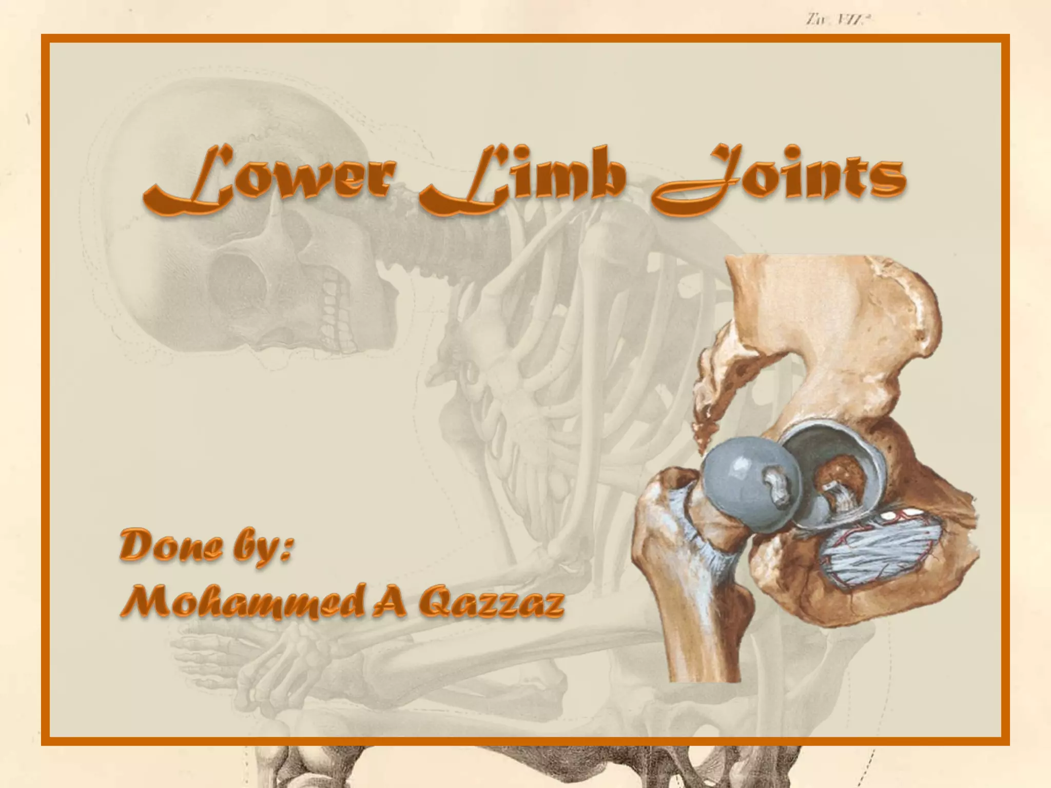
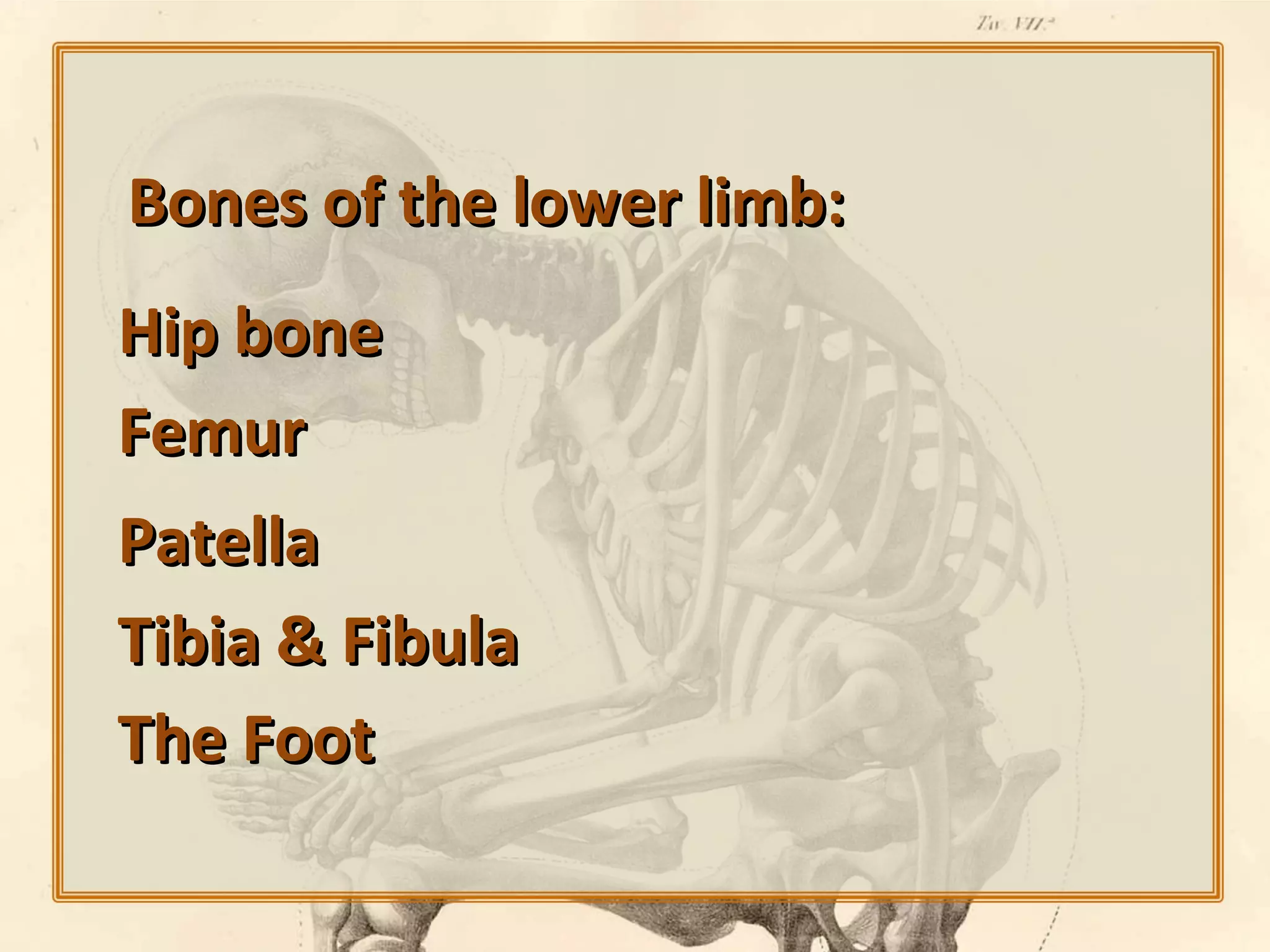
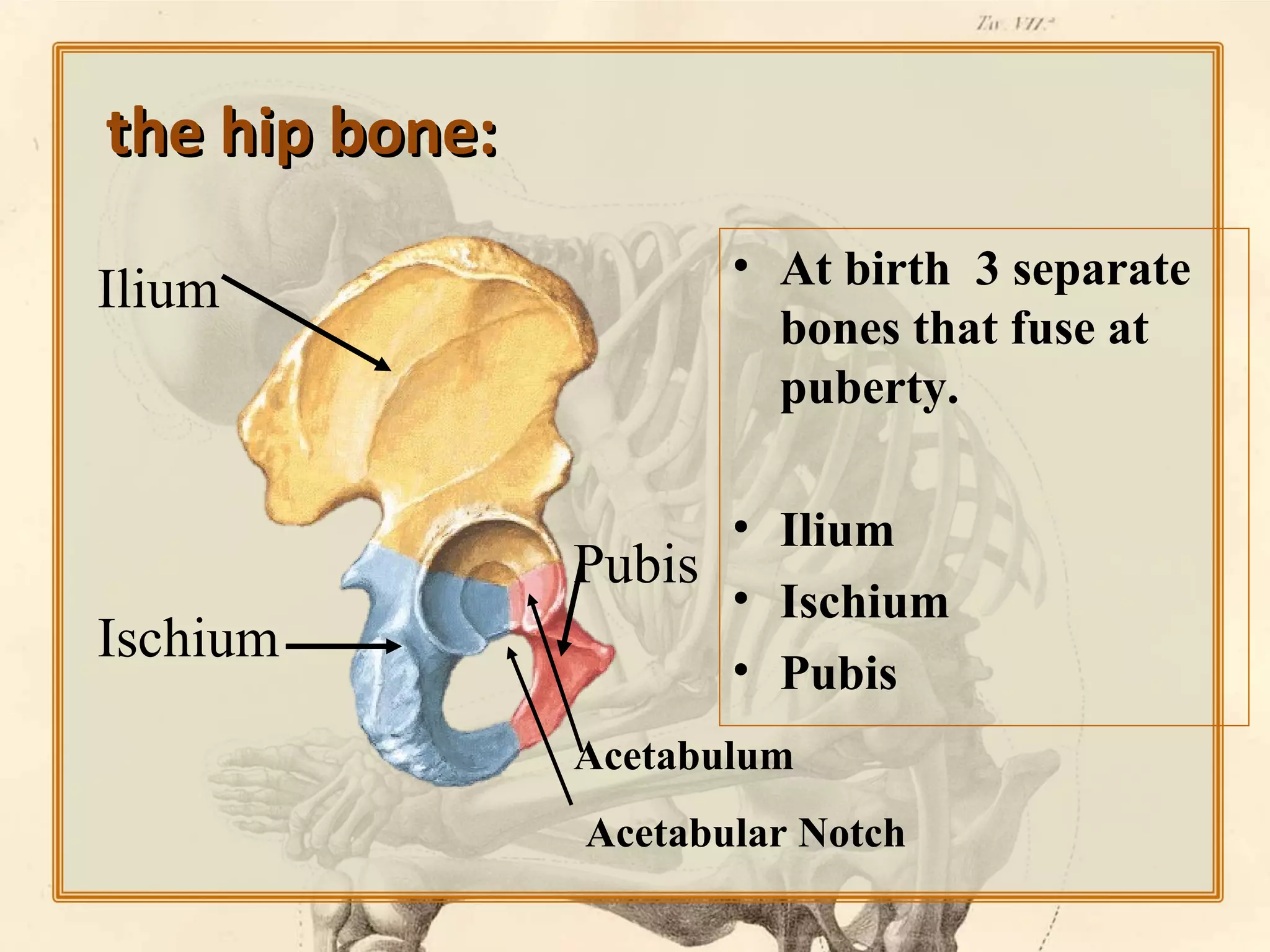
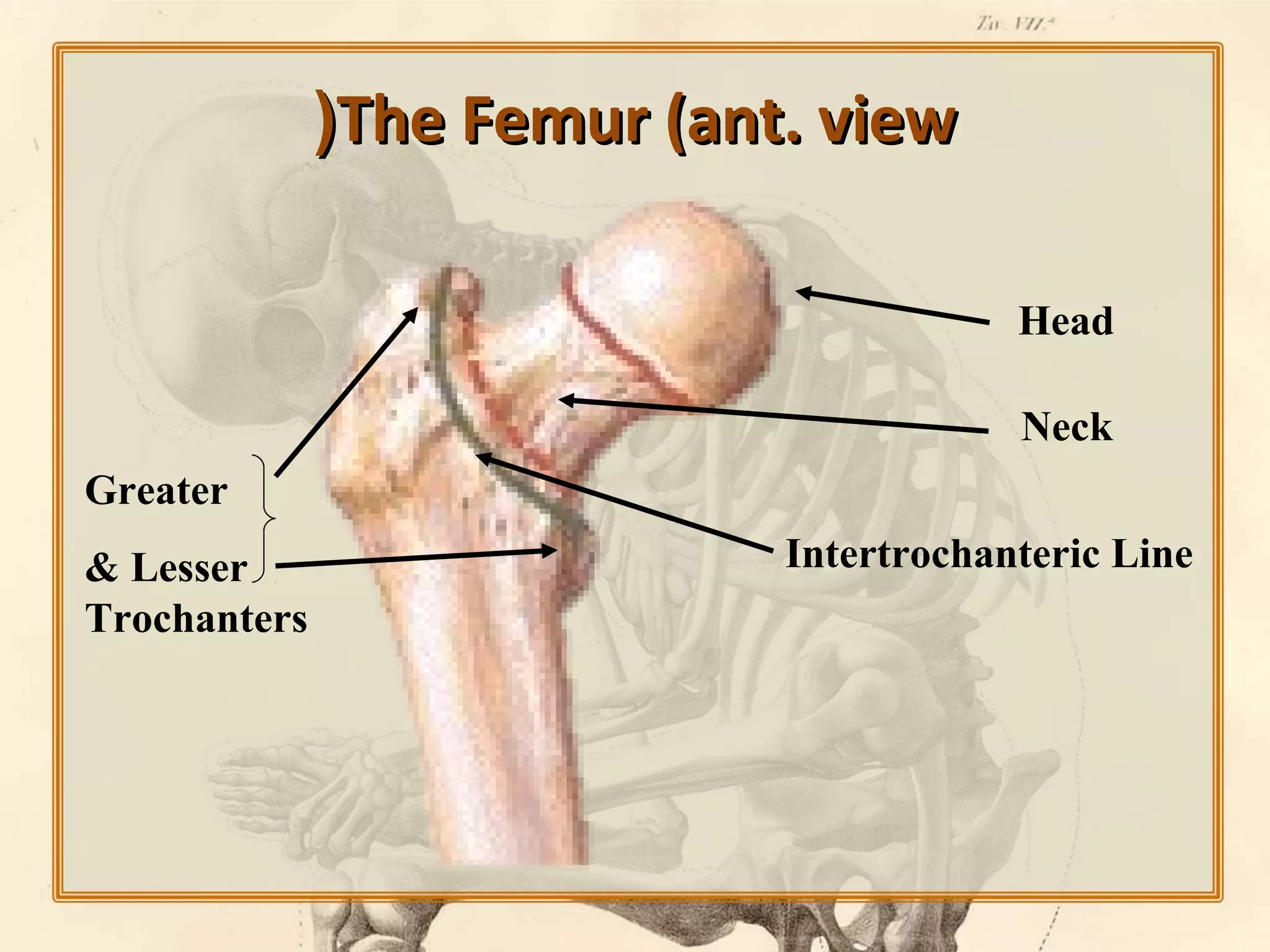
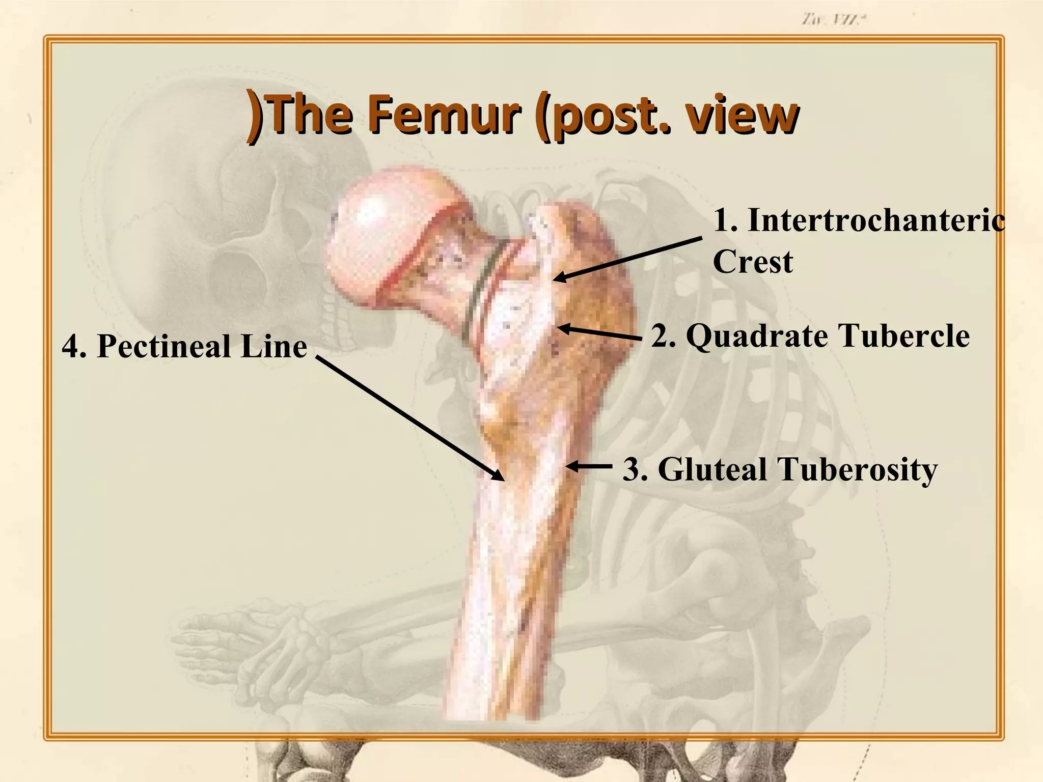
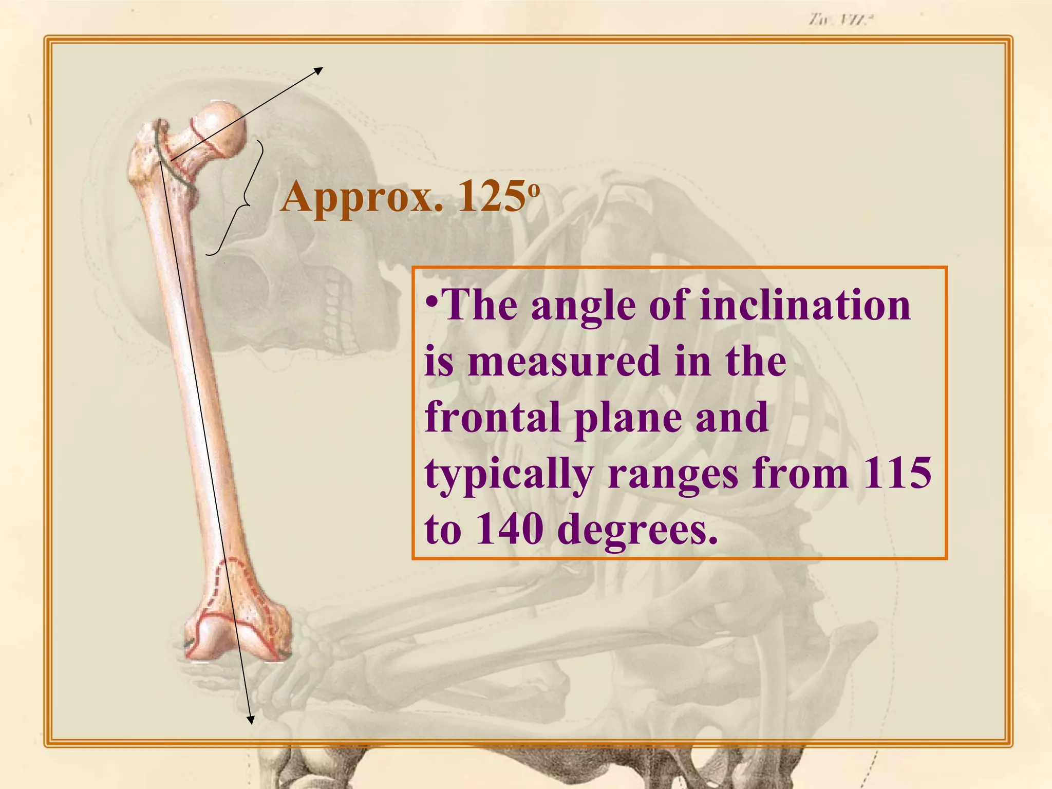
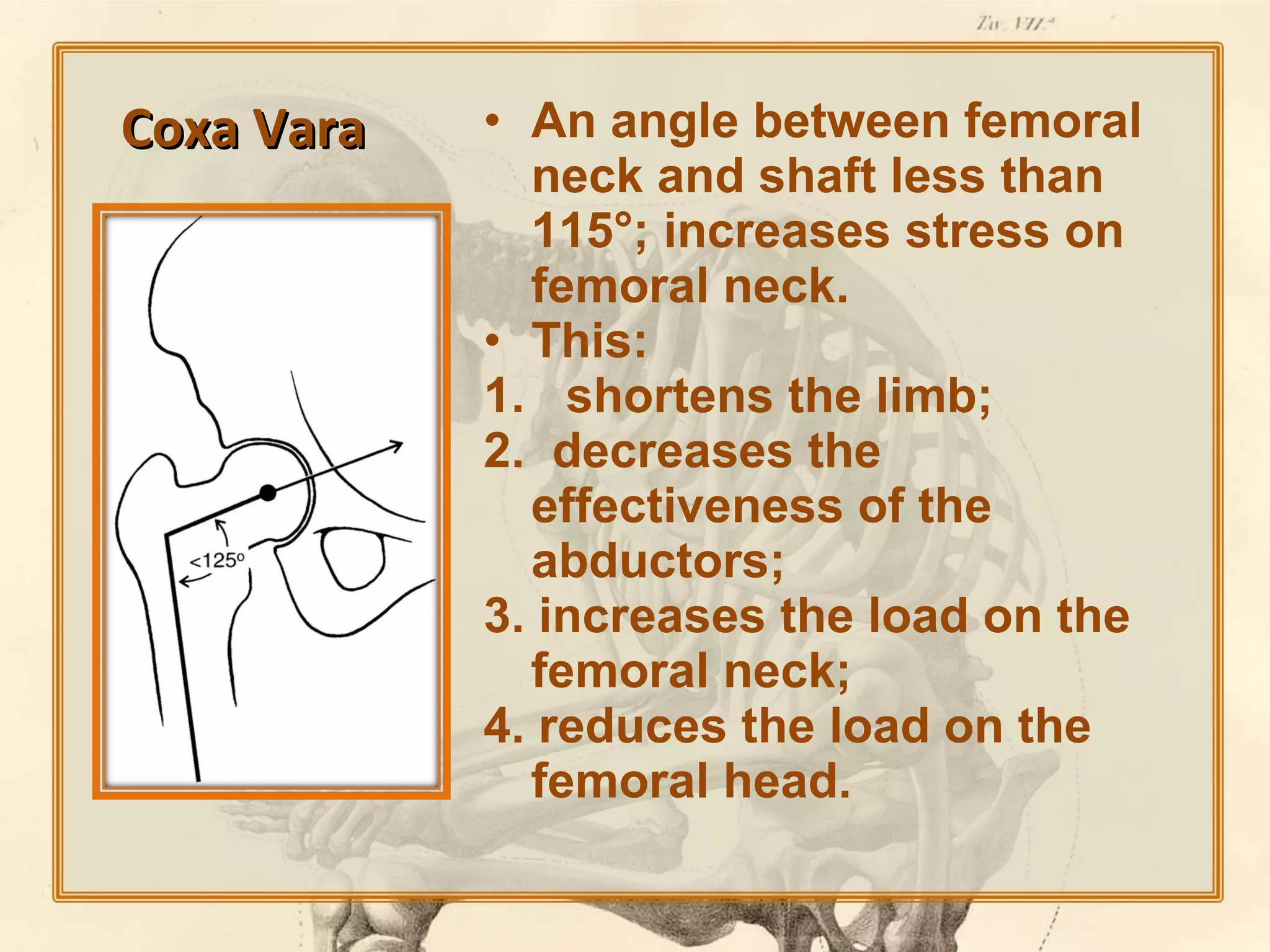
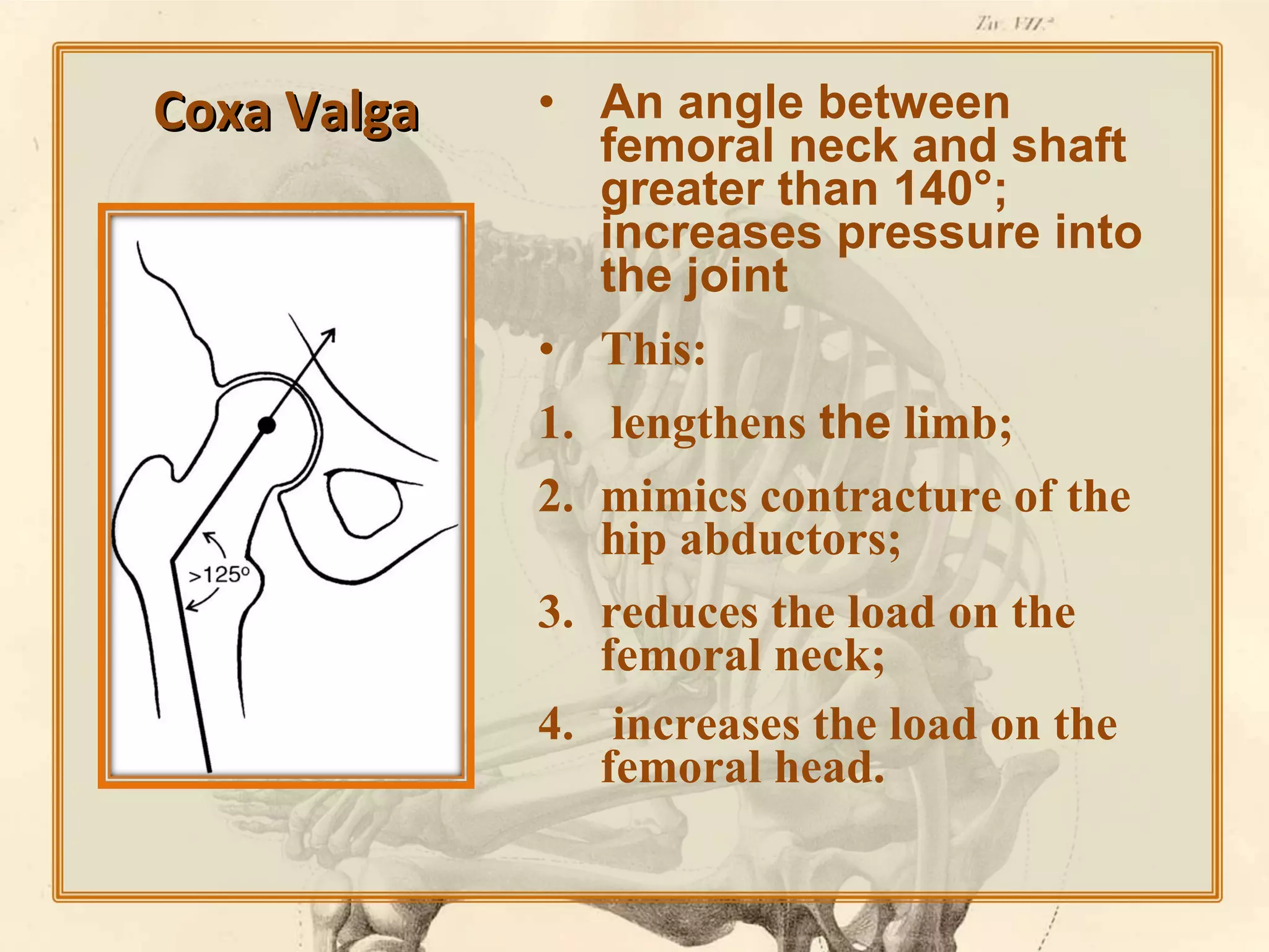
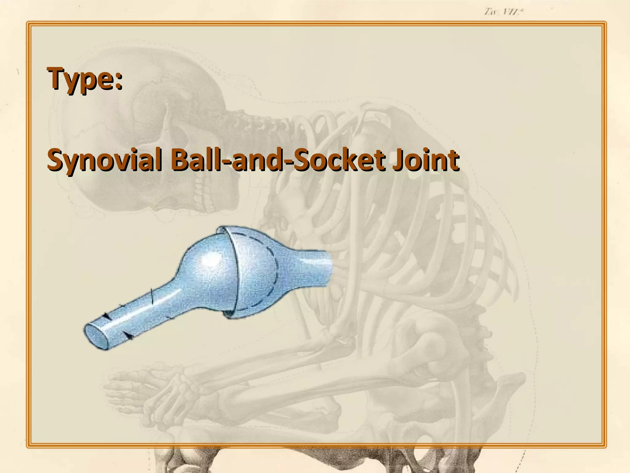
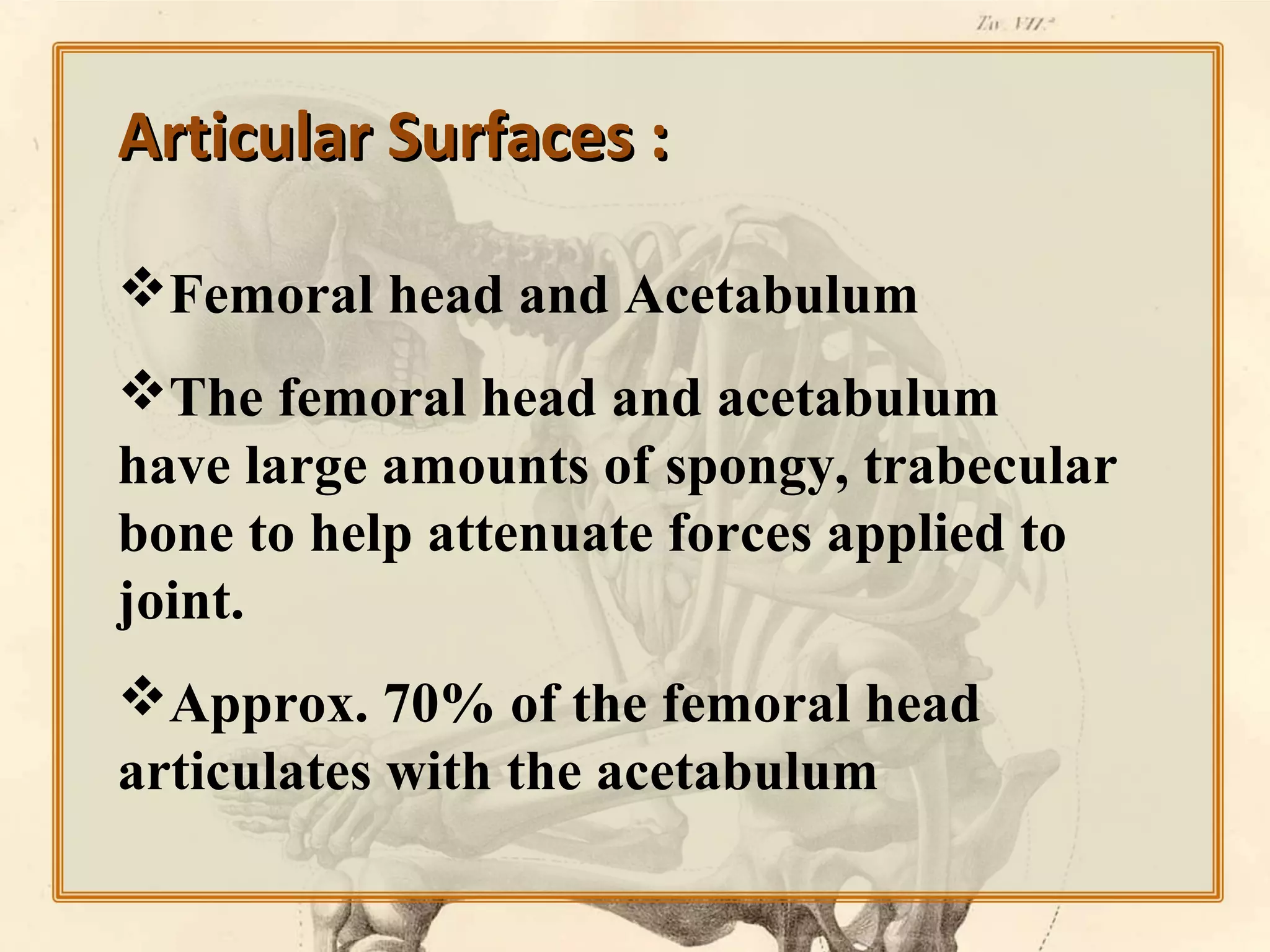
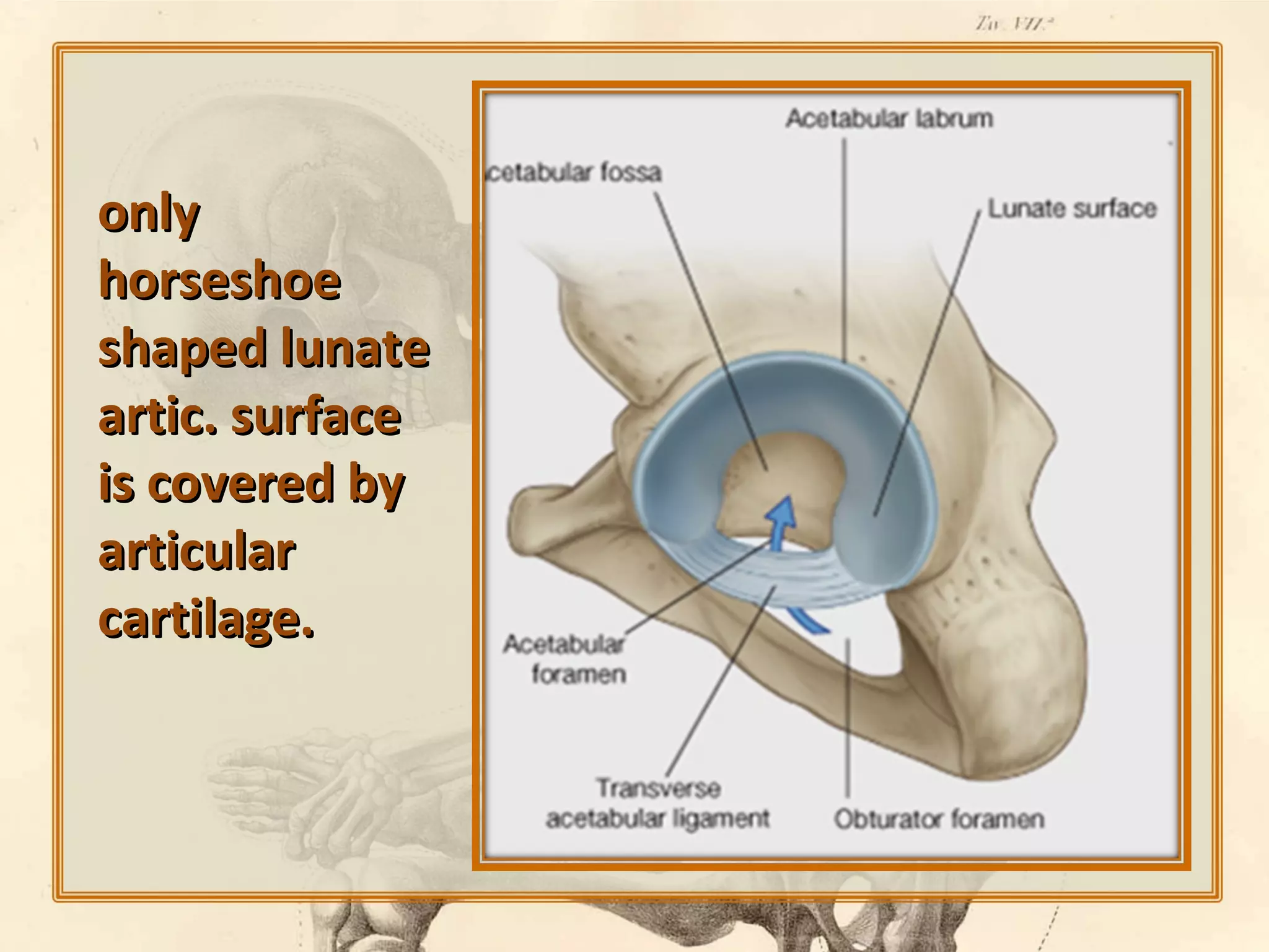
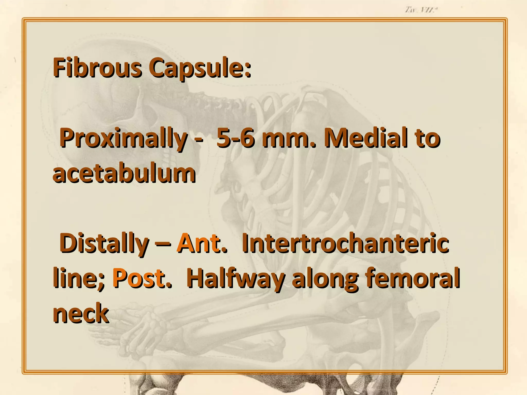
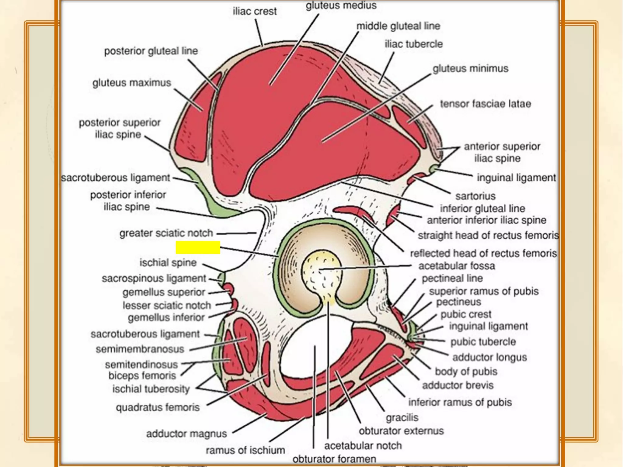
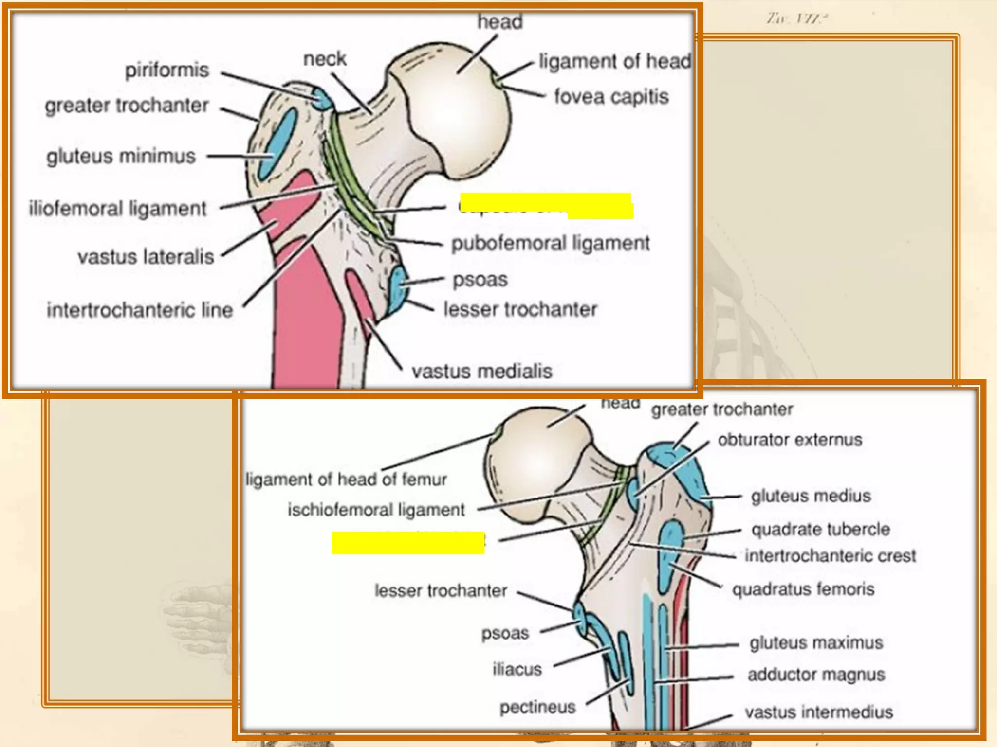
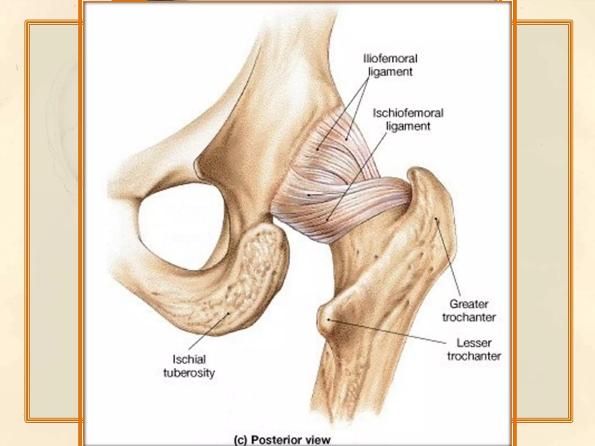
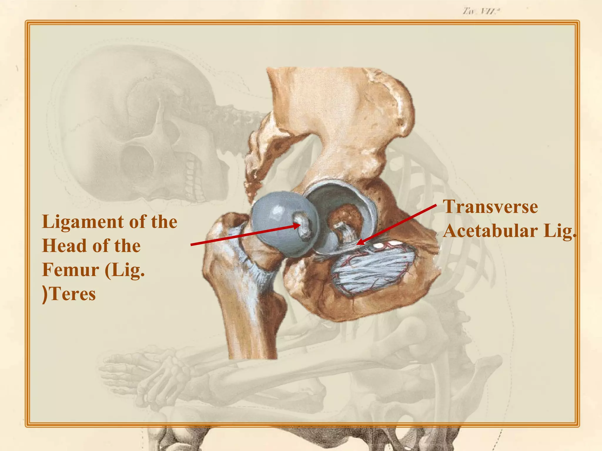
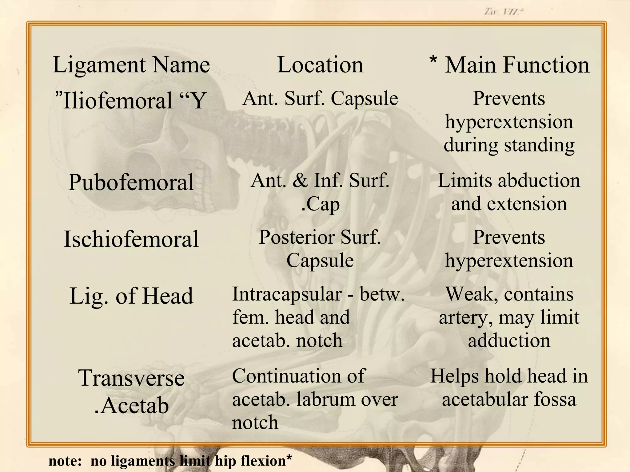
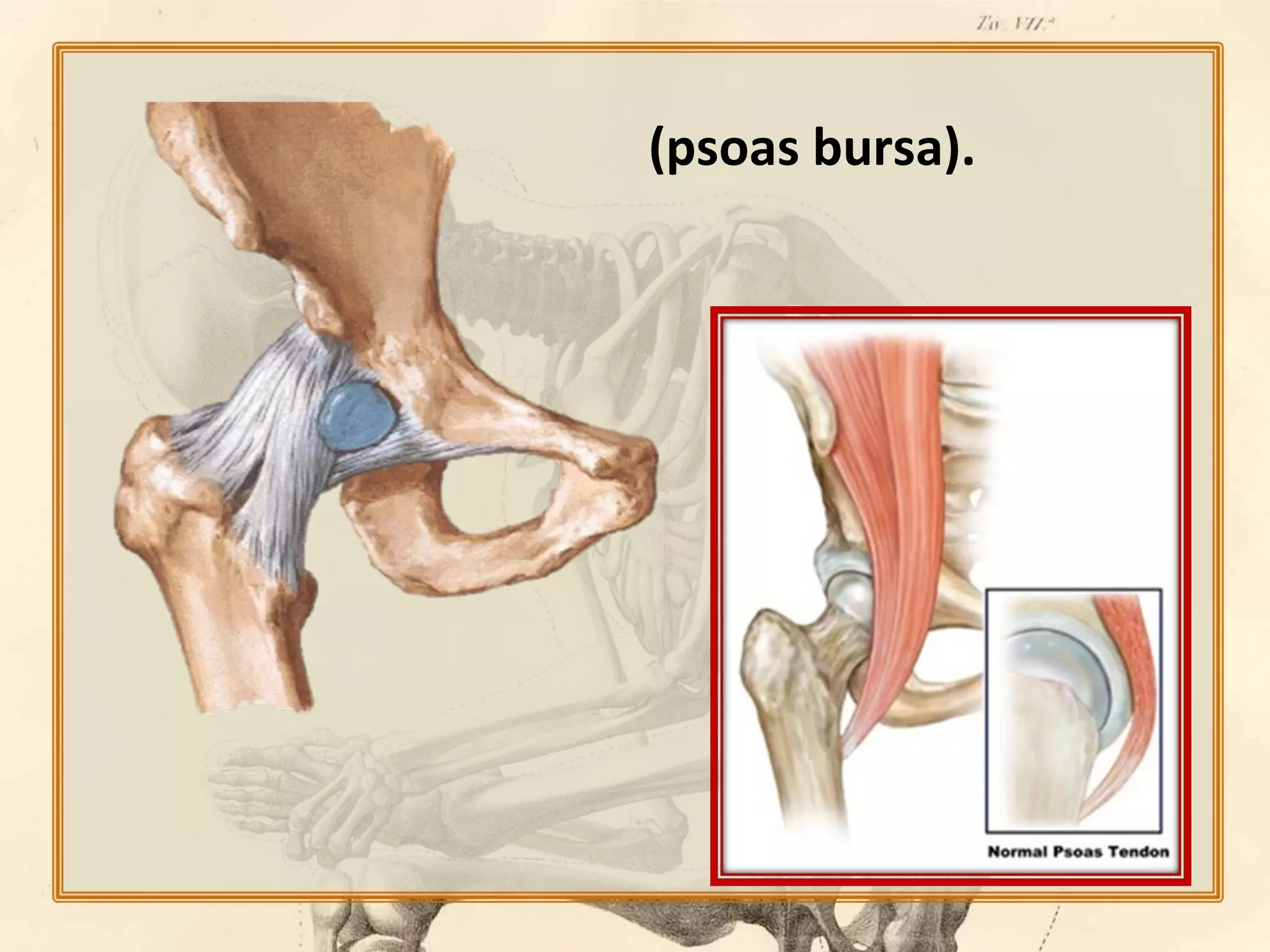
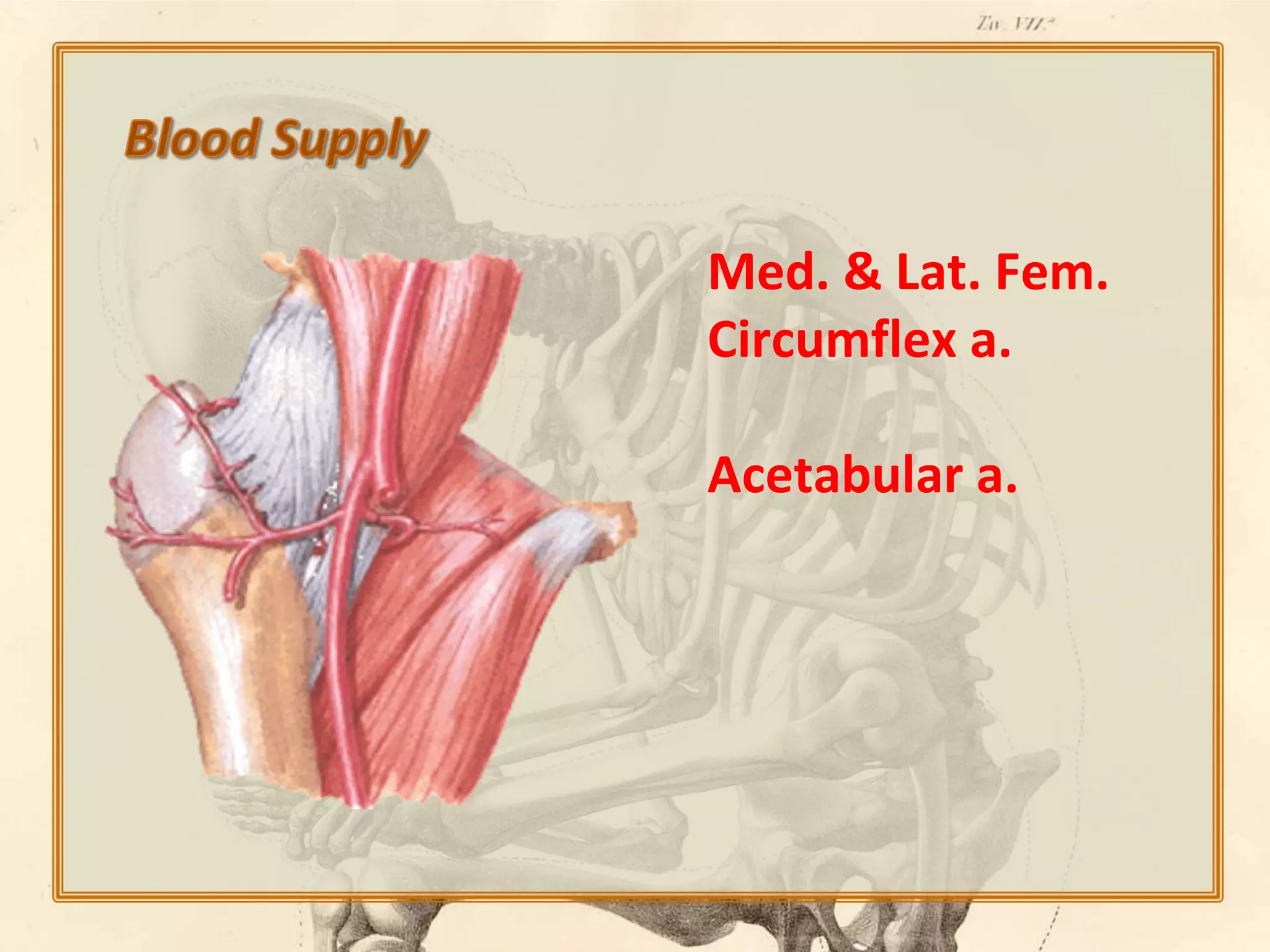
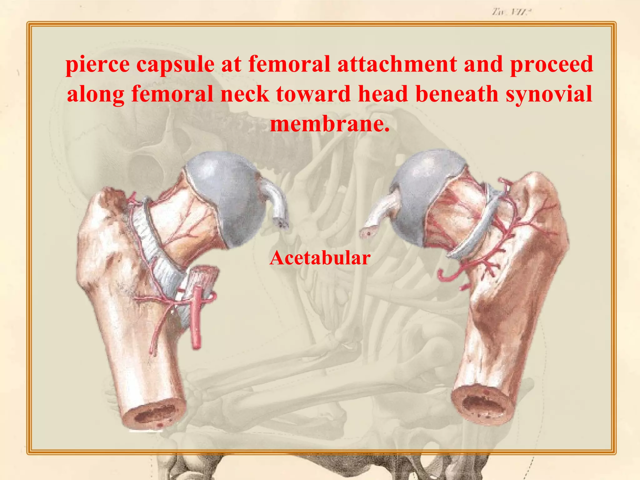
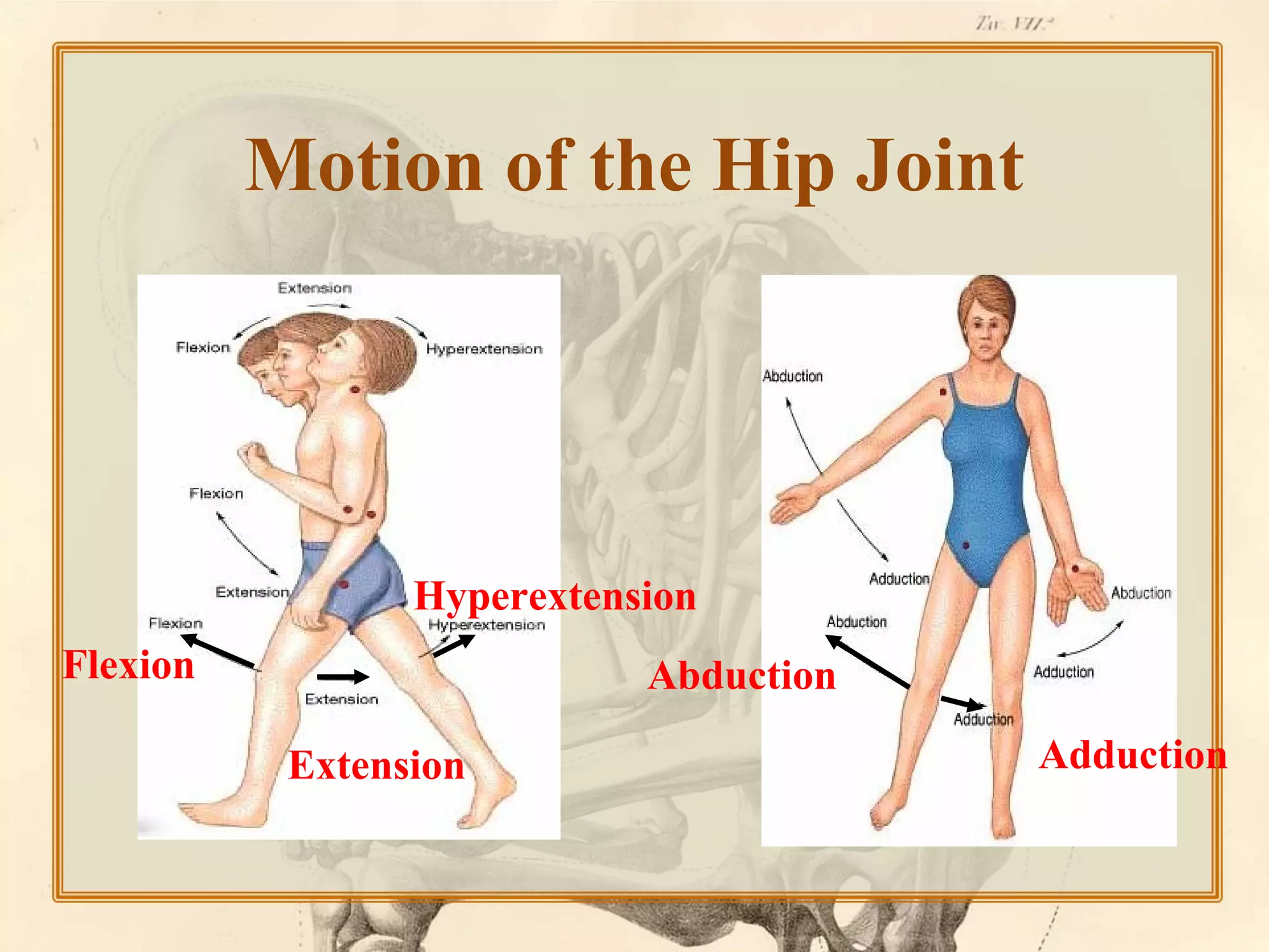
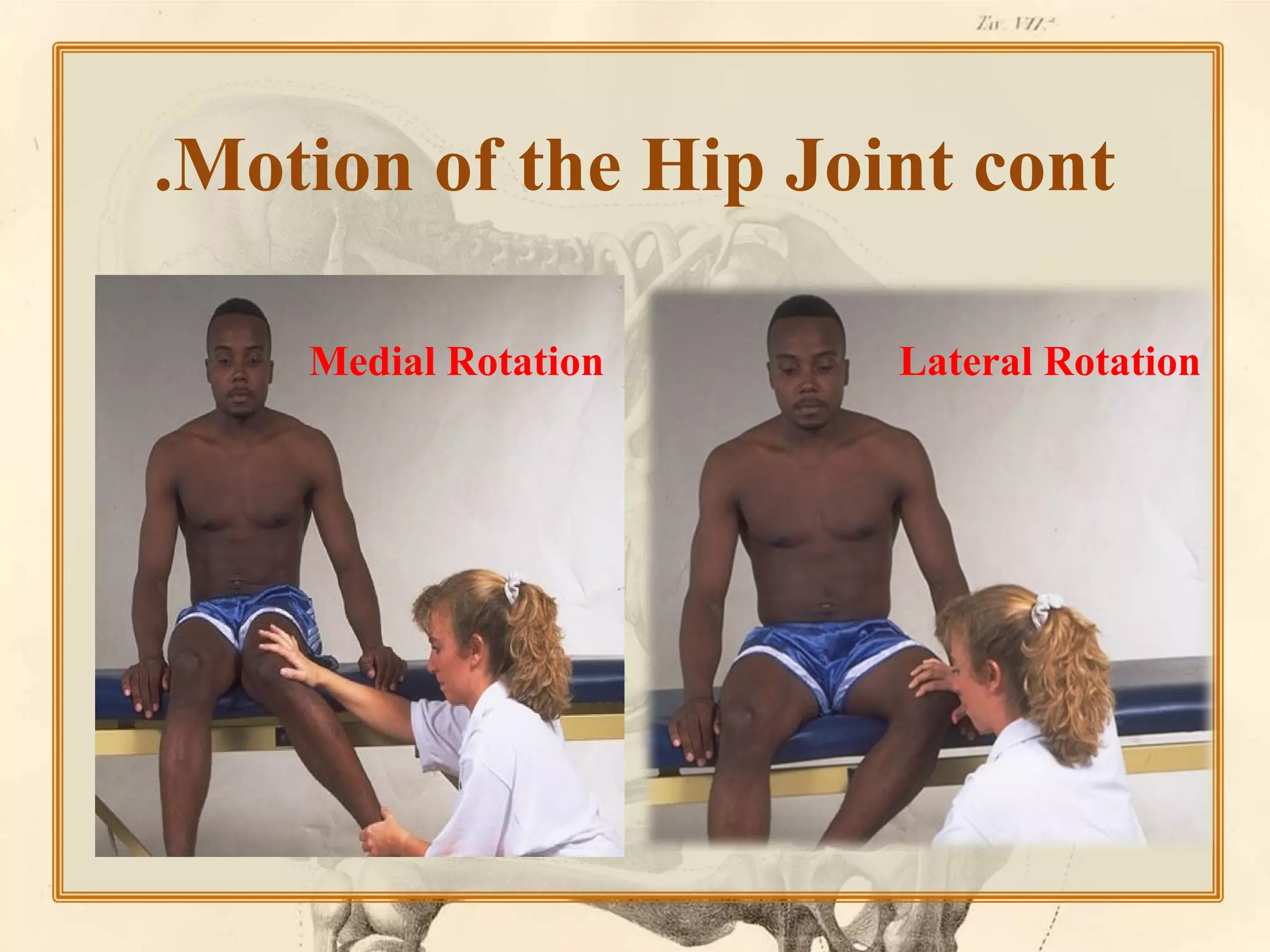
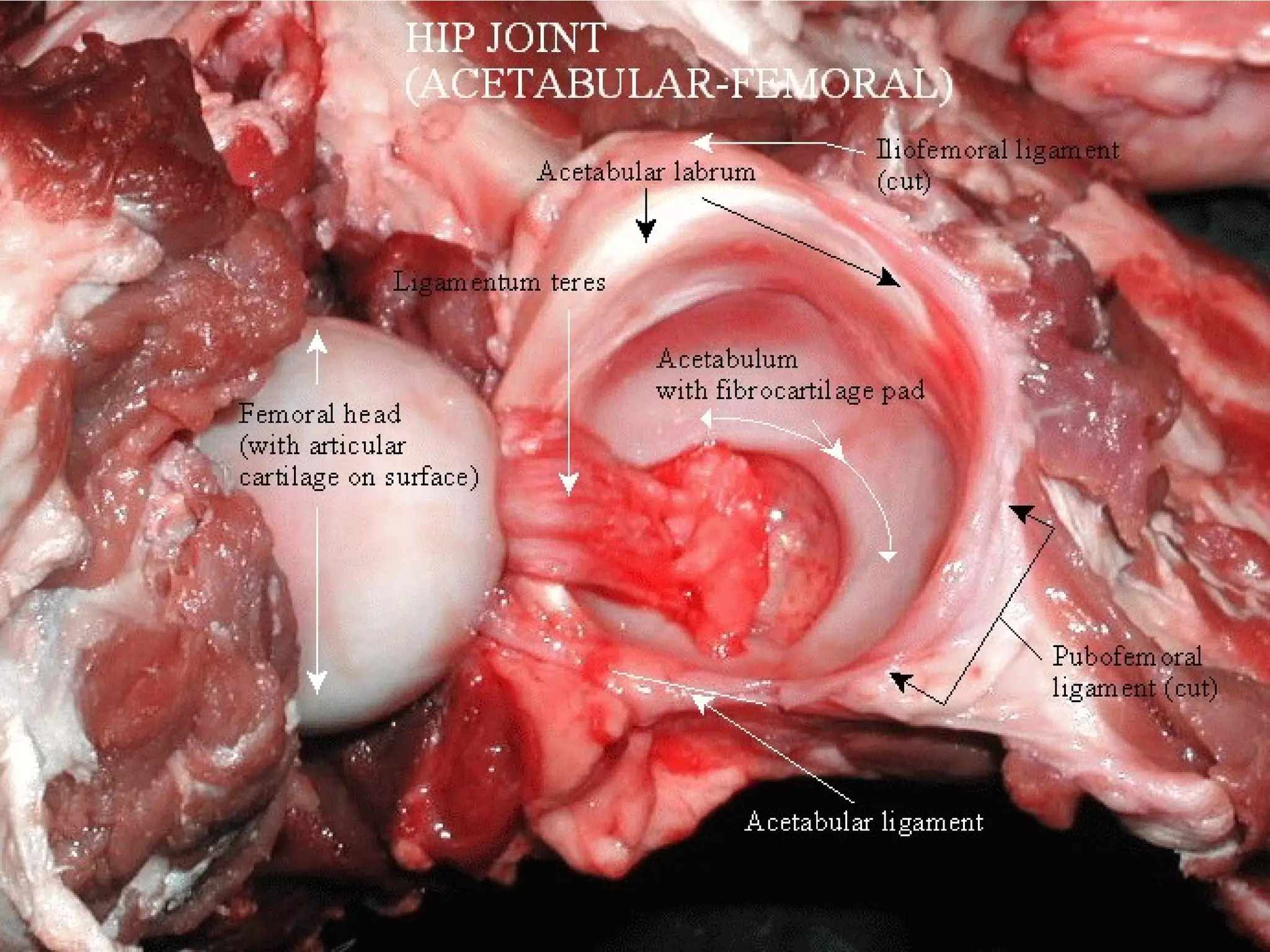
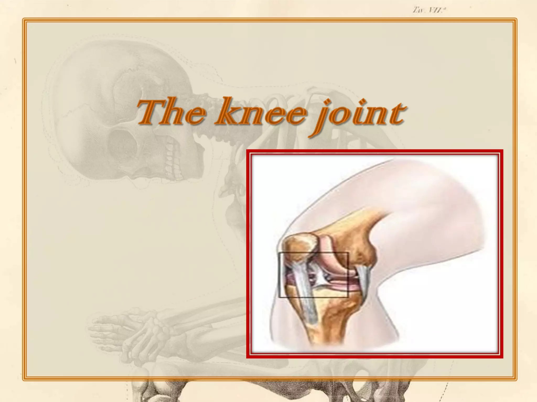
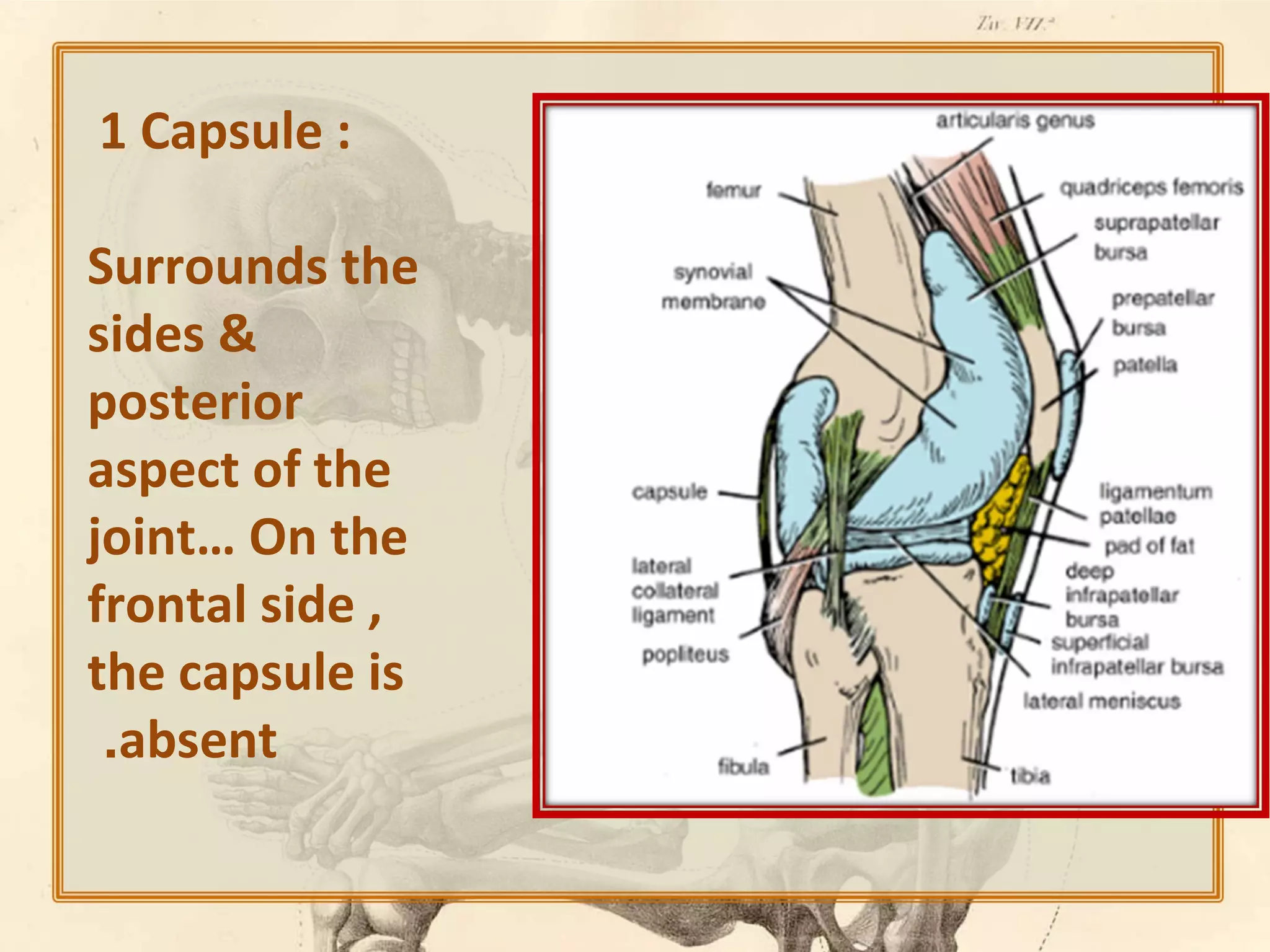
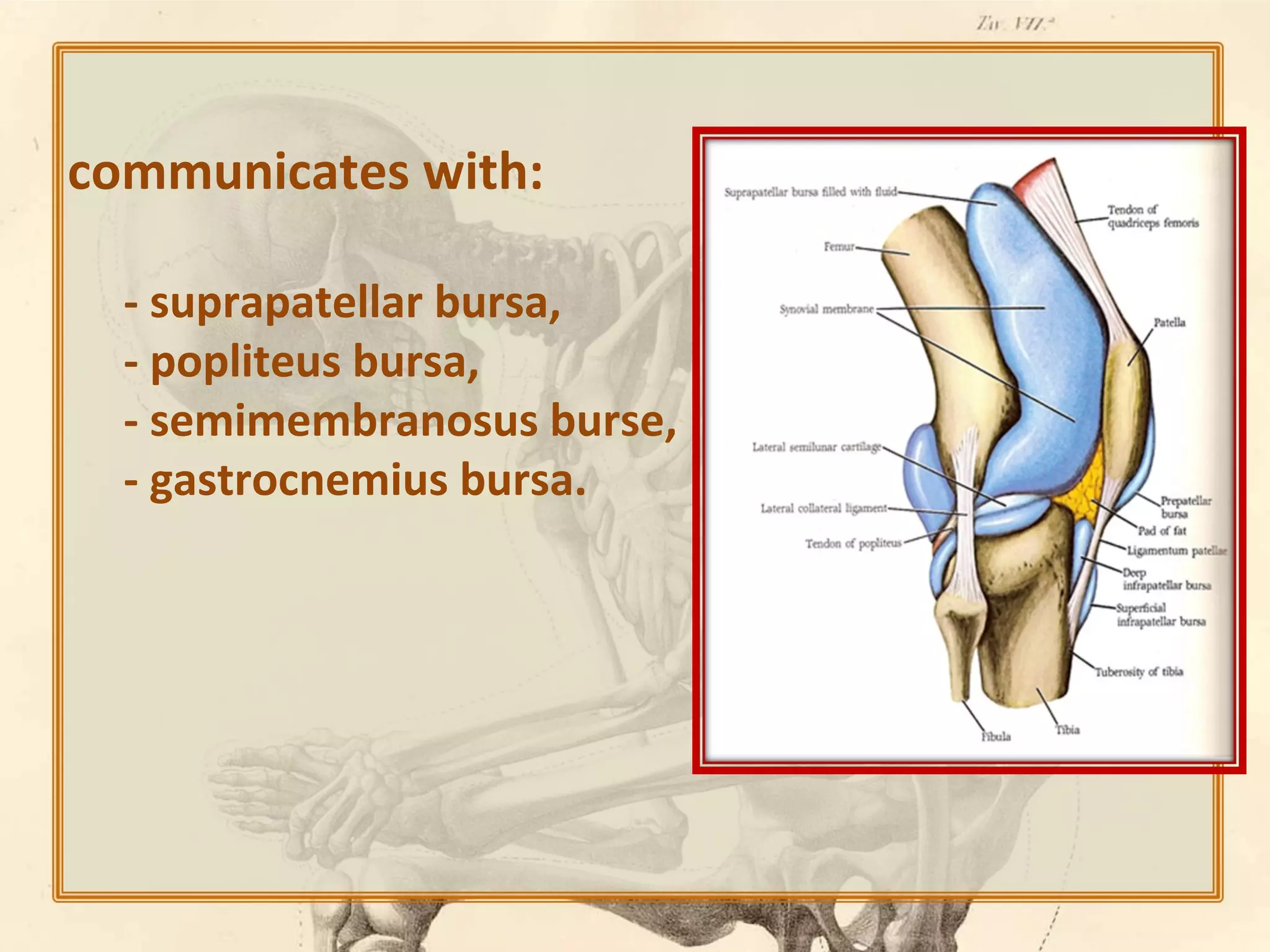
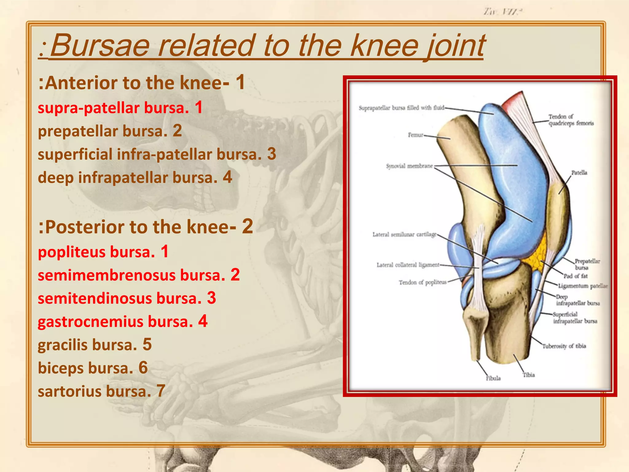
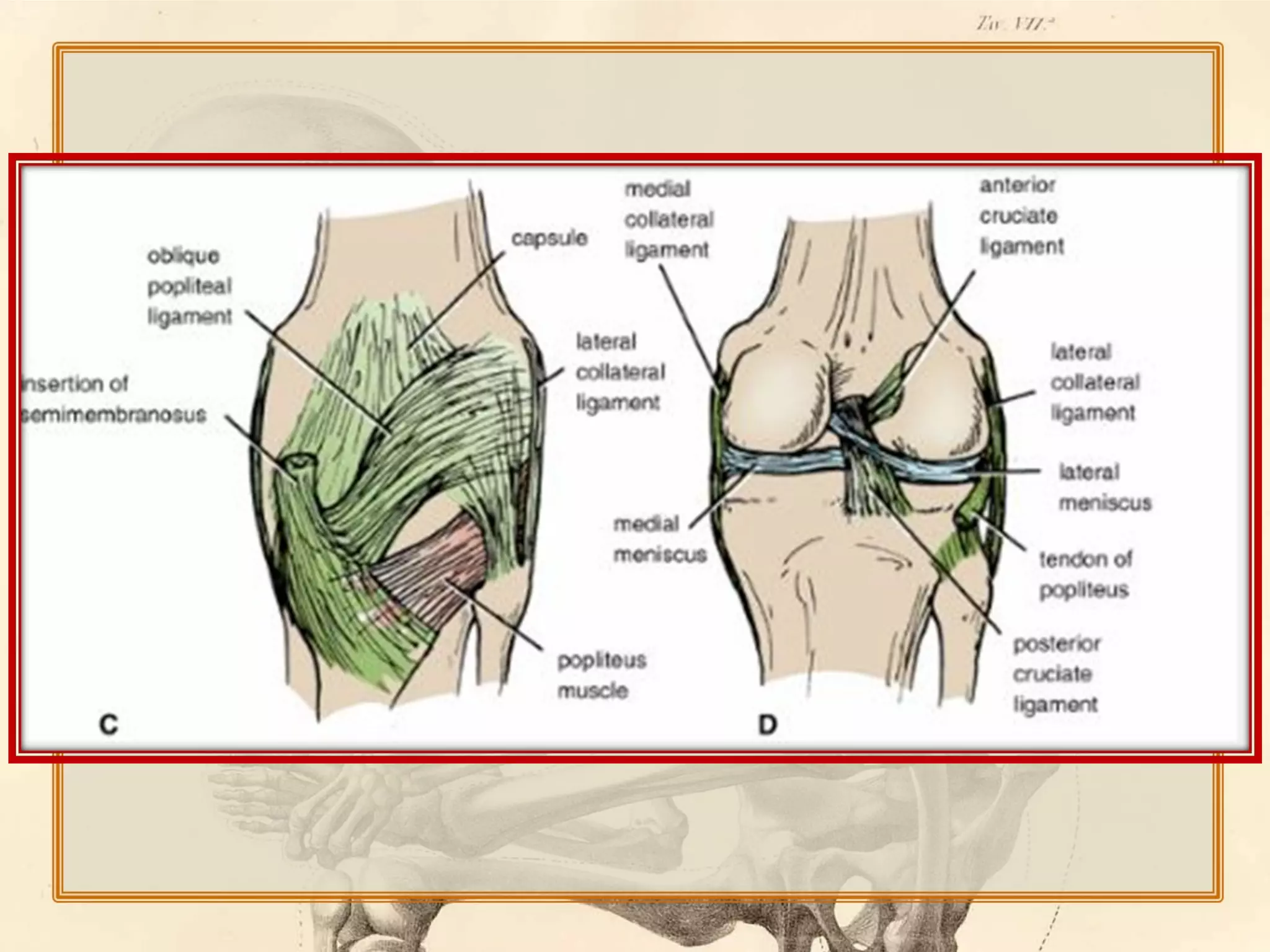
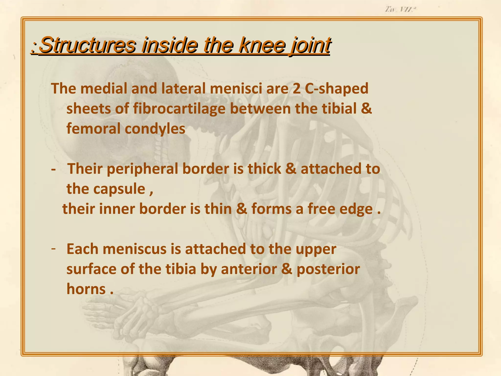
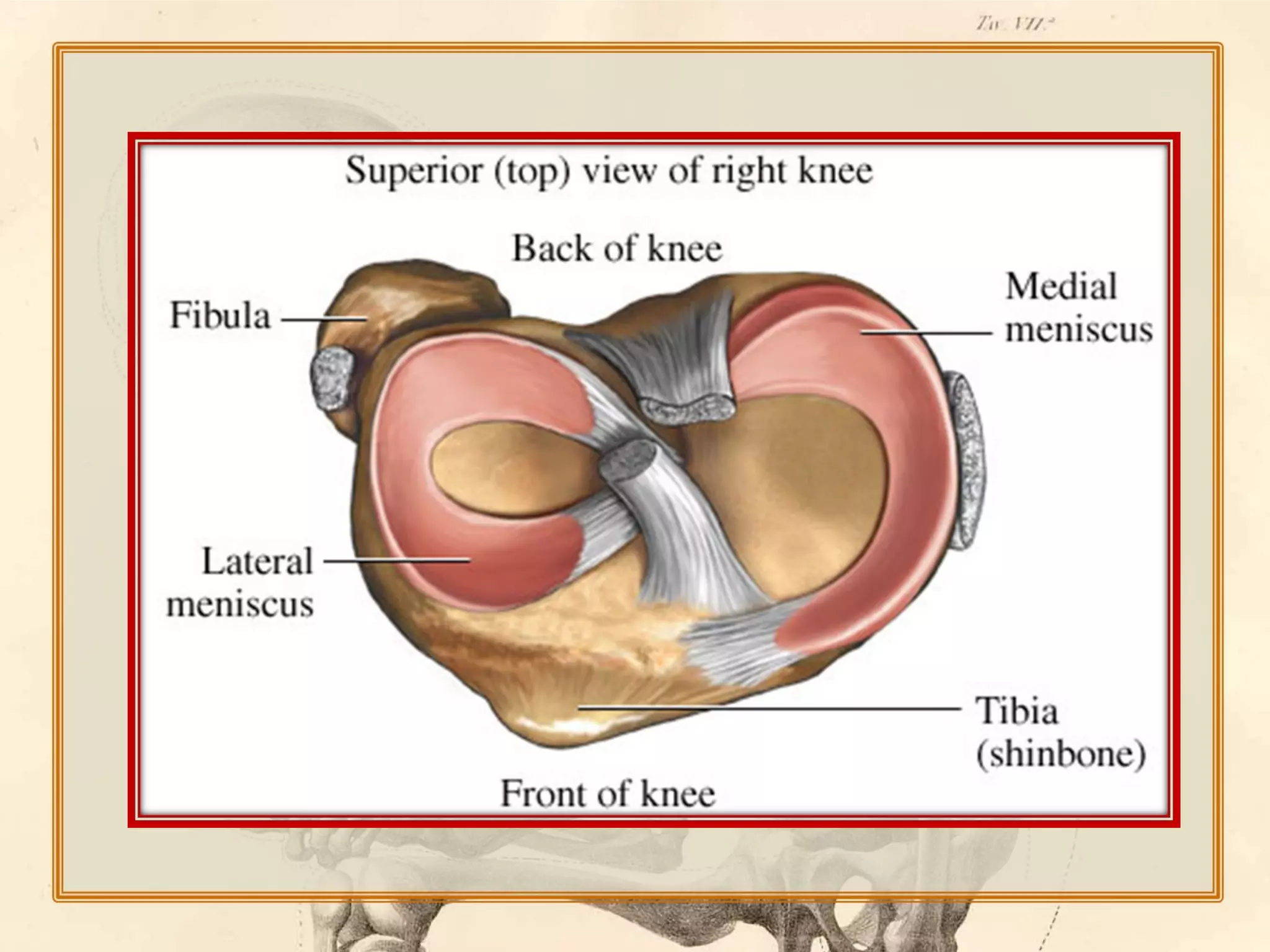
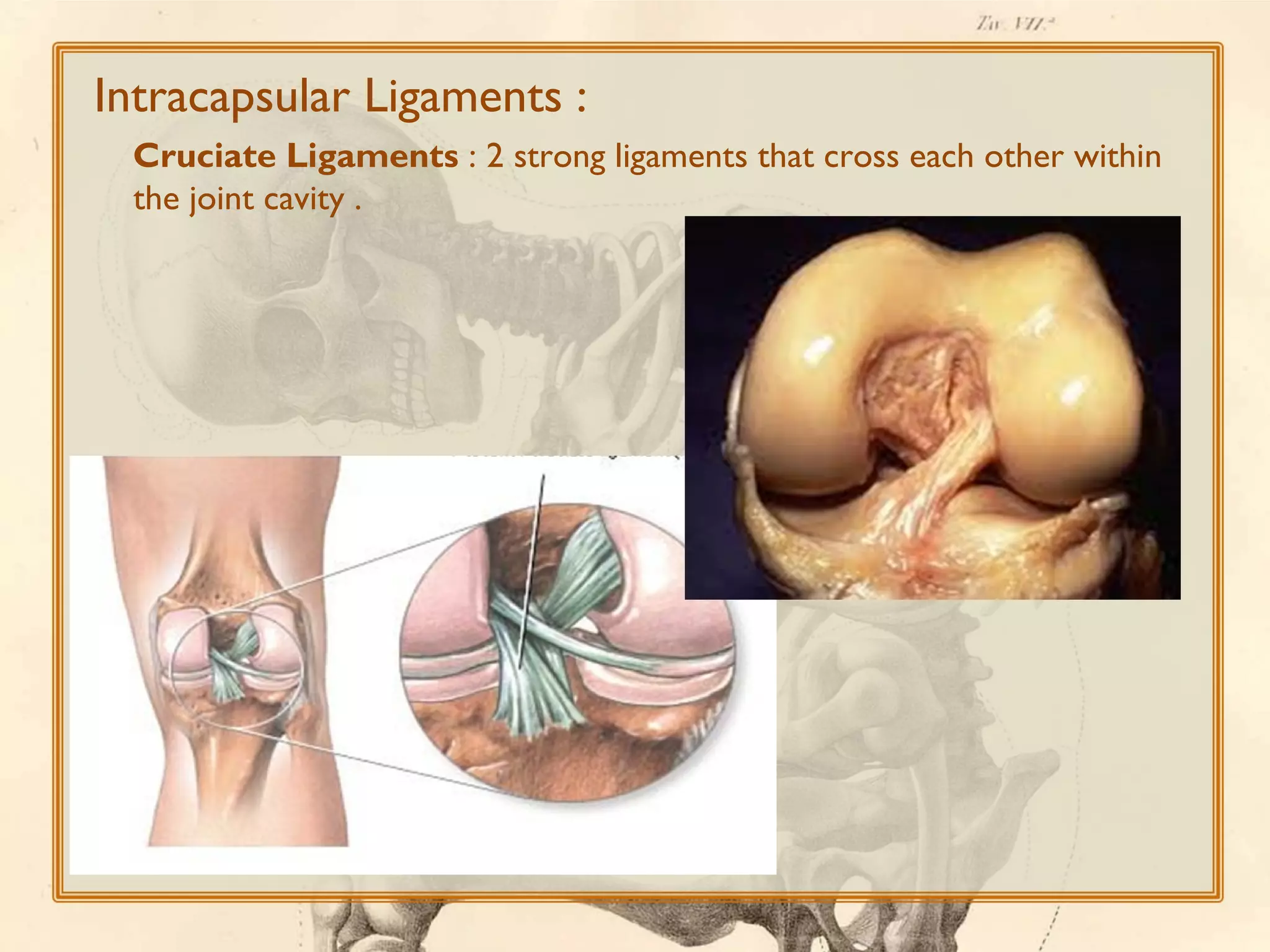
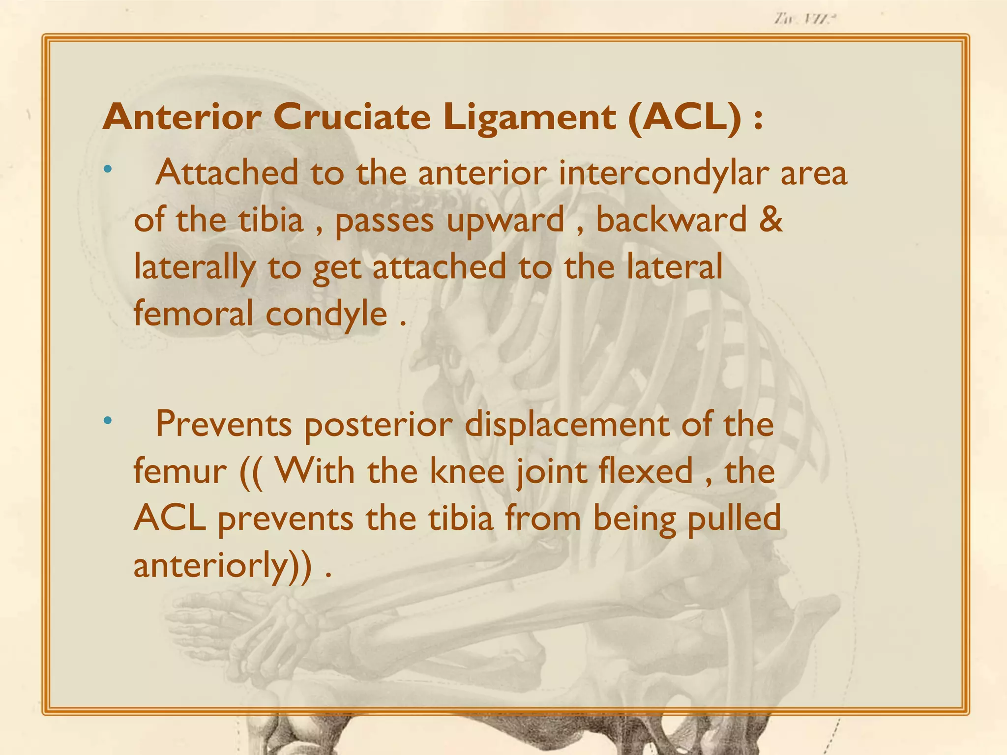
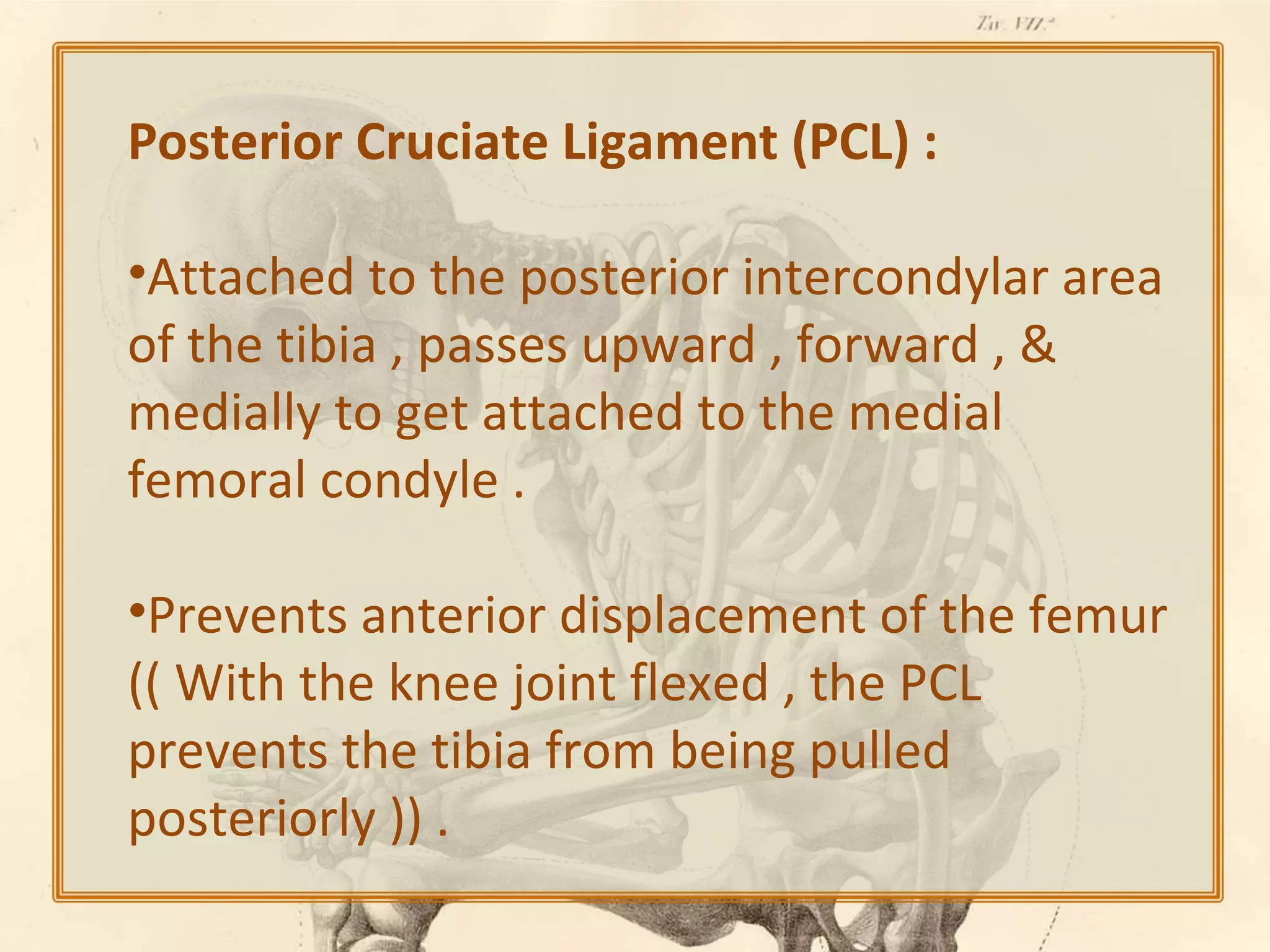
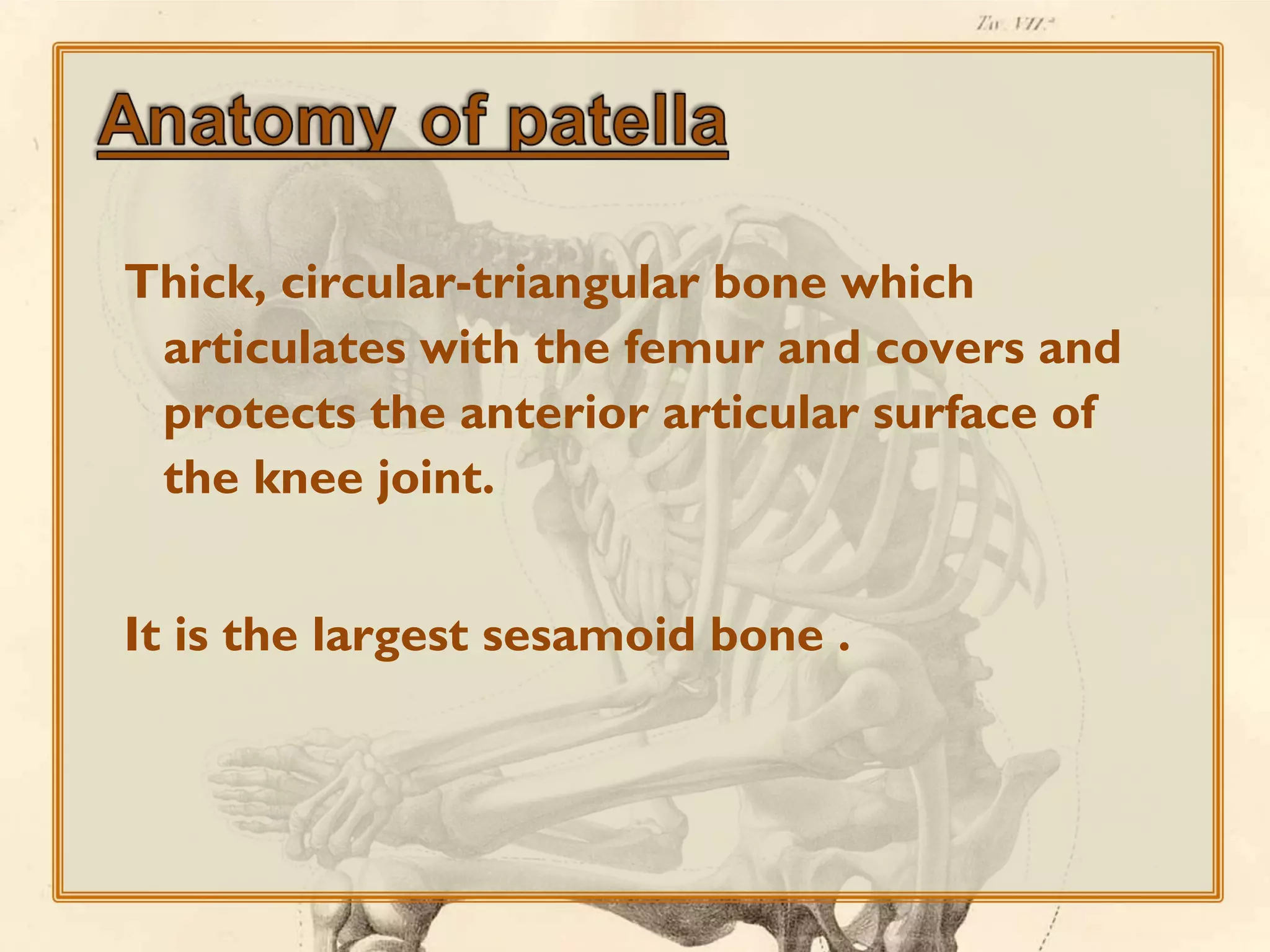
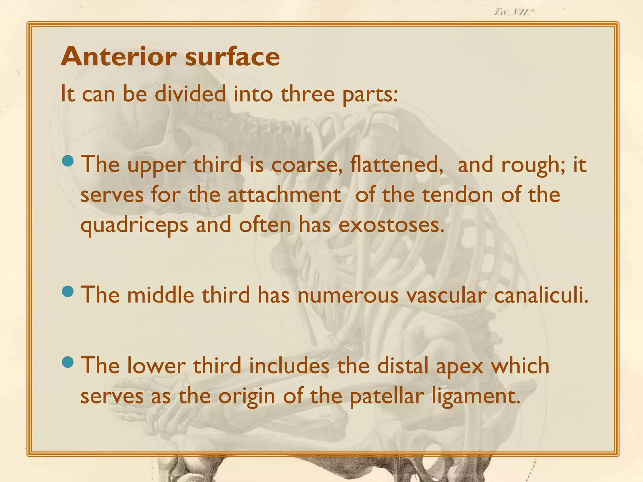
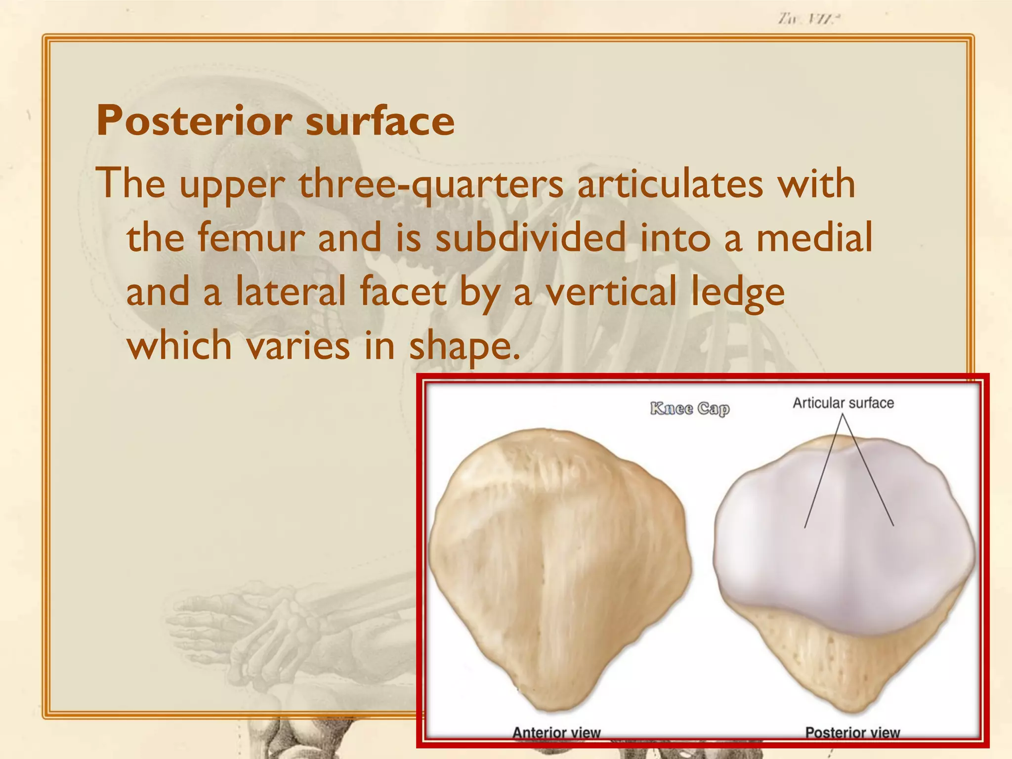
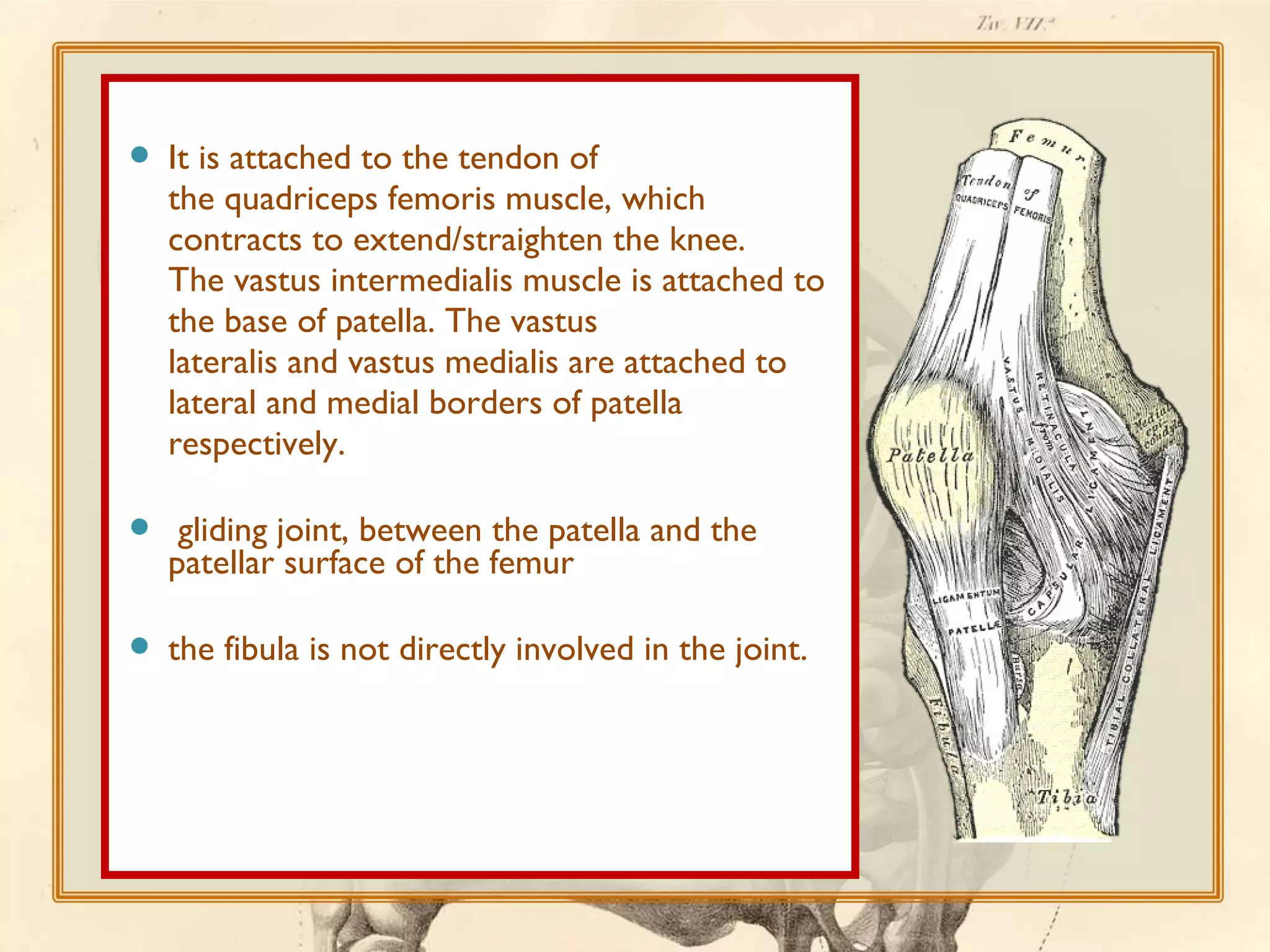
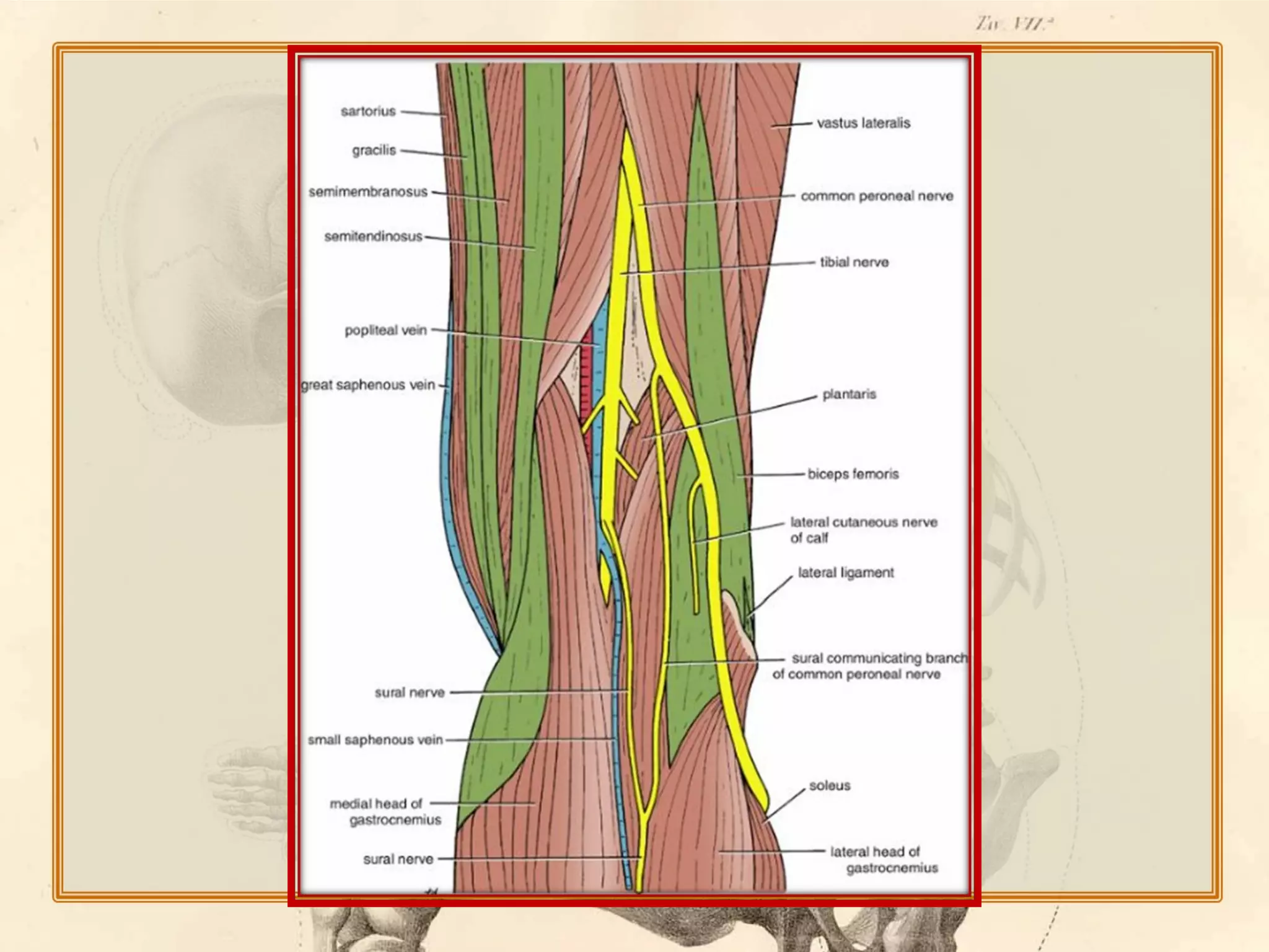
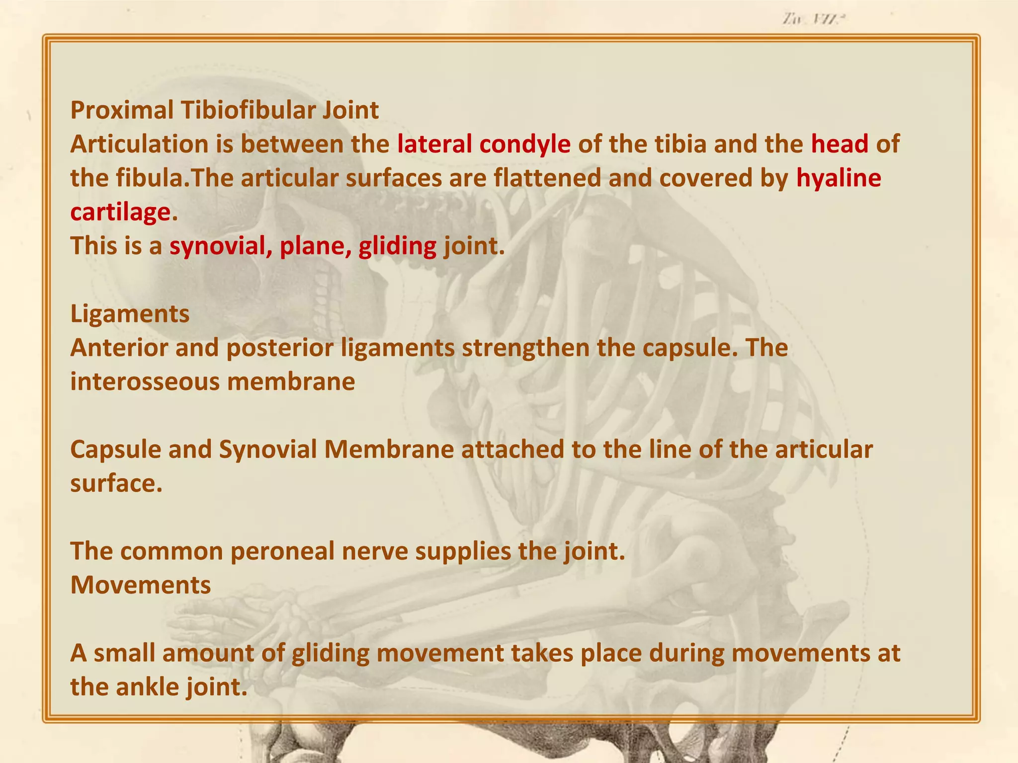
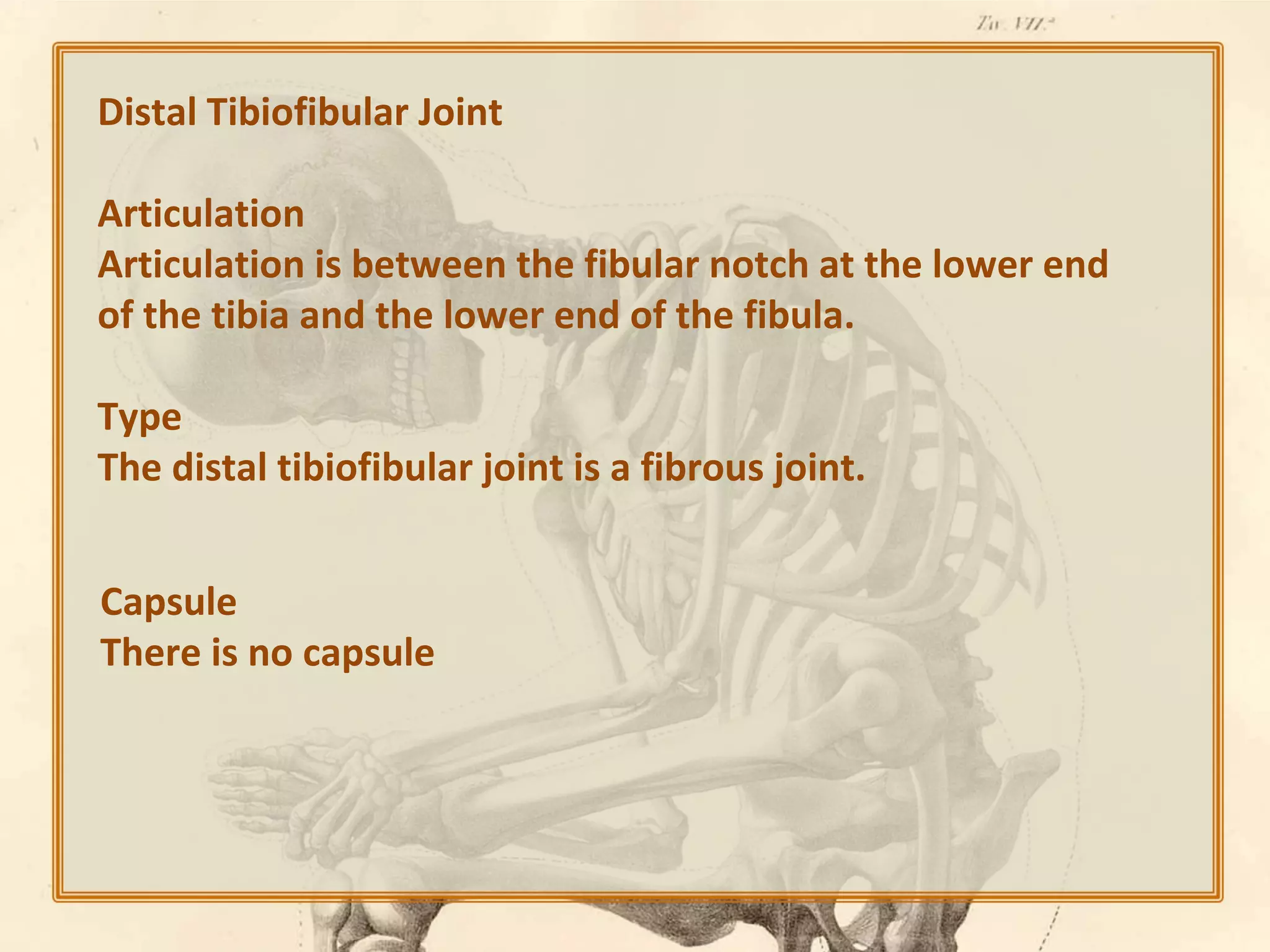
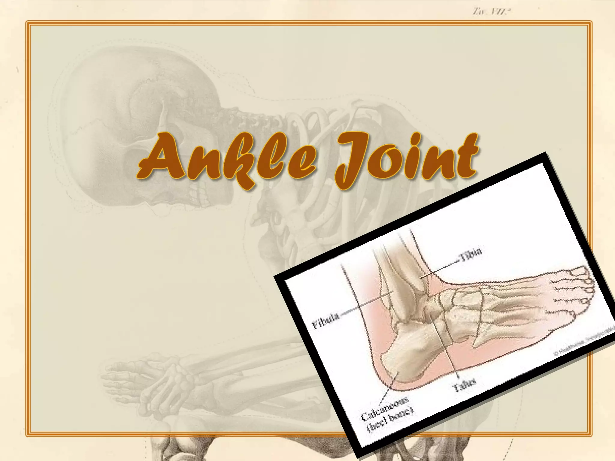
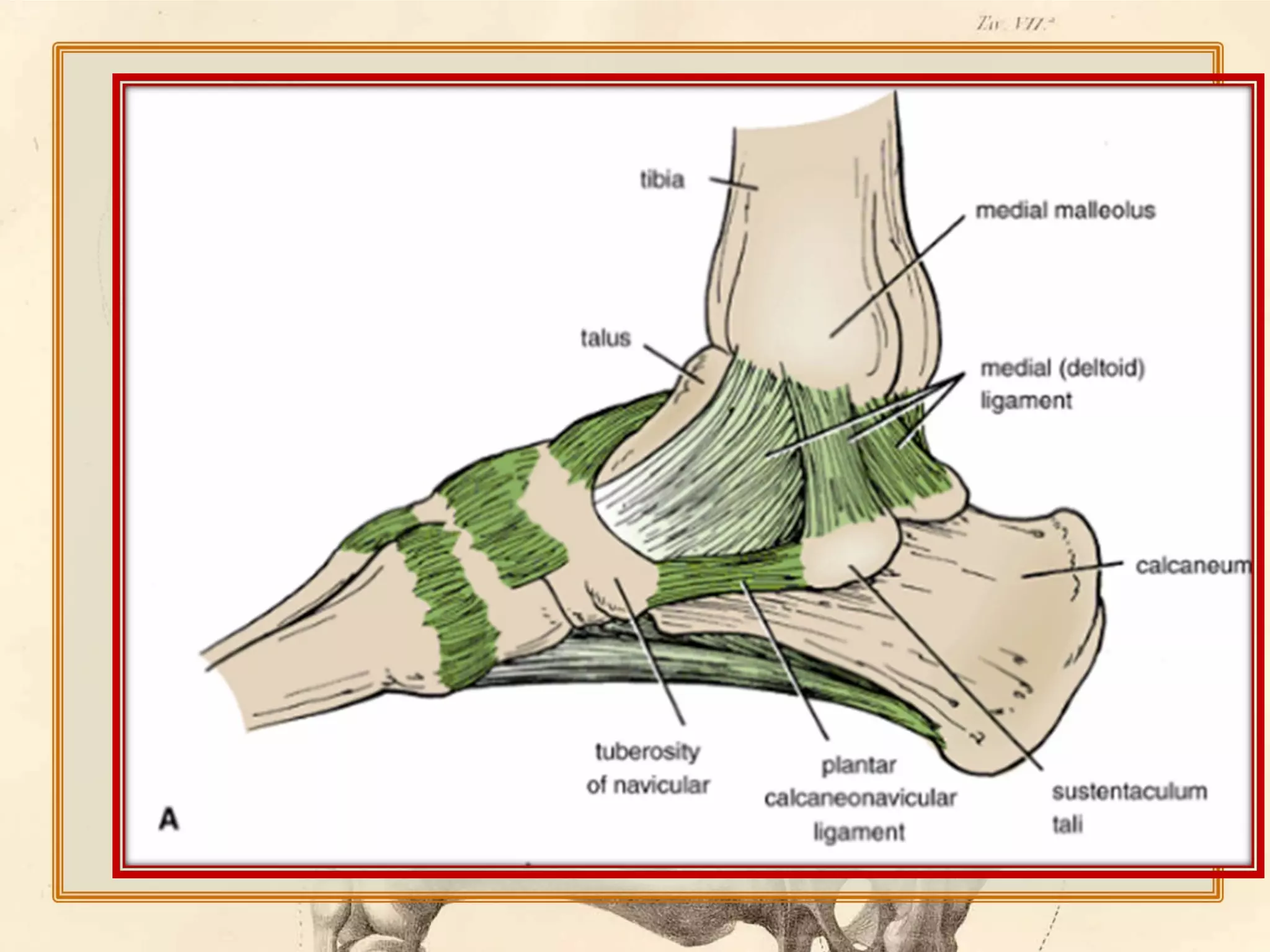
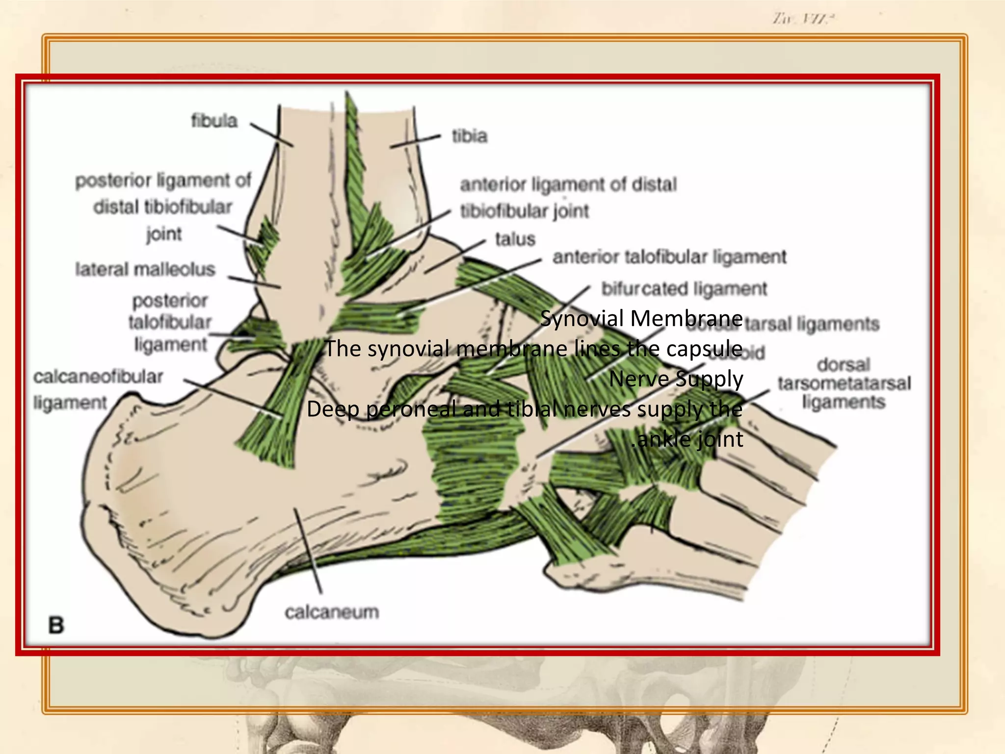
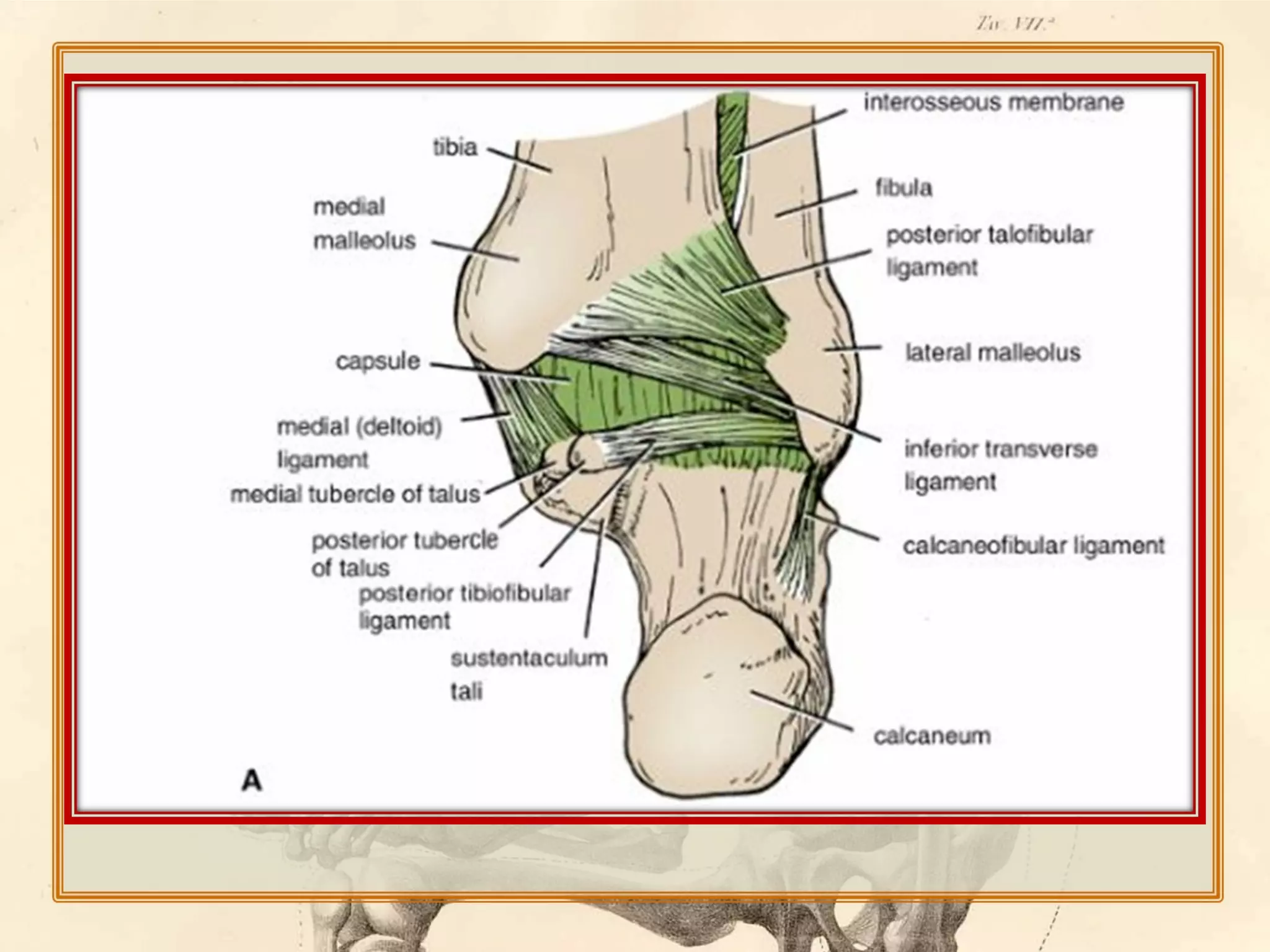
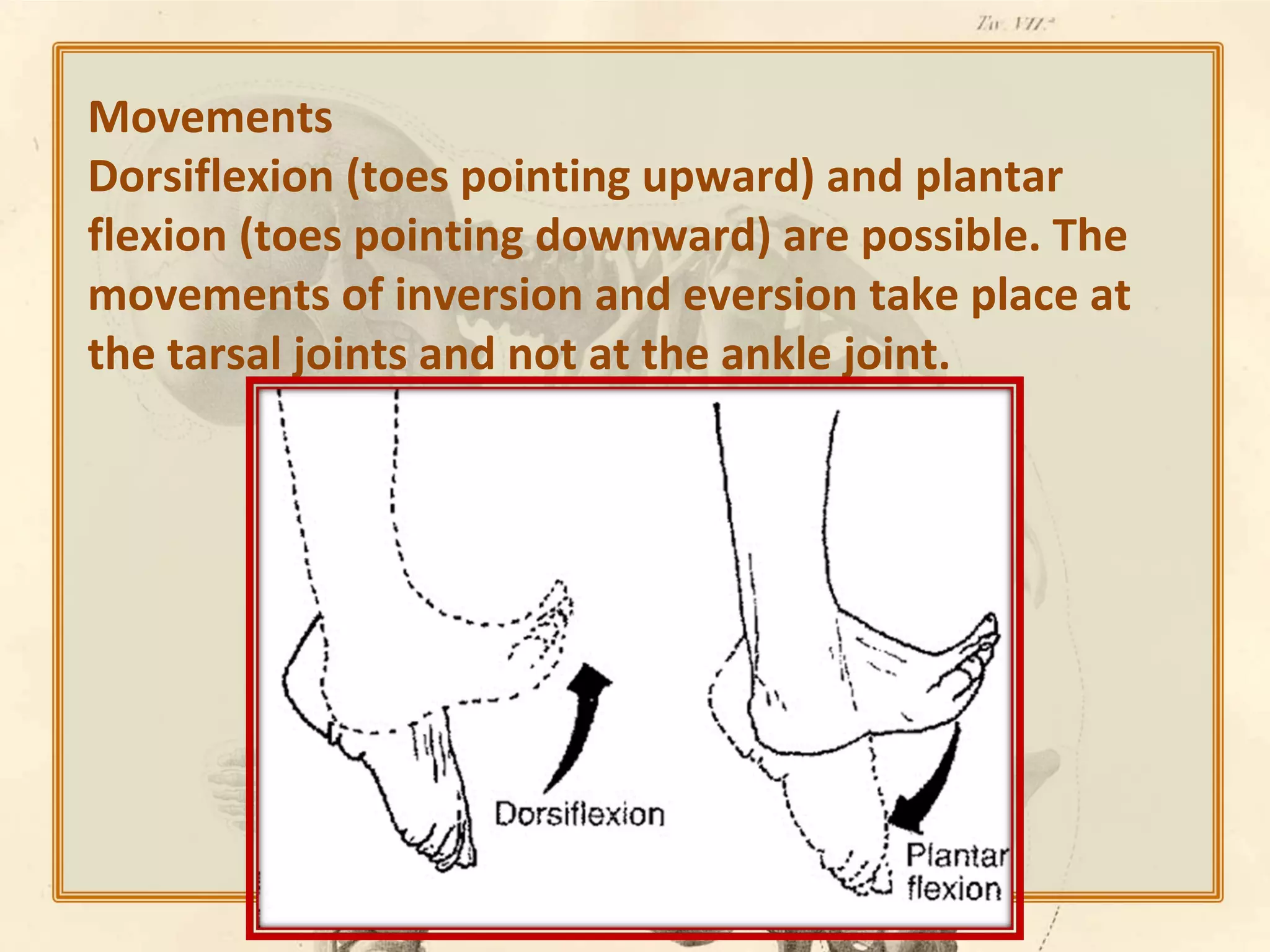
A joint is the location at which
bones connect. They are
constructed to allow movement
(except for skull bones) and
provide mechanical support, and
are classified structurally and
functionally
Structural classification :
fibrous joint
cartilaginous joint
synovial joint
Functional classification:
Synarthrosis
Amphiarthrosis
diarthrosis
Bones of the lower limb:
Hip bone
Femur
Patella
Tibia & Fibula
The Foot
the hip bone:
Ilium

Ischium

• At birth 3 separate
bones that fuse at
puberty.
• Ilium
Pubis
• Ischium
• Pubis
Acetabulum
Acetabular Notch
(The Femur (ant. view
Head
Neck
Greater
& Lesser
Trochanters

Intertrochanteric Line
(The Femur (post. view
1. Intertrochanteric
Crest
4. Pectineal Line

2. Quadrate Tubercle

3. Gluteal Tuberosity
Approx. 125o
•The angle of inclination
is measured in the
frontal plane and
typically ranges from 115
to 140 degrees.
Coxa Vara

• An angle between femoral
neck and shaft less than
115°; increases stress on
femoral neck.
• This:
1. shortens the limb;
2. decreases the
effectiveness of the
abductors;
3. increases the load on the
femoral neck;
4. reduces the load on the
femoral head.
Coxa Valga

• An angle between
femoral neck and shaft
greater than 140°;
increases pressure into
the joint
• This:
1. lengthens the limb;
2. mimics contracture of the
hip abductors;
3. reduces the load on the
femoral neck;
4. increases the load on the
femoral head.
Type:
Synovial Ball-and-Socket Joint
Articular Surfaces :
Femoral head and Acetabulum
The femoral head and acetabulum
have large amounts of spongy, trabecular
bone to help attenuate forces applied to
joint.
Approx. 70% of the femoral head
articulates with the acetabulum
only
horseshoe
shaped lunate
artic. surface
is covered by
articular
cartilage.
Fibrous Capsule:
Proximally - 5-6 mm. Medial to
acetabulum
Distally – Ant. Intertrochanteric
line; Post. Halfway along femoral
neck
Ligament of the
Head of the
Femur (Lig.
(Teres

Transverse
Acetabular Lig.
Location

* Main Function

Ant. Surf. Capsule

Prevents
hyperextension
during standing

Pubofemoral

Ant. & Inf. Surf.
.Cap

Limits abduction
and extension

Ischiofemoral

Posterior Surf.
Capsule

Prevents
hyperextension

Lig. of Head

Intracapsular - betw.
fem. head and
acetab. notch

Weak, contains
artery, may limit
adduction

Transverse
.Acetab

Continuation of
acetab. labrum over
notch

Helps hold head in
acetabular fossa

Ligament Name
”Iliofemoral “Y

note: no ligaments limit hip flexion*
(psoas bursa).
Med. & Lat. Fem.
Circumflex a.
Acetabular a.
pierce capsule at femoral attachment and proceed
along femoral neck toward head beneath synovial
membrane.

Acetabular
Motion of the Hip Joint

Hyperextension
Flexion

Abduction
Extension

Adduction
.Motion of the Hip Joint cont
Medial Rotation

Lateral Rotation
- The Knee Joint is the largest & most complicated joint
in the body .
- It consists of 3 Joints within a single synovial cavity :
 Medial Condylar Joint : Between the medial condyle
“of the femur” & the medial condyle “of the tibia” .
 Latral Condylar Joint : Between the lateral condyle “of
the femur” & the lateral condyle “of the tibia” .
 Patellofemoral Joint : Between the patella & the
patellar surface of the femur .
- The fibula is NOT directly involved in the joint .
1 Capsule :
Surrounds the
sides &
posterior
aspect of the
joint… On the
frontal side ,
the capsule is
.absent
On each side of
the patella ,
the capsule is
strengthened
by the tendons
of Vastus
Lateralis &
Vastus Medialis
.
communicates with:
- suprapatellar bursa,
- popliteus bursa,
- semimembranosus burse,
- gastrocnemius bursa.
:Bursae related to the knee joint
:Anterior to the knee- 1
supra-patellar bursa. 1
prepatellar bursa. 2
superficial infra-patellar bursa. 3
deep infrapatellar bursa. 4

:Posterior to the knee- 2
popliteus bursa. 1
semimembrenosus bursa. 2
semitendinosus bursa. 3
gastrocnemius bursa. 4
gracilis bursa. 5
biceps bursa. 6
sartorius bursa. 7
2) Ligaments :
Extracapsular Ligaments :
- Ligamentum Patellea
((a continuation of the Quariceps
Femoris muscle ))
- Lateral Collateral Lig.
- Medial Collateral Lig.
- Oblique Popliteal Lig
(( derived from the
Semimembranosus muscle )).
:Structures inside the knee joint
The medial and lateral menisci are 2 C-shaped
sheets of fibrocartilage between the tibial &
femoral condyles
- Their peripheral border is thick & attached to
the capsule ,
their inner border is thin & forms a free edge .
- Each meniscus is attached to the upper
surface of the tibia by anterior & posterior
horns .
Intracapsular Ligaments :
Cruciate Ligaments : 2 strong ligaments that cross each other within
the joint cavity .
Anterior Cruciate Ligament (ACL) :
• Attached to the anterior intercondylar area
of the tibia , passes upward , backward &
laterally to get attached to the lateral
femoral condyle .
•

Prevents posterior displacement of the
femur (( With the knee joint flexed , the
ACL prevents the tibia from being pulled
anteriorly)) .
Posterior Cruciate Ligament (PCL) :
•Attached to the posterior intercondylar area
of the tibia , passes upward , forward , &
medially to get attached to the medial
femoral condyle .
•Prevents anterior displacement of the femur
(( With the knee joint flexed , the PCL
prevents the tibia from being pulled
posteriorly )) .
Thick, circular-triangular bone which
articulates with the femur and covers and
protects the anterior articular surface of
the knee joint.
It is the largest sesamoid bone .
Anterior surface
It can be divided into three parts:
The

upper third is coarse, flattened, and rough; it
serves for the attachment of the tendon of the
quadriceps and often has exostoses.

The
The

middle third has numerous vascular canaliculi.

lower third includes the distal apex which
serves as the origin of the patellar ligament.
Posterior surface
The upper three-quarters articulates with
the femur and is subdivided into a medial
and a lateral facet by a vertical ledge
which varies in shape.


It is attached to the tendon of
the quadriceps femoris muscle, which
contracts to extend/straighten the knee.
The vastus intermedialis muscle is attached to
the base of patella. The vastus
lateralis and vastus medialis are attached to
lateral and medial borders of patella
respectively.



gliding joint, between the patella and the
patellar surface of the femur



the fibula is not directly involved in the joint.
Popliteal Fossa

diamond-shaped intermuscular space situated at the back of
the knee.The fossa contains
the popliteal vessels,
the small saphenous vein,
the common peroneal and tibial nerves,
the posterior cutaneous nerve of the thigh,
the genicular branch of the obturator nerve,
connective tissue, and lymph nodes.
Boundaries
•Laterally: The biceps femoris above and the
lateral head of the gastrocnemius and
plantaris below
•Medially: The semimembranosus and
semitendinosus above and the medial head of
the gastrocnemius below
Proximal Tibiofibular Joint
Articulation is between the lateral condyle of the tibia and the head of
the fibula.The articular surfaces are flattened and covered by hyaline
cartilage.
This is a synovial, plane, gliding joint.
Ligaments
Anterior and posterior ligaments strengthen the capsule. The
interosseous membrane
Capsule and Synovial Membrane attached to the line of the articular
surface.
The common peroneal nerve supplies the joint.
Movements
A small amount of gliding movement takes place during movements at
the ankle joint.
Distal Tibiofibular Joint
Articulation
Articulation is between the fibular notch at the lower end
of the tibia and the lower end of the fibula.
Type
The distal tibiofibular joint is a fibrous joint.
Capsule
There is no capsule
Ankle Joint
The ankle joint consists of a deep socket formed
by the lower ends of the tibia and fibula, into
which is fitted the upper part of the body of the
.talus

Articulation
Articulation is between the lower end of
the tibia, the two malleoli, and the body of
the talus.
Type
The ankle is a synovial hinge joint.
Capsule
The capsule encloses the joint and is
attached to the bones near their articular
margins.
Ligaments
The medial, or deltoid, ligament
The lateral ligament is weaker than the medial
ligament and consists of three bands.
The anterior talofibular ligament
The calcaneofibular ligament
The posterior talofibular ligament
Synovial Membrane
.The synovial membrane lines the capsule
Nerve Supply
Deep peroneal and tibial nerves supply the
.ankle joint
Synovial Membrane
.The synovial membrane lines the capsule
Nerve Supply
Deep peroneal and tibial nerves supply the ankle
.joint
Movements
Dorsiflexion (toes pointing upward) and plantar
flexion (toes pointing downward) are possible. The
movements of inversion and eversion take place at
the tarsal joints and not at the ankle joint.
Lower limb joints

Lower limb joints

  • 2.
    A joint isthe location at which bones connect. They are constructed to allow movement (except for skull bones) and provide mechanical support, and are classified structurally and functionally
  • 3.
    Structural classification : fibrousjoint cartilaginous joint synovial joint
  • 4.
  • 5.
    Bones of thelower limb: Hip bone Femur Patella Tibia & Fibula The Foot
  • 6.
    the hip bone: Ilium Ischium •At birth 3 separate bones that fuse at puberty. • Ilium Pubis • Ischium • Pubis Acetabulum Acetabular Notch
  • 7.
    (The Femur (ant.view Head Neck Greater & Lesser Trochanters Intertrochanteric Line
  • 8.
    (The Femur (post.view 1. Intertrochanteric Crest 4. Pectineal Line 2. Quadrate Tubercle 3. Gluteal Tuberosity
  • 9.
    Approx. 125o •The angleof inclination is measured in the frontal plane and typically ranges from 115 to 140 degrees.
  • 10.
    Coxa Vara • Anangle between femoral neck and shaft less than 115°; increases stress on femoral neck. • This: 1. shortens the limb; 2. decreases the effectiveness of the abductors; 3. increases the load on the femoral neck; 4. reduces the load on the femoral head.
  • 11.
    Coxa Valga • Anangle between femoral neck and shaft greater than 140°; increases pressure into the joint • This: 1. lengthens the limb; 2. mimics contracture of the hip abductors; 3. reduces the load on the femoral neck; 4. increases the load on the femoral head.
  • 13.
  • 14.
    Articular Surfaces : Femoralhead and Acetabulum The femoral head and acetabulum have large amounts of spongy, trabecular bone to help attenuate forces applied to joint. Approx. 70% of the femoral head articulates with the acetabulum
  • 15.
    only horseshoe shaped lunate artic. surface iscovered by articular cartilage.
  • 16.
    Fibrous Capsule: Proximally -5-6 mm. Medial to acetabulum Distally – Ant. Intertrochanteric line; Post. Halfway along femoral neck
  • 20.
    Ligament of the Headof the Femur (Lig. (Teres Transverse Acetabular Lig.
  • 21.
    Location * Main Function Ant.Surf. Capsule Prevents hyperextension during standing Pubofemoral Ant. & Inf. Surf. .Cap Limits abduction and extension Ischiofemoral Posterior Surf. Capsule Prevents hyperextension Lig. of Head Intracapsular - betw. fem. head and acetab. notch Weak, contains artery, may limit adduction Transverse .Acetab Continuation of acetab. labrum over notch Helps hold head in acetabular fossa Ligament Name ”Iliofemoral “Y note: no ligaments limit hip flexion*
  • 22.
  • 24.
    Med. & Lat.Fem. Circumflex a. Acetabular a.
  • 25.
    pierce capsule atfemoral attachment and proceed along femoral neck toward head beneath synovial membrane. Acetabular
  • 26.
    Motion of theHip Joint Hyperextension Flexion Abduction Extension Adduction
  • 27.
    .Motion of theHip Joint cont Medial Rotation Lateral Rotation
  • 30.
    - The KneeJoint is the largest & most complicated joint in the body . - It consists of 3 Joints within a single synovial cavity :  Medial Condylar Joint : Between the medial condyle “of the femur” & the medial condyle “of the tibia” .  Latral Condylar Joint : Between the lateral condyle “of the femur” & the lateral condyle “of the tibia” .  Patellofemoral Joint : Between the patella & the patellar surface of the femur . - The fibula is NOT directly involved in the joint .
  • 34.
    1 Capsule : Surroundsthe sides & posterior aspect of the joint… On the frontal side , the capsule is .absent
  • 35.
    On each sideof the patella , the capsule is strengthened by the tendons of Vastus Lateralis & Vastus Medialis .
  • 36.
    communicates with: - suprapatellarbursa, - popliteus bursa, - semimembranosus burse, - gastrocnemius bursa.
  • 37.
    :Bursae related tothe knee joint :Anterior to the knee- 1 supra-patellar bursa. 1 prepatellar bursa. 2 superficial infra-patellar bursa. 3 deep infrapatellar bursa. 4 :Posterior to the knee- 2 popliteus bursa. 1 semimembrenosus bursa. 2 semitendinosus bursa. 3 gastrocnemius bursa. 4 gracilis bursa. 5 biceps bursa. 6 sartorius bursa. 7
  • 38.
    2) Ligaments : ExtracapsularLigaments : - Ligamentum Patellea ((a continuation of the Quariceps Femoris muscle )) - Lateral Collateral Lig. - Medial Collateral Lig. - Oblique Popliteal Lig (( derived from the Semimembranosus muscle )).
  • 40.
    :Structures inside theknee joint The medial and lateral menisci are 2 C-shaped sheets of fibrocartilage between the tibial & femoral condyles - Their peripheral border is thick & attached to the capsule , their inner border is thin & forms a free edge . - Each meniscus is attached to the upper surface of the tibia by anterior & posterior horns .
  • 42.
    Intracapsular Ligaments : CruciateLigaments : 2 strong ligaments that cross each other within the joint cavity .
  • 43.
    Anterior Cruciate Ligament(ACL) : • Attached to the anterior intercondylar area of the tibia , passes upward , backward & laterally to get attached to the lateral femoral condyle . • Prevents posterior displacement of the femur (( With the knee joint flexed , the ACL prevents the tibia from being pulled anteriorly)) .
  • 44.
    Posterior Cruciate Ligament(PCL) : •Attached to the posterior intercondylar area of the tibia , passes upward , forward , & medially to get attached to the medial femoral condyle . •Prevents anterior displacement of the femur (( With the knee joint flexed , the PCL prevents the tibia from being pulled posteriorly )) .
  • 45.
    Thick, circular-triangular bone which articulates withthe femur and covers and protects the anterior articular surface of the knee joint. It is the largest sesamoid bone .
  • 46.
    Anterior surface It canbe divided into three parts: The upper third is coarse, flattened, and rough; it serves for the attachment of the tendon of the quadriceps and often has exostoses. The The middle third has numerous vascular canaliculi. lower third includes the distal apex which serves as the origin of the patellar ligament.
  • 47.
    Posterior surface The upperthree-quarters articulates with the femur and is subdivided into a medial and a lateral facet by a vertical ledge which varies in shape.
  • 48.
     It is attachedto the tendon of the quadriceps femoris muscle, which contracts to extend/straighten the knee. The vastus intermedialis muscle is attached to the base of patella. The vastus lateralis and vastus medialis are attached to lateral and medial borders of patella respectively.  gliding joint, between the patella and the patellar surface of the femur  the fibula is not directly involved in the joint.
  • 49.
    Popliteal Fossa diamond-shaped intermuscularspace situated at the back of the knee.The fossa contains the popliteal vessels, the small saphenous vein, the common peroneal and tibial nerves, the posterior cutaneous nerve of the thigh, the genicular branch of the obturator nerve, connective tissue, and lymph nodes.
  • 50.
    Boundaries •Laterally: The bicepsfemoris above and the lateral head of the gastrocnemius and plantaris below •Medially: The semimembranosus and semitendinosus above and the medial head of the gastrocnemius below
  • 52.
    Proximal Tibiofibular Joint Articulationis between the lateral condyle of the tibia and the head of the fibula.The articular surfaces are flattened and covered by hyaline cartilage. This is a synovial, plane, gliding joint. Ligaments Anterior and posterior ligaments strengthen the capsule. The interosseous membrane Capsule and Synovial Membrane attached to the line of the articular surface. The common peroneal nerve supplies the joint. Movements A small amount of gliding movement takes place during movements at the ankle joint.
  • 53.
    Distal Tibiofibular Joint Articulation Articulationis between the fibular notch at the lower end of the tibia and the lower end of the fibula. Type The distal tibiofibular joint is a fibrous joint. Capsule There is no capsule
  • 55.
    Ankle Joint The anklejoint consists of a deep socket formed by the lower ends of the tibia and fibula, into which is fitted the upper part of the body of the .talus Articulation Articulation is between the lower end of the tibia, the two malleoli, and the body of the talus.
  • 57.
    Type The ankle isa synovial hinge joint. Capsule The capsule encloses the joint and is attached to the bones near their articular margins.
  • 58.
    Ligaments The medial, ordeltoid, ligament The lateral ligament is weaker than the medial ligament and consists of three bands. The anterior talofibular ligament The calcaneofibular ligament The posterior talofibular ligament
  • 60.
    Synovial Membrane .The synovialmembrane lines the capsule Nerve Supply Deep peroneal and tibial nerves supply the .ankle joint
  • 62.
    Synovial Membrane .The synovialmembrane lines the capsule Nerve Supply Deep peroneal and tibial nerves supply the ankle .joint
  • 63.
    Movements Dorsiflexion (toes pointingupward) and plantar flexion (toes pointing downward) are possible. The movements of inversion and eversion take place at the tarsal joints and not at the ankle joint.

Editor's Notes