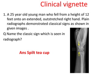
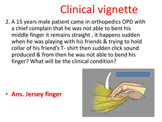
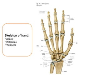
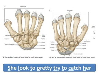
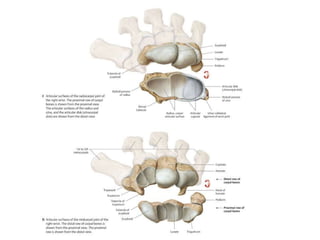
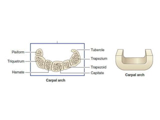
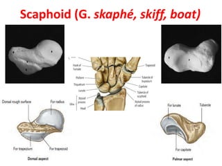
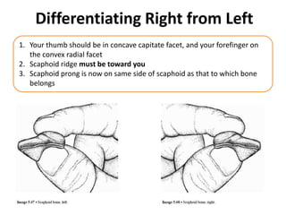
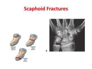
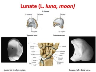
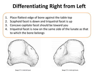
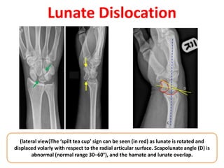
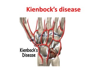
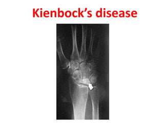
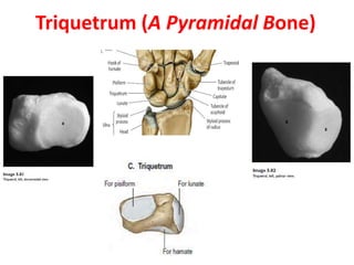
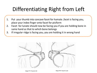
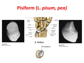
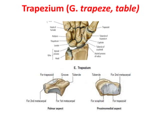
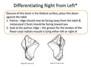
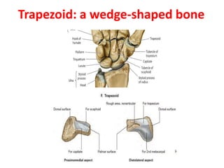
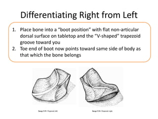
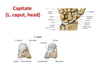
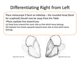
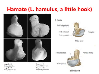
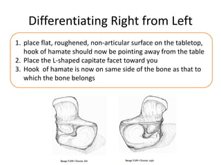
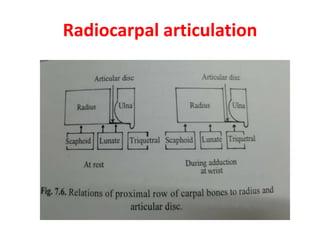
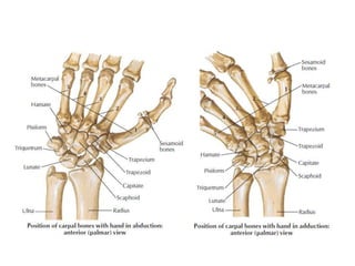
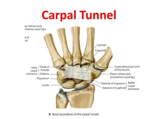
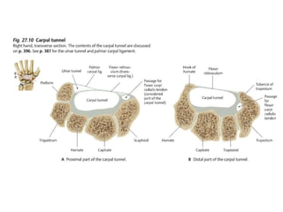
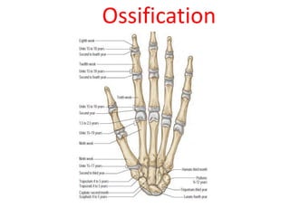
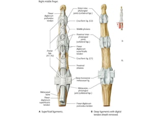
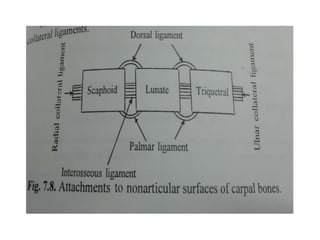
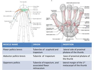
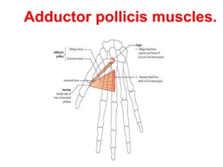
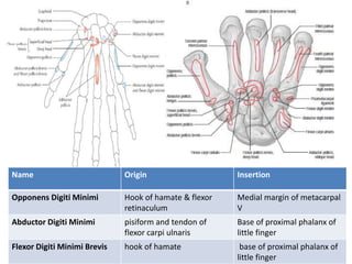
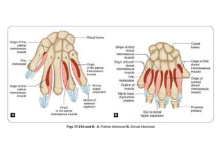
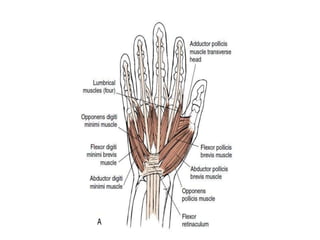
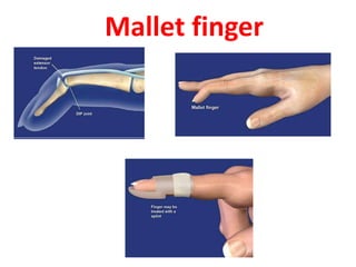
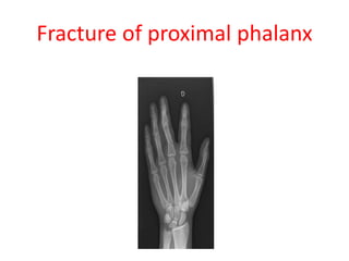
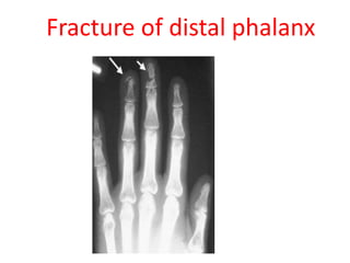
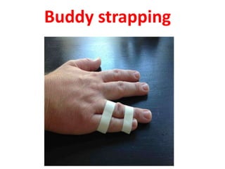
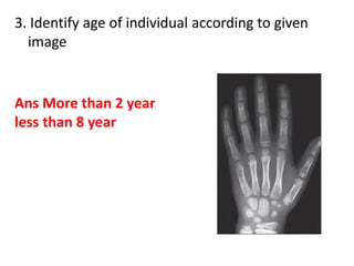
Carpal & Phalanges
Presenter: Rahul Sharma
Objectives
• Introduction
• Carpal bone
• Characteristic features
• Muscular attachment
• Ossification
• Phalanges
• Characteristic features
• Muscular attachment
• Ossification
• Clinical aspect
• Clinical vignette
Skeleton of hand:
•Carpals
•Metacarpal
•Phalanges
She look to pretty try to catch her
Scaphoid (G. skaphé, skiff, boat)
Differentiating Right from Left
1. Your thumb should be in concave capitate facet, and your forefinger on
the convex radial facet
2. Scaphoid ridge must be toward you
3. Scaphoid prong is now on same side of scaphoid as that to which bone
belongs
Scaphoid Fractures
Lunate (L. luna, moon)
Differentiating Right from Left
1. Place flattest edge of bone against the table top
2. Scaphoid facet is down and triquetral facet is up
3. Concave capitate facet should be toward you
4. triquetral facet is now on the same side of the lunate as that
to which the bone belongs
Lunate Dislocation
(lateral view)The ‘spilt tea cup’ sign can be seen (in red) as lunate is rotated and
displaced volarly with respect to the radial articular surface. Scapolunate angle (D) is
abnormal (normal range 30–60°), and the hamate and lunate overlap.
Kienbock’s disease
Kienbock’s disease
Triquetrum (A Pyramidal Bone)
Differentiating Right from Left
1. Put your thumb into concave facet for hamate ,facet is facing you,
place your index finger onto facet for pisiform
2. Facet for lunate should now be facing you if you are holding bone in
same hand as that to which bone belongs
3. If irregular ridge is facing you, you are holding it in wrong hand
Pisiform (L. pisum, pea)
Trapezium (G. trapeze, table)
Differentiating Right from Left*
Dorsum of this bone is the flattest surface, place this down
against the table
1. Palmar ridge should now be facing away from the table &
metacarpal 2 facet should be facing toward you
2. look at the palmar ridge – the groove for the tendon of the
flexor carpi radialis muscle is lying either left or right of
Trapezoid: a wedge-shaped bone
Differentiating Right from Left
1. Place bone into a “boot position” with flat non-articular
dorsal surface on tabletop and the “V-shaped” trapezoid
groove toward you
2. Toe end of boot now points toward same side of body as
that which the bone belongs
Capitate
(L. caput, head)
Differentiating Right from Left
Place metacarpal 3 facet on tabletop – the rounded head (facet
for scaphoid) should now be away from the Table
•Place capitate line toward you
(a) Head leans toward the same side as that which bone belongs
(b) Capitate line travels upwards toward same side as that which bone
belongs
Hamate (L. hamulus, a little hook)
Differentiating Right from Left
1. place flat, roughened, non-articular surface on the tabletop,
hook of hamate should now be pointing away from the table
2. Place the L-shaped capitate facet toward you
3. Hook of hamate is now on same side of the bone as that to
which the bone belongs
Radiocarpal articulation
Carpal Tunnel
Ossification
• Cartilagenous at birth then start to ossify
• Each carpal bone ossified from 1 centre
• 1st – Capitate , last - pisiform
Medico legal Importance
• Ossifications of 8 carpals have a definite
relation with age and can be considered as a
good indicator for age assessment and skeletal
maturation in pediatric age groups.
• Carpals are the most important bones in
determining skeletal age
Phalanges
Parts of phalanx
Characteristic features
Ossification
Muscle Attachment
MUSCLE NAME ORIGIN INSERTION
Flexor pollicis brevis Tubercles of scaphoid and
trapezium
lateral side of proximal
phalanx of the thumb.
Abductor pollicis brevis Tubercle of trapezium base of proximal phalanx of
the thumb
Oppenens pollicis Tubercle of trapezium, and
associated flexor
retinaculum
lateral margin of the 1st
metacarpal of the thumb
Adductor pollicis muscles.
Name Origin Insertion
Opponens Digiti Minimi Hook of hamate & flexor
retinaculum
Medial margin of metacarpal
V
Abductor Digiti Minimi pisiform and tendon of
flexor carpi ulnaris
Base of proximal phalanx of
little finger
Flexor Digiti Minimi Brevis hook of hamate base of proximal phalanx of
little finger
Mallet finger
Jersey finger
Fracture of proximal phalanx
Fracture of distal phalanx
Buddy strapping
MCQs
Q.1) Which carpal bone is not appear on x ray of
below 8 year old children?
A) Hamate
B) Pisiform
C) Capitate
D) Triquetrum
Ans. B
MCQs
A 16 year old girl jump from a 4ft wall, she hit her wrist in
extended position while landing, now her radial side palmar
projection of wrist swollen and having pain during brushing teeth
and in wrist flexion. Which of the most likely muscle tendon injured
at radial site of wrist .
a) Flexor carpi radialis
b) Abductor digiti mini
c) Flexor pollicis brevis
d) Oppenens pollicis
Flexor carpi radialis
Clinical vignette
1. A 25 year old young man who fell from a height of 12
feet onto an extended, outstretched right hand. Plain
radiographs demonstrated classical signs as shown in
given images .
Q Name the classic sign which is seen in
radiograph?
Ans Spilt tea cup
Clinical vignette
2. A 15 years male patient came in orthopedics OPD with
a chief complain that he was not able to bent his
middle finger it remains straight , it happens sudden
when he was playing with his friends & trying to hold
collar of his friend’s T- shirt then sudden click sound
produced & from then he was not able to bend his
finger? What will be the clinical condition?
• Ans. Jersey finger
3. Identify age of individual according to given
image
Ans More than 2 year
less than 8 year
REFERENCES
• Gray's Anatomy_ The Anatomical Basis of
Clinical Practice, 41e
• Thime Atlas of Anatomy
• Human Osteology and Skeletal Radiology_ An
Atlas and Guide by Carrie Allen

Carpal & Phalanges.pptx

  • 1.
  • 2.
    Objectives • Introduction • Carpalbone • Characteristic features • Muscular attachment • Ossification • Phalanges • Characteristic features • Muscular attachment • Ossification • Clinical aspect • Clinical vignette
  • 3.
  • 4.
    She look topretty try to catch her
  • 8.
  • 9.
    Differentiating Right fromLeft 1. Your thumb should be in concave capitate facet, and your forefinger on the convex radial facet 2. Scaphoid ridge must be toward you 3. Scaphoid prong is now on same side of scaphoid as that to which bone belongs
  • 10.
  • 11.
  • 12.
    Differentiating Right fromLeft 1. Place flattest edge of bone against the table top 2. Scaphoid facet is down and triquetral facet is up 3. Concave capitate facet should be toward you 4. triquetral facet is now on the same side of the lunate as that to which the bone belongs
  • 13.
    Lunate Dislocation (lateral view)The‘spilt tea cup’ sign can be seen (in red) as lunate is rotated and displaced volarly with respect to the radial articular surface. Scapolunate angle (D) is abnormal (normal range 30–60°), and the hamate and lunate overlap.
  • 14.
  • 15.
  • 16.
  • 17.
    Differentiating Right fromLeft 1. Put your thumb into concave facet for hamate ,facet is facing you, place your index finger onto facet for pisiform 2. Facet for lunate should now be facing you if you are holding bone in same hand as that to which bone belongs 3. If irregular ridge is facing you, you are holding it in wrong hand
  • 18.
  • 19.
  • 20.
    Differentiating Right fromLeft* Dorsum of this bone is the flattest surface, place this down against the table 1. Palmar ridge should now be facing away from the table & metacarpal 2 facet should be facing toward you 2. look at the palmar ridge – the groove for the tendon of the flexor carpi radialis muscle is lying either left or right of
  • 21.
  • 22.
    Differentiating Right fromLeft 1. Place bone into a “boot position” with flat non-articular dorsal surface on tabletop and the “V-shaped” trapezoid groove toward you 2. Toe end of boot now points toward same side of body as that which the bone belongs
  • 23.
  • 24.
    Differentiating Right fromLeft Place metacarpal 3 facet on tabletop – the rounded head (facet for scaphoid) should now be away from the Table •Place capitate line toward you (a) Head leans toward the same side as that which bone belongs (b) Capitate line travels upwards toward same side as that which bone belongs
  • 25.
    Hamate (L. hamulus,a little hook)
  • 26.
    Differentiating Right fromLeft 1. place flat, roughened, non-articular surface on the tabletop, hook of hamate should now be pointing away from the table 2. Place the L-shaped capitate facet toward you 3. Hook of hamate is now on same side of the bone as that to which the bone belongs
  • 27.
  • 29.
  • 31.
    Ossification • Cartilagenous atbirth then start to ossify • Each carpal bone ossified from 1 centre • 1st – Capitate , last - pisiform
  • 32.
    Medico legal Importance •Ossifications of 8 carpals have a definite relation with age and can be considered as a good indicator for age assessment and skeletal maturation in pediatric age groups. • Carpals are the most important bones in determining skeletal age
  • 33.
  • 34.
  • 35.
  • 36.
  • 39.
  • 40.
    MUSCLE NAME ORIGININSERTION Flexor pollicis brevis Tubercles of scaphoid and trapezium lateral side of proximal phalanx of the thumb. Abductor pollicis brevis Tubercle of trapezium base of proximal phalanx of the thumb Oppenens pollicis Tubercle of trapezium, and associated flexor retinaculum lateral margin of the 1st metacarpal of the thumb
  • 41.
  • 42.
    Name Origin Insertion OpponensDigiti Minimi Hook of hamate & flexor retinaculum Medial margin of metacarpal V Abductor Digiti Minimi pisiform and tendon of flexor carpi ulnaris Base of proximal phalanx of little finger Flexor Digiti Minimi Brevis hook of hamate base of proximal phalanx of little finger
  • 45.
  • 46.
  • 47.
  • 48.
  • 49.
  • 50.
    MCQs Q.1) Which carpalbone is not appear on x ray of below 8 year old children? A) Hamate B) Pisiform C) Capitate D) Triquetrum Ans. B
  • 51.
    MCQs A 16 yearold girl jump from a 4ft wall, she hit her wrist in extended position while landing, now her radial side palmar projection of wrist swollen and having pain during brushing teeth and in wrist flexion. Which of the most likely muscle tendon injured at radial site of wrist . a) Flexor carpi radialis b) Abductor digiti mini c) Flexor pollicis brevis d) Oppenens pollicis Flexor carpi radialis
  • 52.
    Clinical vignette 1. A25 year old young man who fell from a height of 12 feet onto an extended, outstretched right hand. Plain radiographs demonstrated classical signs as shown in given images . Q Name the classic sign which is seen in radiograph? Ans Spilt tea cup
  • 53.
    Clinical vignette 2. A15 years male patient came in orthopedics OPD with a chief complain that he was not able to bent his middle finger it remains straight , it happens sudden when he was playing with his friends & trying to hold collar of his friend’s T- shirt then sudden click sound produced & from then he was not able to bend his finger? What will be the clinical condition? • Ans. Jersey finger
  • 54.
    3. Identify ageof individual according to given image Ans More than 2 year less than 8 year
  • 55.
    REFERENCES • Gray's Anatomy_The Anatomical Basis of Clinical Practice, 41e • Thime Atlas of Anatomy • Human Osteology and Skeletal Radiology_ An Atlas and Guide by Carrie Allen