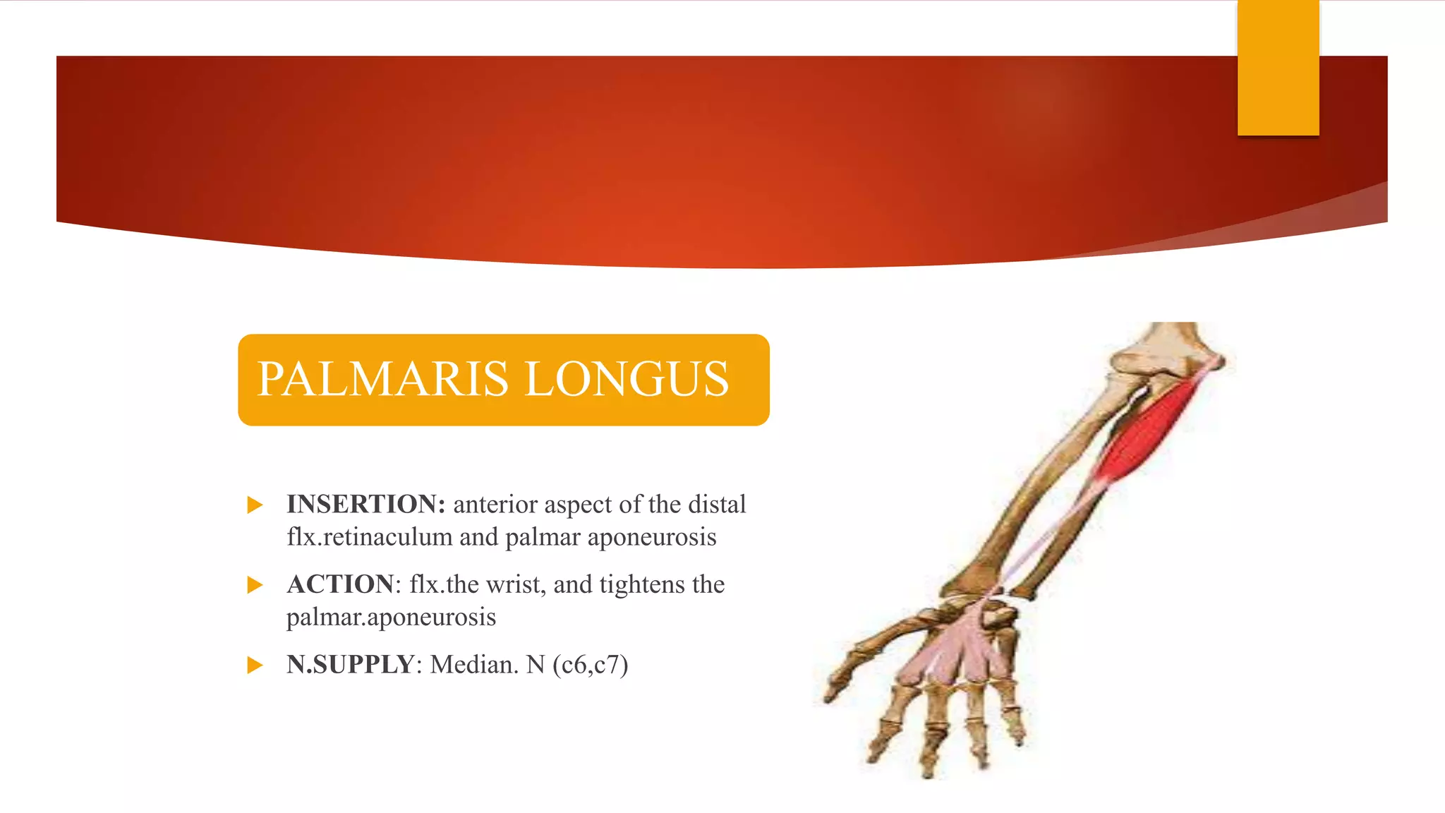
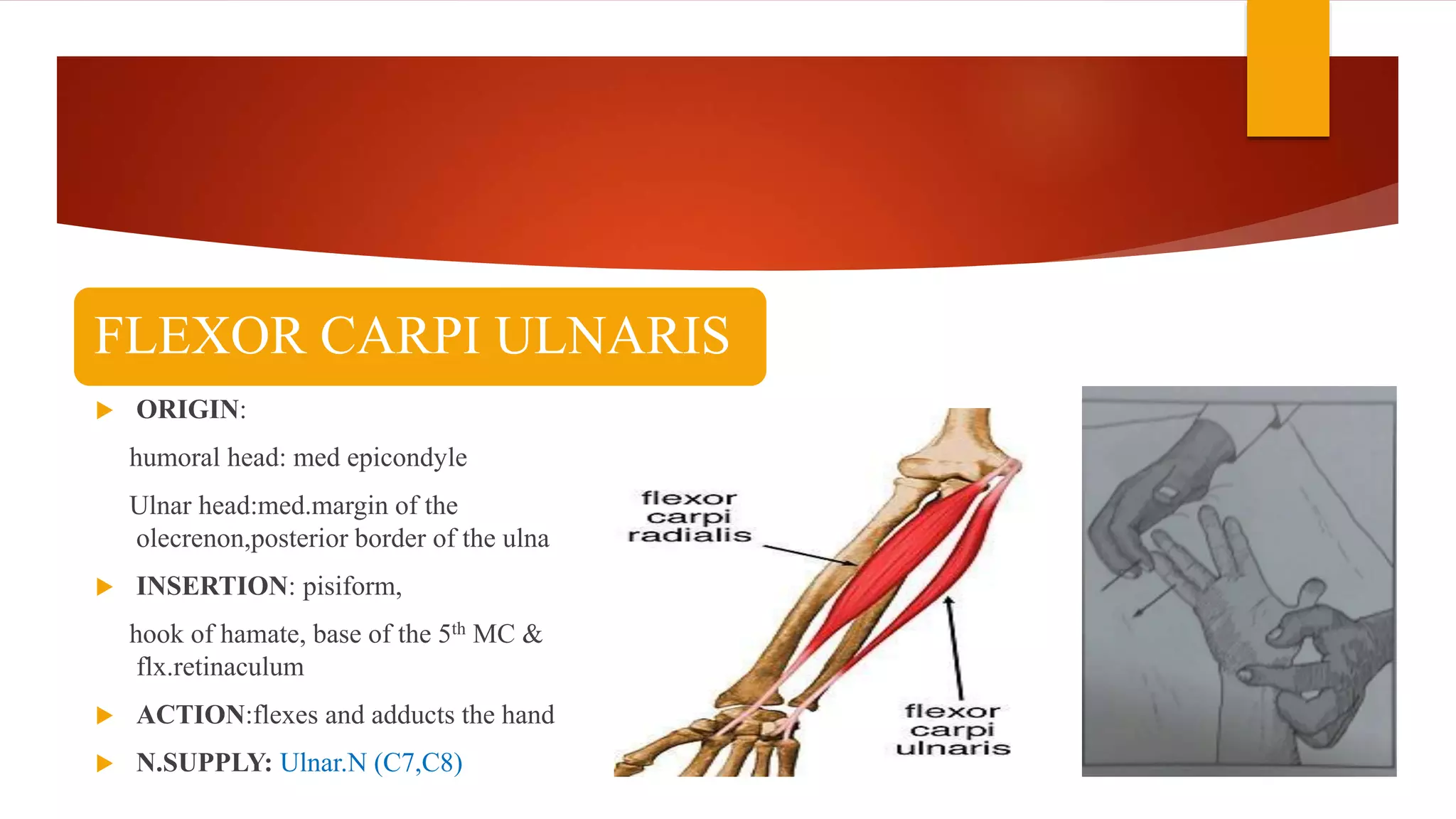
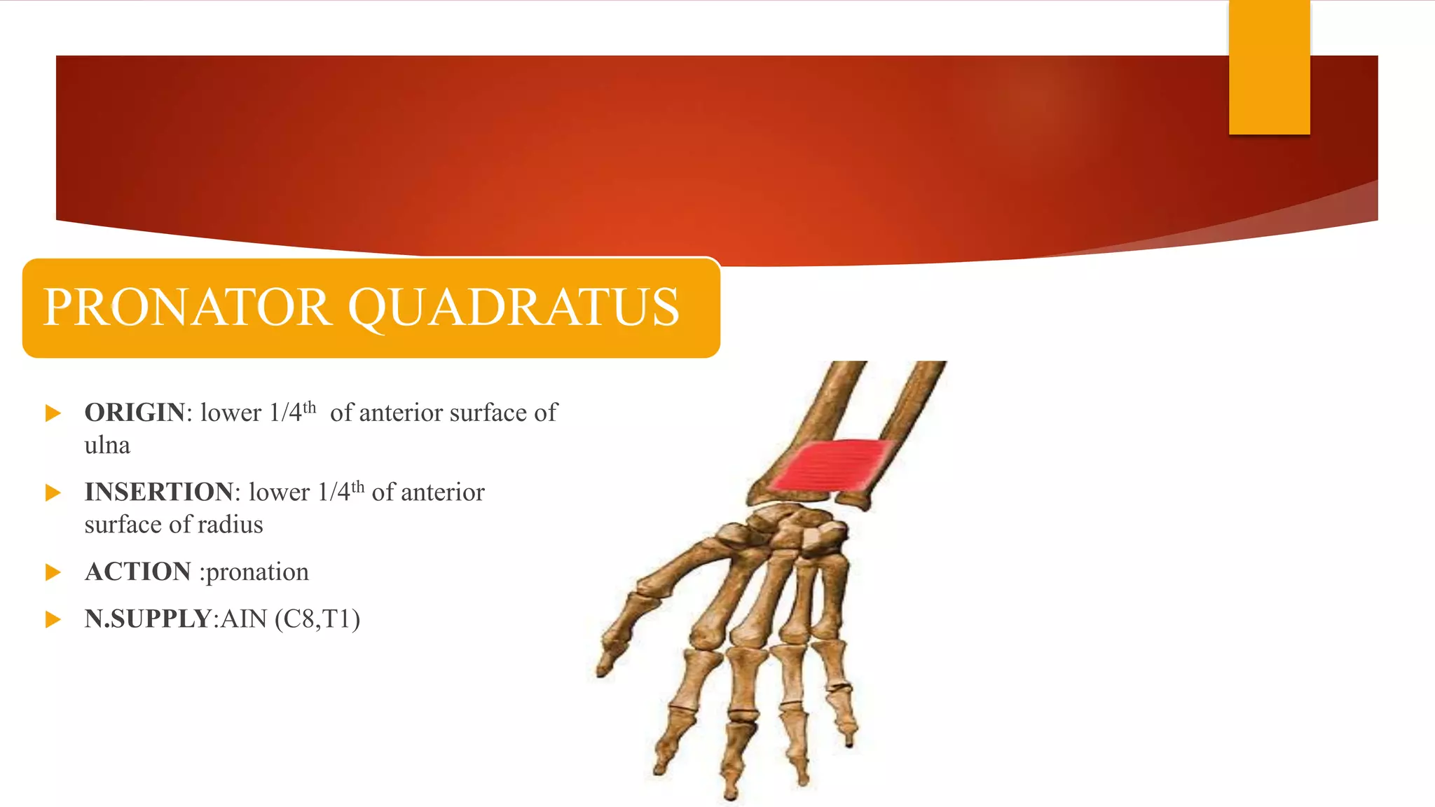
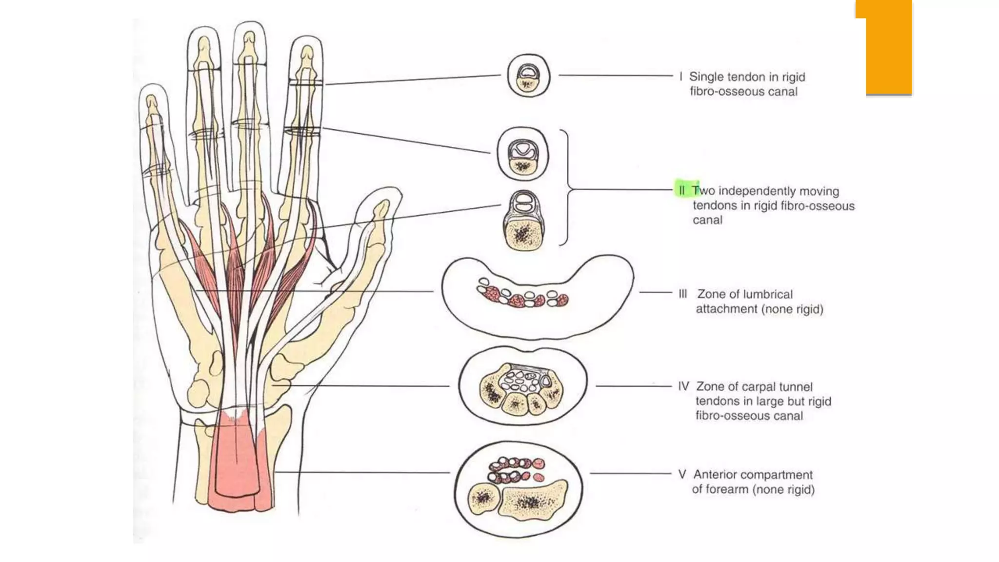
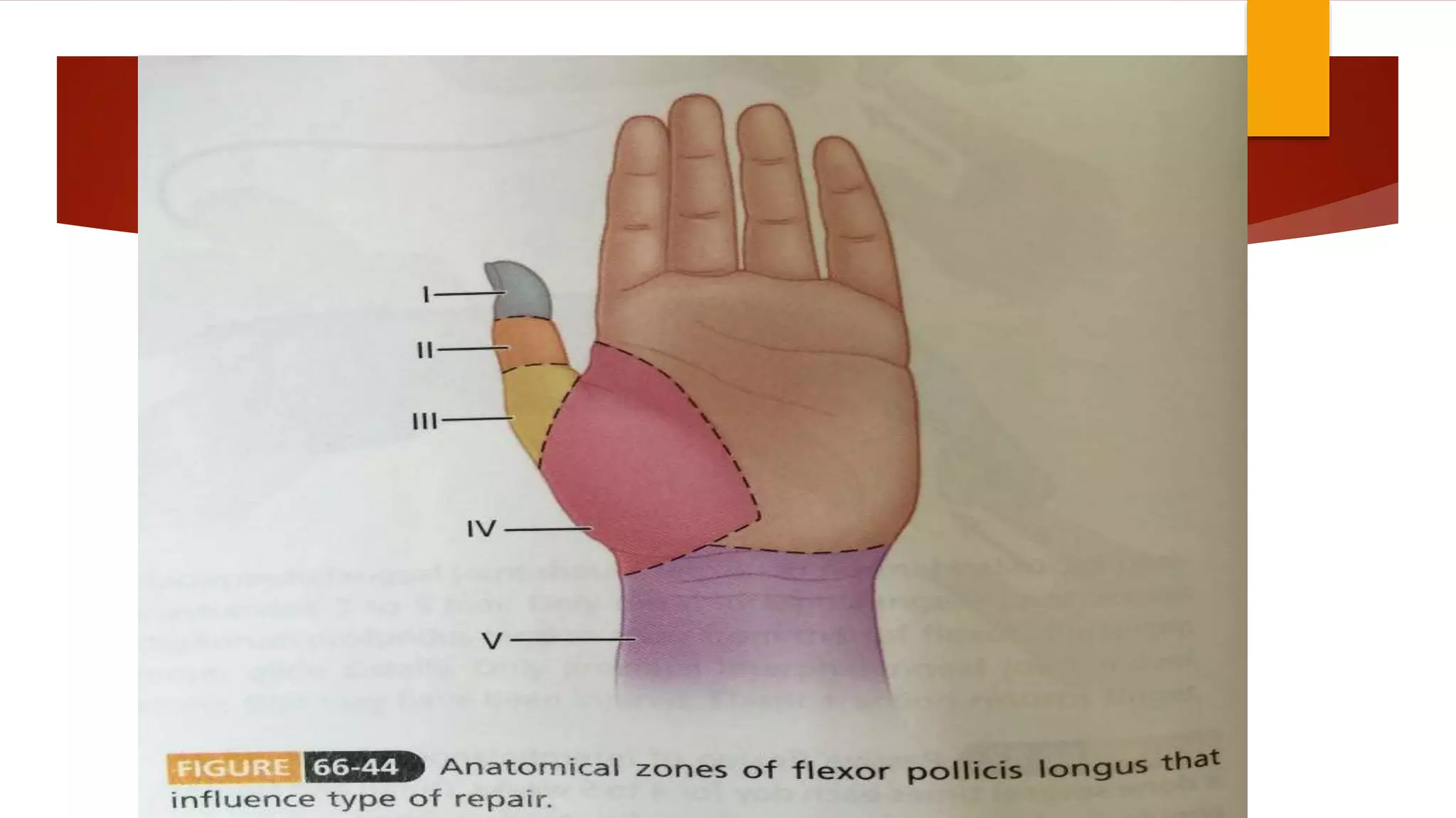
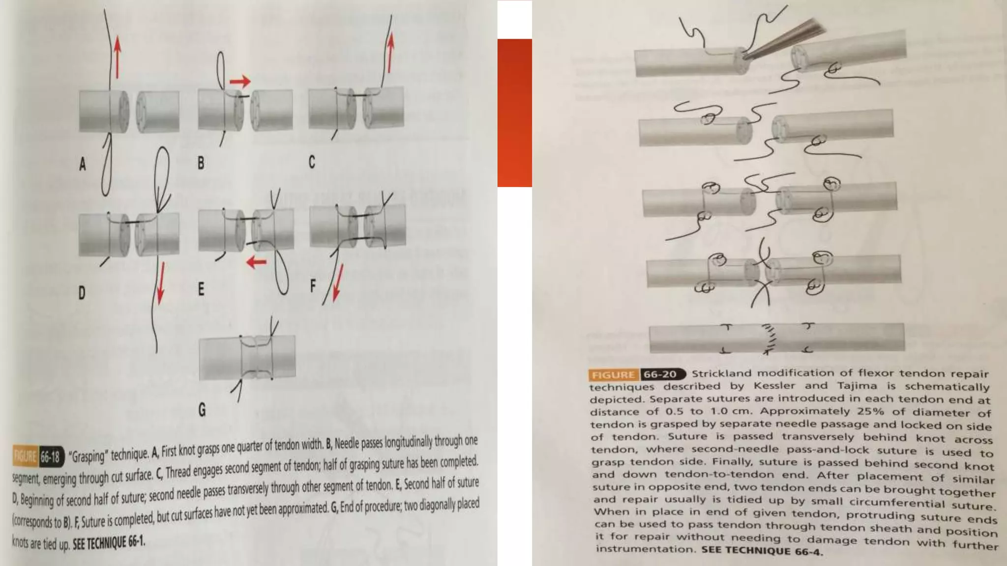
SRI SIDDHARTHA MEDICAL COLLEGE,TUMKUR
DEPARTMENT OF ORTHOPAEDICS
Topic:
Flexor Tendon Injury, Repair & Rehabilitation
Moderator:
Dr. J.K Reddy
HOD & Professor
Dept. of Orthopaedics
Presenter:
Dr. Jaipalsinh Mahida
Jr. Resident M.S Ortho
Dept. of Orthopaedics
INTRODUCTION
 Despite advancement in our understanding of flexor tendon biology, repair &
rehabilitation, a successful outcome after intrasynovial flexor tendon injury can be difficult
to achieve.
 This requires a thorough understanding of the biologic principles of tendon injury &
healing, a detailed knowledge of normal and pathologic flexor tendon anatomy, an
attention to meticulous surgical technique, a careful postoperative rehabilitation protocol &
surgeon, therapist & patient who are motivated to enact that protocol.
ANATOMY
 MUSCLES OF FOREARM
 Anterior Compartment:
 Has a common flexor origin from medial epicondyle humerus
 Divided in 2 compartments
1. SUPERFICIAL
a) Pronator Teres
b) Flexor Carpi Ulnaris
c) Palmaris Longus
d) Flexor Carpi Radialis
e) Flexor Digitorum Superficialis (sublimus)
2. DEEP:
a) Flexor Digitorum Profundus
b) Flexor Pollicis Longus
c) Pronator Quadratus
PRONATOR TERES
 Insertion: midway long the lateral
surface of the radius
 Action: pronation,flexion of forearm
 N.Supply:Median.N (C6c7)
FLEXOR CARPI RADIALIS
 INSERTION:base of the 2nd mc bone
and slip to the bace of the 3rd MC bone
 ACTION: flexion and abduction of
wrist
 N.SUPPLY: Median.N (C6 , C7)
PALMARIS LONGUS
 INSERTION: anterior aspect of the distal
flx.retinaculum and palmar aponeurosis
 ACTION: flx.the wrist, and tightens the
palmar.aponeurosis
 N.SUPPLY: Median. N (c6,c7)
FLEXOR CARPI ULNARIS
 ORIGIN:
humoral head: med epicondyle
Ulnar head:med.margin of the
olecrenon,posterior border of the ulna
 INSERTION: pisiform,
hook of hamate, base of the 5th MC &
flx.retinaculum
 ACTION:flexes and adducts the hand
 N.SUPPLY: Ulnar.N (C7,C8)
FLEXOR DIGITORUM SUPERFICIALIS
 ORIGIN:
humeroulnar head: med.epicondyle of the
humerus, coronoid process
Radial head:
sup.half of anterior aspect of the radius
 INSERTION : bodies of the Middle
phalanges of the medial 4 digits
 ACTION: flx.of all joints it crosses
 N.SUPPLY :Median .N (C7,C8,T1)
FLEXOR DIGITORUM PROFUNDUS
 ORIGIN:prox.3/4 of the medial and anterior
aspect of the ulna and from interosseous memb.
 INSERTION:base of The Diatal phalanges of
the medial 4 digits
 ACTION: flx.DIP,,PIP,MP, wrist
 N.SUPPLY:
Medial-ulnar.N (C8,t1)
Lateral-AIN Of Median.N (C8,t1)
FLEXOR POLLICIS LONGUS
 ORIGIN: upper 3/4 of anterior surface of
radius
 INSERTION: base of distal phalanx of the
thumb
 ACTION: flexion of proximal & distal
phalnx of the thumb
 N.SUPPLY:AIN (C7,C8,T1)
PRONATOR QUADRATUS
 ORIGIN: lower 1/4th of anterior surface of
ulna
 INSERTION: lower 1/4th of anterior
surface of radius
 ACTION :pronation
 N.SUPPLY:AIN (C8,T1)
 At the level of the metacarpal neck, the tendons enter the synovial sheath, at which point the FDS splits into
halves that course dorsally around the FDP tendon. These halves then rejoin at the Camper chiasm, then split
again to insert independently on the middle phalanx.
 The flexor tendon sheath begins at the level of the metacarpal neck, and consists of 5 annular and 3 cruciate
pulleys.
 The A1, A3, and A5 pulleys arise from the volar plates of the metacarpophalangeal (MCP), proximal
interphalangeal (PIP), and distal interphalangeal (DIP) joints, respectively.
 The A2 and A4 pulleys arise from the volar portion of the proximal and middle phalanges.
 The first cruciate pulley (C1) is between A2 and A3, C2 is between A3 and A4, and C3 is between A4 and
A5.
PULLEYS
 The flexor pollicis longus (FPL) has a unique anatomy. The muscle originates from the
middle and proximal aspect of the distal radius and is innervated by the anterior interosseous
nerve
 The FPL tendon courses through the carpal tunnel in a dorsal and radial position. The pulley
system of the FPL consists of 2 annular pulleys, the A1 and A2 pulleys, which originate
from the volar plates of the MCP and interphalangeal (IP) joints, respectively
 The diagonally oriented oblique pulley is critical to maintaining normal FPL function and is
located over the proximal phalanx between the 2 annular pulleys.
 TENDON NUTRITION:
 Mainly from 2 basic sources
1. The synovial fluid produced within tenosynovial sheath
2. Blood supply provided through longitudinal insertions, & vincular circulation.
 Each tendon has long and short vinculum, which are supplied by a transverse “ladder
branch” of the digital arteries
 Each vinculum enters the tendon on its dorsal surface, resulting in the dorsal aspect of the
tendon being the most well vascularized.
 The dorsal aspect of the tendon is also the optimal biomechanical location for suture
placement during tendon repair. Sutures placed in this location may interfere with the
blood supply.
 between the insertion of the vinculae, each tendon has a hypovascular watershed zone. In
this area, the tendons rely on passive diffusion through the sheath. The hypovascular FDS
tendon is located in the region of the camper chiasm, between the vinculum breve
superficialis (VBS) and vinculum longum (VLS) .
 The FDP has 2 hypovascular zones: proximally in region of A2 between the intrinsic
longitudinal vessels and the vinculum longum profundus (VLP), and distally between the
VLP & vinculum breve profundus(VBP)
 FLEXOR ZONES OF HAND : 5 zones
 ZONE I: contains only FDP tendons
 Extends from just distal to insertion of sublimis tendon to site of insertion of FDP tendon.
 ZONE II: critical area of pulleys (bunnell’s “no man’s land”)
 Contains both FDP & FDS tendons
 Between distal palmar crease & insertion of sublimis tendon.
 Tang subdivided zone II in 4 parts
1. IIa
2. IIb
3. IIc : it is area in which satisfactory functional recovery is most difficult to achieve after 10 repair of both FDP
& FDS tendons.
4. IId
 ZONE III: comprises area of lumbrical origine
 Between distal margin of transverse carpal ligament & beginning of critical area of pulleys or 1st
annulus.
 ZONE IV:
 Zone covered by transverse carpal ligament
 ZONE V:
 Zone proximal to transverse carpal ligament and include forearm
 FLEXOR ZONES OF THUMB : 5 zones
 ZONE I:
 Area at interphalangeal joint & insertion of flexor pollicis longus
 ZONE II:
 Includes fibroosseous sheath extending just proximal to metacarpal head & MCP joint.
 ZONE III:
 Area of metacarpal beneath thenar muscles
 ZONE IV:
 Corresponds to carpal tunnel
 ZONE V:
 Distal forearm just proximal to wrist.
PATIENT EVALUATION:
 The initial evaluation of a patient with a flexor tendon injury is important for
• formulating a diagnosis
• establishing a treatment plan
• counseling the patient regarding expected outcomes.
 The mechanism of injury has implications for
• quality of the tendon
• status of the surrounding soft tissues.
 A sharp laceration is more likely to have a cleanly cut tendon end and less soft tissue
damage than a crushing or avulsion type mechanism.
 The position of the finger at the time of injury can help predict the location and lengths
of the underlying tendon stumps
• A finger that was held in flexion at the time of laceration can be expected to have a
shorter, more distally located stump and may require more substantial distal wound
extension
 Observation can provide many useful details about the nature of the injury. Any soft
tissue defects or deformities suggesting underlying bony injury should be noted
 In the absence of any concomitant bone or joint Injuries, a digit with lacerated FDP and
FDS tendons will assume an extended position (Fig. 1)
• In this scenario, the tenodesis effect, which would typically cause flexion of the digit, will not occur.
 Each flexor tendon must be examined
independently
 Stabilizing the middle phalanx of
the injured digit and asking the
patient to actively flex the Dip
joint isolates the FDP.
 The FDS is examined by holding
the adjacent digits in extension and
asking the patient to flex the
injured digit at the PIP Joint.
 In thumb, to check the integrity of flexor pollicis longus tendon
• MCP joint of thumb is stabilized
• If tendon is divided – flexion at interphalangeal joint is absent.
 If a wound is located at level of wrist, joint of finger can be actively flexed even though tendon to that
finger is severed
• Result of intercommunication of flexor profundus tendon at wrist, particularly in little & ring finger
 A neurovascular examination is performed before administration of local anesthesia or sedation.
 The digit is examined for color, turgor, and capillary refill time.
 Both ulnar and radial digital neurovascular bundles are examined and assessed for digital pulses
 Radiographs of the injured digit are routinely obtained to ensure there are no foreign bodies or underlying
bony abnormalities.
 TENDON HEALING:
 Believed to occur through activity of extrinsic & intrinsic mechanisms
 Occurs in 3 phases
1. Inflammatory : 48 to 72 hrs
2. Fibroblastic : 5 days to 4 weeks
3. Remodeling : 4 weeks to about 3.5 months
 Extrinsic mechanism occurs through activity of peripheral fibroblasts & seems to be dominant
mechanism contributing to formation of scar & adhesions.
 Intrinsic healing seems to occur through activity of fibroblastic derived from tendon.
BASIC TENDON TECHNIQUES
 During tendon suturing, handling should be gentle & delicate, causing as little reaction & scaring as
possible.
 Pinching & grasping of uninjured surfaces should be avoided, as it can contribute to adhesions formation.
 Strickland stressed 6 characteristics of an ideal tendon repair:
1. Easy placement of sutures in tendon
2. Secure suture knots
3. Smooth juncture of tendon ends
4. Minimal gapping at repair site
5. Minimal interference with tendon vascularity
6. Sufficient strength throughout healing to permit application of early motion stress to tendon.
 SUTURE MATERIAL:
 Although monofilament stainless steel has highest tensile strength,
 Difficult to handle
 Tends to pull through the tendon limits use in fingers
 Makes a large knot
 Can be used satisfactorily in distal forearm
 Catgut & polyglycolic acid group(dexon, vicryl) becomes weak too early after surgery
 Synthetic sutures of caprolactum family & nylon maintain their resistance to disrupting forces longer
than polypropylene (prolene) & polyester suture.
 Polydioxanone has been shown to be strong as polypropylene.
 Comparison of polyglycolide-trimethylene carbonate & polydioxanone found
 Polydioxanone repairs maintained better strength over 28 days.
 Monofilament nylon permitted earlier gap formation & failure of the repair compared with braided polyester.
 In biomechanical study
 Braided polyethylene & braided stainless steel wire are most suitable mechanically.
 Braided polyester was intermediate.
 Monofilament nylon & polypropylene were least satisfactory.
 In cadaver biomechanical analysis
 Intratendendious, crimped, single-strand, multifilament stainless steel device (teno fix) compared favorably
with 4-strand cruciate repairs.
 Intratendendious device group had lower rupture rate & compared favorable in grip, pinch strength.
 SUTURE CONFIGURATIONS:
 Various tendon repair types can be divided into 3 groups:
1. Group 1: exemplified by simple sutures; suture pull is
parallel to tendon collagen bundle, transmitting stress of
repair directly to opposing tendon ends.
2. Group 2: exemplified by bunnell suture; stress is transmitted
directly across juncture by suture material & depends on
strength of suture.
3. Group 3: exemplified by pulvertaft technique (fish mouth
weave); suture are placed perpendicular to tendon collagen
bundles & applied stress.
 Interrupted sutures were found to weakest & unsuitable in most tendon repair.
 Fish-mouth or end weave repairs are strongest & most suitable for tendon graft & tendon transfer
junctures in distal forearm & palm, where intermediate suture configurations(bunnell, kessler) didn’t
differ significantly in strength.
 4-strand, 6- strand & 8-strand core sutures
 create stronger repairs
 Reduce possibility of gap formation
 Permit greater active forces to be applied to repaired tendons, allowing earlier active motion.
 END-TO-END SUTURES:
 DOUBLE RIGHT-ANGLED SUTURES:
 FISH-MOUTH END-TO-END SUTURE (PULVERTAFT):
 TENDON-TO-BONE ATTACHMENT:
 SUTURE ANCHOR TENDON ATTACHMENT:
 TIMING OF FLEXOR TENDON REPAIR:
 If wound is caused by sharp object such as knife is reasonably clean, can be repaired at time of primary
wound closure.
 Primary tendon repair is done within first 12 hours of injury and can be extended to within 24 hrs of
injury in rare situations.
 Delayed primary repair is done within 24 hrs to approx. 10 days.
 10 to 14 days - secondary repair.
 After about 4 weeks – late secondary repair
 Secondary repair is indicated when tendon injury is associated with complicating factors ( extensive
crushing with bony comminution near level of tendon injury, severe neurovascular injury, severe joint
injury, skin loss) that could compromise end result.
 PRINCIPLES OF TENDON REPAIR:
 The resistance to gap formation at the repair site and the ultimate strength of the repair are interrelated
 influenced by the number of core suture strands, the core suture configuration, the core suture size,
and the addition of a peripheral epitendinous suture.
 additional factors influence the strength of a repair including material properties of the suture,
presence of locking sutures, number of knots, tension of the core sutures, and depth of suture
purchase
 The number of core suture strands across the repair is the single most important factor in the strength of
repair
 multiple strands crossing the repair site have greater tensile strength than those with fewer strands
 . Four strands repairs have been shown to be superior to 2-strand repairs both at time zero and during
the first 6 weeks after repair
 Although these multistrand repairs do have increased bulk, this has not been shown to have an
adverse impact on tendon gliding
 primary limitation to these repairs is the surgeon's ability to effectively place the core suture while
minimizing trauma to the tendon and surrounding sheath, which can cause adhesions
 different core, suture configuration can be simplified into 3 components
1. longitudinal component
2. transverse component
3. link connecting them
 The Kessler 2-strand suture technique has been known as the workhorse for flexor tendon repair
and has been modified many times since its introduction.
 The original Kessler 2-strand suture technique consists of a continuous loop anchored at 4 comers
however, this technique has encountered problems with tendon deformation under load due to load
transmission from the longitudinal components to the transverse components.
 When an axial load is placed across the repair site, the longitudinal components can lengthen
while the transverse components narrow
o To avoid this, a double-Kessler technique results in a 4-strand repair with significantly smaller gap
formation.
 The cruciate repair technique eliminates the transverse component and therefore can better withstand
axial forces.
 Without a transverse component, the cruciate technique tends to fall via suture pullout at higher ultimate
tensile strengths
 other strong repairs, such as the augmented Becker repair (Massachusetts General Hospital [MGH]
repair) and its modifications, have been developed and appear to not weaken during dynamic,
nonlinear testing.
 In cyclical testing, the MGH repair technique had significantly higher force for 2-mm gap, higher
ultimate load to failure, and increased stiffness compared with the modified Kessler suture
 size of suture purchase has been shown to effect repair strength
 depth of purchase of the core suture In the tendon should be at least 7 mm, and ideally greater than
10mm, to avoid significant weakening of the repair.
 Another method of increasing the strength of repair is by locking of the suture loops
 Suture loop tightens around bundles of fibers as longitudinal tension is applied.
 Locking loops have been shown to have greater time-zero tensile strength than grasping loops where
this tightening does not occur.
 The locking loop can be ensured by placing the vertical component of the suture deep to the transverse
component of the suture, relative to the cut end of the tendon.
 The ideal size of a locking loop should involve 25% of the tendon's width to maximize repair strength.
 caliber of core Suture is an important determinant of repair strength.
 Sutures of increased caliber have greater strength to failure than sutures of lesser caliber.
 Larger suture is more likely to fail by pullout than smaller-caliber suture ,which is more likely to fail by
rupture.
 Although larger suture is favorable, sutures thicker than & 3-0 in size should be avoided as they can
increase the bulk of the repair.
 Following core suture placement, the addition of a peripheral epitendinous suture is recommended
 It helps to decrease bulk by smoothing out the repair site
 addition of peripheral suture itself contributes up to 50% of ultimate load to failure and potentially up to
50 N of strength.
 Several epitendinous configurations have been suggested, Including simple running, cross-stitch,
interlocking cross-stitch, and Interlocking horizontal mattress
 simple Running peripheral suture should be placed 2 mm from the cut end of the tendon.
 The current recommendation for repair is to use a 4-strand or greater core suture
combined with a running epitendinous suture
 If an epitendinous suture is not used, a 6-strand core repair is recommended with slight
tensioning of the core sutures to prevent gapping.
 It should be noted that the repair strength of the core stitch and epitendinous suture are not
summative and can fail independently.
 BIOLOGY OF TENDON REPAIR:
 Several growth factor have been recognized to increase after tendon injury, which may
contribute to both tendon healing and scar formation, include
1. vascular endothelial grow factor,
2. Insulin like growth factor
3. platelet derived growth factor (PDGF)
4. basic fibroblast growth factor (bFGF)
5. transforming growth factor-beta
 Fibronectin and integrins are upregulated during the early postoperative period in
tendons undergoing early passive mobilization.
 In vitro studies on canine fibroblasts have revealed a dose- dependent effect of flexor
tendon cell proliferation from synergy of PDGF-BB & bFGF.
 Tang and colleagues used leghorn chicken FDP tendons and an adeno-associated viral
vector to transport bFGF genes directly onto the cut tendon ends, resulting in
significantly increased strength at 2 and 4 weeks & also significantly fewer adhesions at
12 weeks compared with controls.
 tendons treated with hyaluronic acid have showed improved gliding mechanics, and
those modified through carbodiimide derivatization (Cd-HA) may even have improved
HA retention.
PRIMARY FLEXOR TENDON REPAIR
 ZONE I:
 Zone I injuries occur distal to the FDS insertion and involve only the FDP tendon.
 Lacerations in which there is less than 1 cm of distal stump remaining attached to the distal phalanx
can be treated with advancement and direct repair of the proximal tendon to the distal phalanx.
 If the tendon must be advanced more than 1 cm, or if there is excessive tension at the repair site, a
"quadriga effect“ can occur.
 digit with the overtensioned tendon will reach maximal flexion before the adjacent uninjured
digits.
 Because the FDP tendons are united by a common muscle belly, the uninjured digits will not be
able to flex further, resulting in loss of motion and grip strength.
 If there is more than 1 cm of distal stump remaining, the tendon ends can be directly repaired
 placement of a 2-strand core suture and direct repair to the base of the distal phalanx
 Disadvantages - potential for Infection, suture rupture, nail deformity, discomfort from pressure on the
nail, and the externally placed-button catching on clothing or objects.
 The most common mechanism of injury in zone I is eccentric contraction of the FDP tendon during
forceful hyperextension of the distal phalanx.
 This is termed a “ Jersey finger“ as it often occurs while an athlete tries to grab another player's jersey.
 Leddy and Packer initially described 3 types of FDP avulsions
1. type I : in which the tendon has retracted Into the palm
2. type II in which the tendon has retracted to the level of the A2 pulley
3. type Ill in which the tendon, still attached to a bony fragment, has retracted to the level of the A4
pulley.
 A type IV was later added to the classification in which there is a fracture of the distal phalanx and the
tendon, which has retracted Into the palm, is no longer attached to this fragment.
 vincular blood supply to types II and Ill remains intact, and these injuries can be repaired even after
considerable delay
 tendons in types I and IV Injures, on the other hand, are separated from the blood supply.
 These require early surgical repair, within 7 to 10 days of injury, to prevent ischemic contracture.
 More recently, suture anchors have been used to repair FDP avulsions
 In cadaver studies, the use of a microsuture anchor combined with a 4-strand modified Becker core suture
has been shown to have pullout strength significantly higher than the dorsal button technique
 Use of suture anchors avoids the potential drawbacks of the dorsal button, and has been shown to yield equal
clinical results
 More recently, Lee described a technique using both suture anchors and a button to secure the FDP tendon
to the distal phalanx.
 This technique demonstrated less gapping and greater strength when compared with more traditional
techniques and may allow for early active postoperative motion
 ZONE II:
 Lacerations in zone II may involve both the FDS and FDP tendons.
 primary repair is generally not suitable for injuries more than 6 weeks old, and repair should ideally be
performed within 7 to 14 days of injury
 Exposure of the tendons can be performed through either a Bruner zigzag incision or a midlateral Incision.
which incorporates the laceration
 The latter approach is typically preferred
 distal stumps can be retrieved by flexion of the digit. The length of the tendon stump will vary depending on
the position of the finger at the time of laceration
 proximal tendon stumps can be delivered into the wound via a proximal-to-distal “ milking“ of the
sheath.
 If the stumps are not visible in the sheath, repeated blind attempts to retrieve the tendon with a clamp
should be avoided
 Tendons should be handled only on their cut ends with a nontoothed forceps. Handling of the outside of
the tendon can lead to scarring and adhesion formation
 Both the FDS and FDP tendons should be repaired if possible.
 The FDP tendon and the FDS, If lacerated before the decussation, are repaired with a 4-strand or greater
core stitch and a running epitendinous suture.
 If lacerated more distally, repair of the Individual FDS slips can be difficult because of their small size
 After repair, patients are placed in a dorsal blocking splint with the wrist in 200 to 300 of flexion, the MP joints
in 600 to 700 flexion, and the IP Joints extended.
 Lalonde advocates the following 4 benefits of this approach
1. avoids late gapping
2. prevents poor fit of repair through pulley
3. allows more pulley preservation via Sheathotomies
4. provides uninterrupted patient education by the physician
REHABILITATION
 Complete immobllizallon after flexor tendon repair is indicated only in limited situations such as
 pediatric patients,
 patients with concomitant bone or soft tissue Injuries requiring immobilization,
 patients who are unable to comply with a protected motion protocol
 In most cases, a protected motion protocol is used postoperatively
 Early motion of flexor tendons improves recovery of tensile strength, decreases adhesions,
improves tendon excursion, and promotes Intrinsic healing
 optimal time for initiating therapy has not been established, but can be as early as the first
postoperative day.
 Experimentally, Gliding resistance has been shown to be lowest on the fifth postoperative day, but the extent
to which this is clinically relevant has not been documented
 Rehabilitation protocols that emphasize higher degrees of excursion of the tendons with
low applied force have been shown to be beneficial.
 Various postoperative protocols exist. The selection of a specific protocol is dependent on
the strength of repair and the compliance of the Patient
 The Kleinert and Duran protocols are Passive motion protocols, which use dorsal
blocking splints to limit wrist and digital extension.
 The Kleinert protocol uses a rubber band attached to the nail plate to effect passive
flexion of the digit. Active extension exercises are performed to the limit of the splint.
 The Duran protocol uses controlled passive motion, alternating DIP flexion, PIP flexion
& full composite flexion
 Recently, protocols that permit early active motion have been increasingly used.
 This can be accomplished by the use of a hinged dorsal blocking splint, which at
baseline holds the wrist in 200 of Flexion, the MP joints in 50°, and the IP joints
extended, but has a removable block that allows the splint to extend 300 at the wrist.
 Patients are instructed to passively flex the digits into the palm.
 The wrist is then gently extended while the digits are actively maintained in flexion.
 This "place and hold“ maximizes intrasynovial excursion while minimizing force at
the repair site
 Given that active motion increases the force across the repair site and risk of rupture,
these protocols require strict patient compliance and a controlled environment.
 In a systemic review of postoperative protocols after tendon repair, passive protocols
had a statistically significant lower risk of rupture but also had a higher risk of decreased
range of motion.
 In a recent prospective randomized study comparing active place-and-hold therapy and
passive-motion therapy, Trumble and colleagues found greater active interphalangeal
joint motion at all time points with early active motion without an increased risk of
rupture.
 Factors associated with poor outcomes include smoking, multiple digit Injury, and
concomitant nerve injury
 It remains unknown whether an injury to a specific digit causes more disability than others,
but active therapy in the small finger has been associated with more ruptures.
 Trumble and colleagues reported significantly better combined active flexion of the injured
digit with less PIP and DIP joint contracture when treated by certified hand therapists for
both active-therapy and passive-therapy groups.
 Matarrese and Hammert recommend Initiating postoperative exercises supervised by hand
therapists within 5 days.
SUMMARY
 Flexor tendon injury remains a challenging problem in hand surgery due to intimate
anatomy of the FDP, FDS & pulley system.
 Repair need to be strong enough to begin early range of motion, while avoiding bulkiness
within confines of pulley system.
 Currently, although many repair configurations are acceptable, it is recommended that at
minimum 4-strand core suture repair with epitendinous suture is used.
 Although every attempts should be made to repair both FDP & FDS tendon, sacrificing one
limb of FDS is acceptable should a repair become too bulky with A2 pulley.
 Venting of pulley system should be done prudently to allow smooth gliding of repaired tendon
while avoiding excessive release.
 Compliance with postoperative motion protocols is vital in securing a successful results.
 Future advances in the biology of tendon healing may aid in better outcomes, but ultimately, the
healing of the tendon repair is dependent on the surgeon, the patient & the therapist.
THANK YOU

Jc flexor tendon injury, repair & rehabilitaion

  • 1.
    SRI SIDDHARTHA MEDICALCOLLEGE,TUMKUR DEPARTMENT OF ORTHOPAEDICS Topic: Flexor Tendon Injury, Repair & Rehabilitation Moderator: Dr. J.K Reddy HOD & Professor Dept. of Orthopaedics Presenter: Dr. Jaipalsinh Mahida Jr. Resident M.S Ortho Dept. of Orthopaedics
  • 2.
    INTRODUCTION  Despite advancementin our understanding of flexor tendon biology, repair & rehabilitation, a successful outcome after intrasynovial flexor tendon injury can be difficult to achieve.  This requires a thorough understanding of the biologic principles of tendon injury & healing, a detailed knowledge of normal and pathologic flexor tendon anatomy, an attention to meticulous surgical technique, a careful postoperative rehabilitation protocol & surgeon, therapist & patient who are motivated to enact that protocol.
  • 3.
    ANATOMY  MUSCLES OFFOREARM  Anterior Compartment:  Has a common flexor origin from medial epicondyle humerus  Divided in 2 compartments 1. SUPERFICIAL a) Pronator Teres b) Flexor Carpi Ulnaris c) Palmaris Longus d) Flexor Carpi Radialis e) Flexor Digitorum Superficialis (sublimus)
  • 4.
    2. DEEP: a) FlexorDigitorum Profundus b) Flexor Pollicis Longus c) Pronator Quadratus
  • 5.
    PRONATOR TERES  Insertion:midway long the lateral surface of the radius  Action: pronation,flexion of forearm  N.Supply:Median.N (C6c7)
  • 6.
    FLEXOR CARPI RADIALIS INSERTION:base of the 2nd mc bone and slip to the bace of the 3rd MC bone  ACTION: flexion and abduction of wrist  N.SUPPLY: Median.N (C6 , C7)
  • 7.
    PALMARIS LONGUS  INSERTION:anterior aspect of the distal flx.retinaculum and palmar aponeurosis  ACTION: flx.the wrist, and tightens the palmar.aponeurosis  N.SUPPLY: Median. N (c6,c7)
  • 8.
    FLEXOR CARPI ULNARIS ORIGIN: humoral head: med epicondyle Ulnar head:med.margin of the olecrenon,posterior border of the ulna  INSERTION: pisiform, hook of hamate, base of the 5th MC & flx.retinaculum  ACTION:flexes and adducts the hand  N.SUPPLY: Ulnar.N (C7,C8)
  • 9.
    FLEXOR DIGITORUM SUPERFICIALIS ORIGIN: humeroulnar head: med.epicondyle of the humerus, coronoid process Radial head: sup.half of anterior aspect of the radius  INSERTION : bodies of the Middle phalanges of the medial 4 digits  ACTION: flx.of all joints it crosses  N.SUPPLY :Median .N (C7,C8,T1)
  • 10.
    FLEXOR DIGITORUM PROFUNDUS ORIGIN:prox.3/4 of the medial and anterior aspect of the ulna and from interosseous memb.  INSERTION:base of The Diatal phalanges of the medial 4 digits  ACTION: flx.DIP,,PIP,MP, wrist  N.SUPPLY: Medial-ulnar.N (C8,t1) Lateral-AIN Of Median.N (C8,t1)
  • 11.
    FLEXOR POLLICIS LONGUS ORIGIN: upper 3/4 of anterior surface of radius  INSERTION: base of distal phalanx of the thumb  ACTION: flexion of proximal & distal phalnx of the thumb  N.SUPPLY:AIN (C7,C8,T1)
  • 12.
    PRONATOR QUADRATUS  ORIGIN:lower 1/4th of anterior surface of ulna  INSERTION: lower 1/4th of anterior surface of radius  ACTION :pronation  N.SUPPLY:AIN (C8,T1)
  • 13.
     At thelevel of the metacarpal neck, the tendons enter the synovial sheath, at which point the FDS splits into halves that course dorsally around the FDP tendon. These halves then rejoin at the Camper chiasm, then split again to insert independently on the middle phalanx.  The flexor tendon sheath begins at the level of the metacarpal neck, and consists of 5 annular and 3 cruciate pulleys.  The A1, A3, and A5 pulleys arise from the volar plates of the metacarpophalangeal (MCP), proximal interphalangeal (PIP), and distal interphalangeal (DIP) joints, respectively.  The A2 and A4 pulleys arise from the volar portion of the proximal and middle phalanges.  The first cruciate pulley (C1) is between A2 and A3, C2 is between A3 and A4, and C3 is between A4 and A5.
  • 14.
  • 17.
     The flexorpollicis longus (FPL) has a unique anatomy. The muscle originates from the middle and proximal aspect of the distal radius and is innervated by the anterior interosseous nerve  The FPL tendon courses through the carpal tunnel in a dorsal and radial position. The pulley system of the FPL consists of 2 annular pulleys, the A1 and A2 pulleys, which originate from the volar plates of the MCP and interphalangeal (IP) joints, respectively  The diagonally oriented oblique pulley is critical to maintaining normal FPL function and is located over the proximal phalanx between the 2 annular pulleys.
  • 18.
     TENDON NUTRITION: Mainly from 2 basic sources 1. The synovial fluid produced within tenosynovial sheath 2. Blood supply provided through longitudinal insertions, & vincular circulation.  Each tendon has long and short vinculum, which are supplied by a transverse “ladder branch” of the digital arteries  Each vinculum enters the tendon on its dorsal surface, resulting in the dorsal aspect of the tendon being the most well vascularized.
  • 19.
     The dorsalaspect of the tendon is also the optimal biomechanical location for suture placement during tendon repair. Sutures placed in this location may interfere with the blood supply.  between the insertion of the vinculae, each tendon has a hypovascular watershed zone. In this area, the tendons rely on passive diffusion through the sheath. The hypovascular FDS tendon is located in the region of the camper chiasm, between the vinculum breve superficialis (VBS) and vinculum longum (VLS) .  The FDP has 2 hypovascular zones: proximally in region of A2 between the intrinsic longitudinal vessels and the vinculum longum profundus (VLP), and distally between the VLP & vinculum breve profundus(VBP)
  • 22.
     FLEXOR ZONESOF HAND : 5 zones  ZONE I: contains only FDP tendons  Extends from just distal to insertion of sublimis tendon to site of insertion of FDP tendon.  ZONE II: critical area of pulleys (bunnell’s “no man’s land”)  Contains both FDP & FDS tendons  Between distal palmar crease & insertion of sublimis tendon.  Tang subdivided zone II in 4 parts 1. IIa 2. IIb 3. IIc : it is area in which satisfactory functional recovery is most difficult to achieve after 10 repair of both FDP & FDS tendons. 4. IId
  • 23.
     ZONE III:comprises area of lumbrical origine  Between distal margin of transverse carpal ligament & beginning of critical area of pulleys or 1st annulus.  ZONE IV:  Zone covered by transverse carpal ligament  ZONE V:  Zone proximal to transverse carpal ligament and include forearm
  • 26.
     FLEXOR ZONESOF THUMB : 5 zones  ZONE I:  Area at interphalangeal joint & insertion of flexor pollicis longus  ZONE II:  Includes fibroosseous sheath extending just proximal to metacarpal head & MCP joint.  ZONE III:  Area of metacarpal beneath thenar muscles  ZONE IV:  Corresponds to carpal tunnel  ZONE V:  Distal forearm just proximal to wrist.
  • 28.
    PATIENT EVALUATION:  Theinitial evaluation of a patient with a flexor tendon injury is important for • formulating a diagnosis • establishing a treatment plan • counseling the patient regarding expected outcomes.  The mechanism of injury has implications for • quality of the tendon • status of the surrounding soft tissues.  A sharp laceration is more likely to have a cleanly cut tendon end and less soft tissue damage than a crushing or avulsion type mechanism.
  • 29.
     The positionof the finger at the time of injury can help predict the location and lengths of the underlying tendon stumps • A finger that was held in flexion at the time of laceration can be expected to have a shorter, more distally located stump and may require more substantial distal wound extension  Observation can provide many useful details about the nature of the injury. Any soft tissue defects or deformities suggesting underlying bony injury should be noted  In the absence of any concomitant bone or joint Injuries, a digit with lacerated FDP and FDS tendons will assume an extended position (Fig. 1) • In this scenario, the tenodesis effect, which would typically cause flexion of the digit, will not occur.
  • 32.
     Each flexortendon must be examined independently  Stabilizing the middle phalanx of the injured digit and asking the patient to actively flex the Dip joint isolates the FDP.  The FDS is examined by holding the adjacent digits in extension and asking the patient to flex the injured digit at the PIP Joint.
  • 33.
     In thumb,to check the integrity of flexor pollicis longus tendon • MCP joint of thumb is stabilized • If tendon is divided – flexion at interphalangeal joint is absent.  If a wound is located at level of wrist, joint of finger can be actively flexed even though tendon to that finger is severed • Result of intercommunication of flexor profundus tendon at wrist, particularly in little & ring finger  A neurovascular examination is performed before administration of local anesthesia or sedation.  The digit is examined for color, turgor, and capillary refill time.  Both ulnar and radial digital neurovascular bundles are examined and assessed for digital pulses  Radiographs of the injured digit are routinely obtained to ensure there are no foreign bodies or underlying bony abnormalities.
  • 34.
     TENDON HEALING: Believed to occur through activity of extrinsic & intrinsic mechanisms  Occurs in 3 phases 1. Inflammatory : 48 to 72 hrs 2. Fibroblastic : 5 days to 4 weeks 3. Remodeling : 4 weeks to about 3.5 months  Extrinsic mechanism occurs through activity of peripheral fibroblasts & seems to be dominant mechanism contributing to formation of scar & adhesions.  Intrinsic healing seems to occur through activity of fibroblastic derived from tendon.
  • 35.
    BASIC TENDON TECHNIQUES During tendon suturing, handling should be gentle & delicate, causing as little reaction & scaring as possible.  Pinching & grasping of uninjured surfaces should be avoided, as it can contribute to adhesions formation.  Strickland stressed 6 characteristics of an ideal tendon repair: 1. Easy placement of sutures in tendon 2. Secure suture knots 3. Smooth juncture of tendon ends 4. Minimal gapping at repair site 5. Minimal interference with tendon vascularity 6. Sufficient strength throughout healing to permit application of early motion stress to tendon.
  • 36.
     SUTURE MATERIAL: Although monofilament stainless steel has highest tensile strength,  Difficult to handle  Tends to pull through the tendon limits use in fingers  Makes a large knot  Can be used satisfactorily in distal forearm  Catgut & polyglycolic acid group(dexon, vicryl) becomes weak too early after surgery  Synthetic sutures of caprolactum family & nylon maintain their resistance to disrupting forces longer than polypropylene (prolene) & polyester suture.  Polydioxanone has been shown to be strong as polypropylene.
  • 37.
     Comparison ofpolyglycolide-trimethylene carbonate & polydioxanone found  Polydioxanone repairs maintained better strength over 28 days.  Monofilament nylon permitted earlier gap formation & failure of the repair compared with braided polyester.  In biomechanical study  Braided polyethylene & braided stainless steel wire are most suitable mechanically.  Braided polyester was intermediate.  Monofilament nylon & polypropylene were least satisfactory.  In cadaver biomechanical analysis  Intratendendious, crimped, single-strand, multifilament stainless steel device (teno fix) compared favorably with 4-strand cruciate repairs.  Intratendendious device group had lower rupture rate & compared favorable in grip, pinch strength.
  • 38.
     SUTURE CONFIGURATIONS: Various tendon repair types can be divided into 3 groups: 1. Group 1: exemplified by simple sutures; suture pull is parallel to tendon collagen bundle, transmitting stress of repair directly to opposing tendon ends. 2. Group 2: exemplified by bunnell suture; stress is transmitted directly across juncture by suture material & depends on strength of suture. 3. Group 3: exemplified by pulvertaft technique (fish mouth weave); suture are placed perpendicular to tendon collagen bundles & applied stress.
  • 39.
     Interrupted sutureswere found to weakest & unsuitable in most tendon repair.  Fish-mouth or end weave repairs are strongest & most suitable for tendon graft & tendon transfer junctures in distal forearm & palm, where intermediate suture configurations(bunnell, kessler) didn’t differ significantly in strength.  4-strand, 6- strand & 8-strand core sutures  create stronger repairs  Reduce possibility of gap formation  Permit greater active forces to be applied to repaired tendons, allowing earlier active motion.
  • 40.
  • 48.
  • 49.
     FISH-MOUTH END-TO-ENDSUTURE (PULVERTAFT):
  • 50.
  • 52.
     SUTURE ANCHORTENDON ATTACHMENT:
  • 53.
     TIMING OFFLEXOR TENDON REPAIR:  If wound is caused by sharp object such as knife is reasonably clean, can be repaired at time of primary wound closure.  Primary tendon repair is done within first 12 hours of injury and can be extended to within 24 hrs of injury in rare situations.  Delayed primary repair is done within 24 hrs to approx. 10 days.  10 to 14 days - secondary repair.  After about 4 weeks – late secondary repair  Secondary repair is indicated when tendon injury is associated with complicating factors ( extensive crushing with bony comminution near level of tendon injury, severe neurovascular injury, severe joint injury, skin loss) that could compromise end result.
  • 54.
     PRINCIPLES OFTENDON REPAIR:  The resistance to gap formation at the repair site and the ultimate strength of the repair are interrelated  influenced by the number of core suture strands, the core suture configuration, the core suture size, and the addition of a peripheral epitendinous suture.  additional factors influence the strength of a repair including material properties of the suture, presence of locking sutures, number of knots, tension of the core sutures, and depth of suture purchase  The number of core suture strands across the repair is the single most important factor in the strength of repair  multiple strands crossing the repair site have greater tensile strength than those with fewer strands  . Four strands repairs have been shown to be superior to 2-strand repairs both at time zero and during the first 6 weeks after repair
  • 55.
     Although thesemultistrand repairs do have increased bulk, this has not been shown to have an adverse impact on tendon gliding  primary limitation to these repairs is the surgeon's ability to effectively place the core suture while minimizing trauma to the tendon and surrounding sheath, which can cause adhesions  different core, suture configuration can be simplified into 3 components 1. longitudinal component 2. transverse component 3. link connecting them
  • 56.
     The Kessler2-strand suture technique has been known as the workhorse for flexor tendon repair and has been modified many times since its introduction.  The original Kessler 2-strand suture technique consists of a continuous loop anchored at 4 comers however, this technique has encountered problems with tendon deformation under load due to load transmission from the longitudinal components to the transverse components.  When an axial load is placed across the repair site, the longitudinal components can lengthen while the transverse components narrow o To avoid this, a double-Kessler technique results in a 4-strand repair with significantly smaller gap formation.
  • 57.
     The cruciaterepair technique eliminates the transverse component and therefore can better withstand axial forces.  Without a transverse component, the cruciate technique tends to fall via suture pullout at higher ultimate tensile strengths  other strong repairs, such as the augmented Becker repair (Massachusetts General Hospital [MGH] repair) and its modifications, have been developed and appear to not weaken during dynamic, nonlinear testing.  In cyclical testing, the MGH repair technique had significantly higher force for 2-mm gap, higher ultimate load to failure, and increased stiffness compared with the modified Kessler suture
  • 58.
     size ofsuture purchase has been shown to effect repair strength  depth of purchase of the core suture In the tendon should be at least 7 mm, and ideally greater than 10mm, to avoid significant weakening of the repair.  Another method of increasing the strength of repair is by locking of the suture loops  Suture loop tightens around bundles of fibers as longitudinal tension is applied.  Locking loops have been shown to have greater time-zero tensile strength than grasping loops where this tightening does not occur.  The locking loop can be ensured by placing the vertical component of the suture deep to the transverse component of the suture, relative to the cut end of the tendon.  The ideal size of a locking loop should involve 25% of the tendon's width to maximize repair strength.
  • 59.
     caliber ofcore Suture is an important determinant of repair strength.  Sutures of increased caliber have greater strength to failure than sutures of lesser caliber.  Larger suture is more likely to fail by pullout than smaller-caliber suture ,which is more likely to fail by rupture.  Although larger suture is favorable, sutures thicker than & 3-0 in size should be avoided as they can increase the bulk of the repair.  Following core suture placement, the addition of a peripheral epitendinous suture is recommended  It helps to decrease bulk by smoothing out the repair site  addition of peripheral suture itself contributes up to 50% of ultimate load to failure and potentially up to 50 N of strength.  Several epitendinous configurations have been suggested, Including simple running, cross-stitch, interlocking cross-stitch, and Interlocking horizontal mattress  simple Running peripheral suture should be placed 2 mm from the cut end of the tendon.
  • 60.
     The currentrecommendation for repair is to use a 4-strand or greater core suture combined with a running epitendinous suture  If an epitendinous suture is not used, a 6-strand core repair is recommended with slight tensioning of the core sutures to prevent gapping.  It should be noted that the repair strength of the core stitch and epitendinous suture are not summative and can fail independently.
  • 61.
     BIOLOGY OFTENDON REPAIR:  Several growth factor have been recognized to increase after tendon injury, which may contribute to both tendon healing and scar formation, include 1. vascular endothelial grow factor, 2. Insulin like growth factor 3. platelet derived growth factor (PDGF) 4. basic fibroblast growth factor (bFGF) 5. transforming growth factor-beta
  • 62.
     Fibronectin andintegrins are upregulated during the early postoperative period in tendons undergoing early passive mobilization.  In vitro studies on canine fibroblasts have revealed a dose- dependent effect of flexor tendon cell proliferation from synergy of PDGF-BB & bFGF.  Tang and colleagues used leghorn chicken FDP tendons and an adeno-associated viral vector to transport bFGF genes directly onto the cut tendon ends, resulting in significantly increased strength at 2 and 4 weeks & also significantly fewer adhesions at 12 weeks compared with controls.  tendons treated with hyaluronic acid have showed improved gliding mechanics, and those modified through carbodiimide derivatization (Cd-HA) may even have improved HA retention.
  • 63.
    PRIMARY FLEXOR TENDONREPAIR  ZONE I:  Zone I injuries occur distal to the FDS insertion and involve only the FDP tendon.  Lacerations in which there is less than 1 cm of distal stump remaining attached to the distal phalanx can be treated with advancement and direct repair of the proximal tendon to the distal phalanx.  If the tendon must be advanced more than 1 cm, or if there is excessive tension at the repair site, a "quadriga effect“ can occur.  digit with the overtensioned tendon will reach maximal flexion before the adjacent uninjured digits.  Because the FDP tendons are united by a common muscle belly, the uninjured digits will not be able to flex further, resulting in loss of motion and grip strength.  If there is more than 1 cm of distal stump remaining, the tendon ends can be directly repaired  placement of a 2-strand core suture and direct repair to the base of the distal phalanx  Disadvantages - potential for Infection, suture rupture, nail deformity, discomfort from pressure on the nail, and the externally placed-button catching on clothing or objects.
  • 66.
     The mostcommon mechanism of injury in zone I is eccentric contraction of the FDP tendon during forceful hyperextension of the distal phalanx.  This is termed a “ Jersey finger“ as it often occurs while an athlete tries to grab another player's jersey.  Leddy and Packer initially described 3 types of FDP avulsions 1. type I : in which the tendon has retracted Into the palm 2. type II in which the tendon has retracted to the level of the A2 pulley 3. type Ill in which the tendon, still attached to a bony fragment, has retracted to the level of the A4 pulley.  A type IV was later added to the classification in which there is a fracture of the distal phalanx and the tendon, which has retracted Into the palm, is no longer attached to this fragment.
  • 67.
     vincular bloodsupply to types II and Ill remains intact, and these injuries can be repaired even after considerable delay  tendons in types I and IV Injures, on the other hand, are separated from the blood supply.  These require early surgical repair, within 7 to 10 days of injury, to prevent ischemic contracture.  More recently, suture anchors have been used to repair FDP avulsions  In cadaver studies, the use of a microsuture anchor combined with a 4-strand modified Becker core suture has been shown to have pullout strength significantly higher than the dorsal button technique  Use of suture anchors avoids the potential drawbacks of the dorsal button, and has been shown to yield equal clinical results  More recently, Lee described a technique using both suture anchors and a button to secure the FDP tendon to the distal phalanx.  This technique demonstrated less gapping and greater strength when compared with more traditional techniques and may allow for early active postoperative motion
  • 69.
     ZONE II: Lacerations in zone II may involve both the FDS and FDP tendons.  primary repair is generally not suitable for injuries more than 6 weeks old, and repair should ideally be performed within 7 to 14 days of injury  Exposure of the tendons can be performed through either a Bruner zigzag incision or a midlateral Incision. which incorporates the laceration  The latter approach is typically preferred  distal stumps can be retrieved by flexion of the digit. The length of the tendon stump will vary depending on the position of the finger at the time of laceration  proximal tendon stumps can be delivered into the wound via a proximal-to-distal “ milking“ of the sheath.  If the stumps are not visible in the sheath, repeated blind attempts to retrieve the tendon with a clamp should be avoided
  • 70.
     Tendons shouldbe handled only on their cut ends with a nontoothed forceps. Handling of the outside of the tendon can lead to scarring and adhesion formation  Both the FDS and FDP tendons should be repaired if possible.  The FDP tendon and the FDS, If lacerated before the decussation, are repaired with a 4-strand or greater core stitch and a running epitendinous suture.  If lacerated more distally, repair of the Individual FDS slips can be difficult because of their small size  After repair, patients are placed in a dorsal blocking splint with the wrist in 200 to 300 of flexion, the MP joints in 600 to 700 flexion, and the IP Joints extended.  Lalonde advocates the following 4 benefits of this approach 1. avoids late gapping 2. prevents poor fit of repair through pulley 3. allows more pulley preservation via Sheathotomies 4. provides uninterrupted patient education by the physician
  • 72.
    REHABILITATION  Complete immobllizallonafter flexor tendon repair is indicated only in limited situations such as  pediatric patients,  patients with concomitant bone or soft tissue Injuries requiring immobilization,  patients who are unable to comply with a protected motion protocol  In most cases, a protected motion protocol is used postoperatively  Early motion of flexor tendons improves recovery of tensile strength, decreases adhesions, improves tendon excursion, and promotes Intrinsic healing  optimal time for initiating therapy has not been established, but can be as early as the first postoperative day.  Experimentally, Gliding resistance has been shown to be lowest on the fifth postoperative day, but the extent to which this is clinically relevant has not been documented
  • 73.
     Rehabilitation protocolsthat emphasize higher degrees of excursion of the tendons with low applied force have been shown to be beneficial.  Various postoperative protocols exist. The selection of a specific protocol is dependent on the strength of repair and the compliance of the Patient  The Kleinert and Duran protocols are Passive motion protocols, which use dorsal blocking splints to limit wrist and digital extension.  The Kleinert protocol uses a rubber band attached to the nail plate to effect passive flexion of the digit. Active extension exercises are performed to the limit of the splint.  The Duran protocol uses controlled passive motion, alternating DIP flexion, PIP flexion & full composite flexion
  • 76.
     Recently, protocolsthat permit early active motion have been increasingly used.  This can be accomplished by the use of a hinged dorsal blocking splint, which at baseline holds the wrist in 200 of Flexion, the MP joints in 50°, and the IP joints extended, but has a removable block that allows the splint to extend 300 at the wrist.  Patients are instructed to passively flex the digits into the palm.  The wrist is then gently extended while the digits are actively maintained in flexion.  This "place and hold“ maximizes intrasynovial excursion while minimizing force at the repair site  Given that active motion increases the force across the repair site and risk of rupture, these protocols require strict patient compliance and a controlled environment.
  • 78.
     In asystemic review of postoperative protocols after tendon repair, passive protocols had a statistically significant lower risk of rupture but also had a higher risk of decreased range of motion.  In a recent prospective randomized study comparing active place-and-hold therapy and passive-motion therapy, Trumble and colleagues found greater active interphalangeal joint motion at all time points with early active motion without an increased risk of rupture.  Factors associated with poor outcomes include smoking, multiple digit Injury, and concomitant nerve injury
  • 80.
     It remainsunknown whether an injury to a specific digit causes more disability than others, but active therapy in the small finger has been associated with more ruptures.  Trumble and colleagues reported significantly better combined active flexion of the injured digit with less PIP and DIP joint contracture when treated by certified hand therapists for both active-therapy and passive-therapy groups.  Matarrese and Hammert recommend Initiating postoperative exercises supervised by hand therapists within 5 days.
  • 81.
    SUMMARY  Flexor tendoninjury remains a challenging problem in hand surgery due to intimate anatomy of the FDP, FDS & pulley system.  Repair need to be strong enough to begin early range of motion, while avoiding bulkiness within confines of pulley system.  Currently, although many repair configurations are acceptable, it is recommended that at minimum 4-strand core suture repair with epitendinous suture is used.  Although every attempts should be made to repair both FDP & FDS tendon, sacrificing one limb of FDS is acceptable should a repair become too bulky with A2 pulley.
  • 82.
     Venting ofpulley system should be done prudently to allow smooth gliding of repaired tendon while avoiding excessive release.  Compliance with postoperative motion protocols is vital in securing a successful results.  Future advances in the biology of tendon healing may aid in better outcomes, but ultimately, the healing of the tendon repair is dependent on the surgeon, the patient & the therapist.
  • 83.