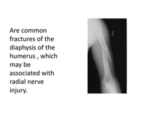
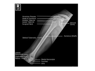
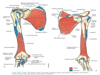
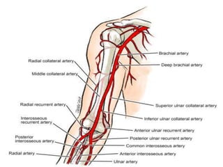
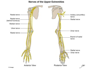
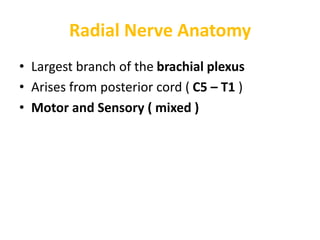
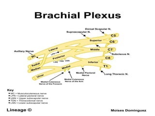
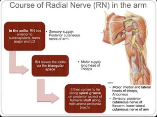
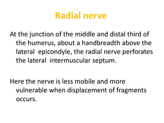
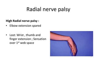
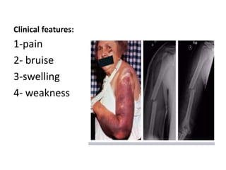
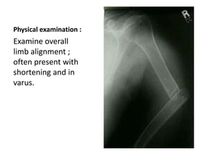
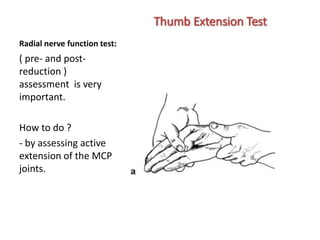
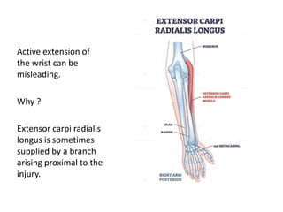
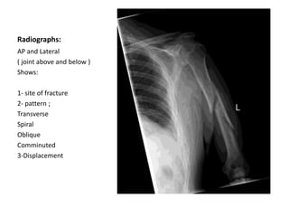
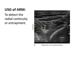
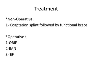
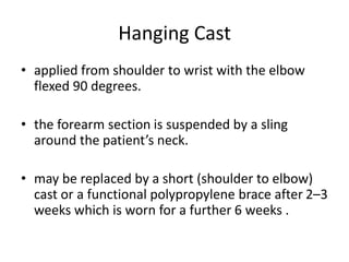
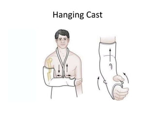
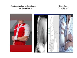
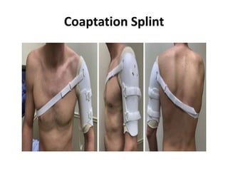
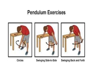
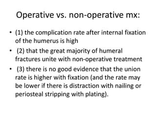
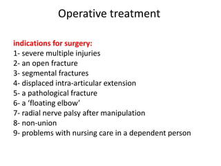
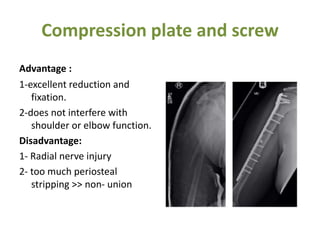
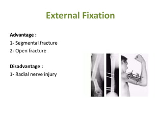
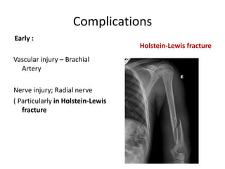
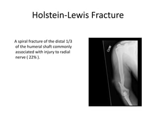
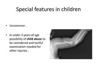
Humeral Shaft Fracture and
Principles of Management
Presented by :
(
Ismael Othman Mahmood (KBMS trainee – 2nd stage
Supervised by :
Dr. Aso Ali Bakir
Are common
fractures of the
diaphysis of the
humerus , which
may be
associated with
radial nerve
injury.
Epidemiology
• Incidence : 3-5 % of all fractures
•
• Demographics :
Bimodal age distribution
Young pt. ; high energy
Elderly pt. ; low energy
Anatomy
• extends from the surgical neck proximally to
the humeral condyles distally.
• Cylindrical shape proximally
• conical in its middle 1/3
• Flattened dramatically in the coronal plane
distally .
• muscles are divided into anterior flexor and
posterior extensor compartments .
Radial Nerve Anatomy
• Largest branch of the brachial plexus
• Arises from posterior cord ( C5 – T1 )
• Motor and Sensory ( mixed )
Radial n.
Courses along spiral groove
14 cm proximal to the lateral epicondyle and 20
cm proximal to the medial epicondyle .
Radial nerve
At the junction of the middle and distal third of
the humerus, about a handbreadth above the
lateral epicondyle, the radial nerve perforates
the lateral intermuscular septum.
Here the nerve is less mobile and more
vulnerable when displacement of fragments
occurs.
Radial nerve palsy
High Radial nerve palsy :
• Elbow extension spared
• Lost: Wrist , thumb and
finger extension ; Sensation
over 1st web space
Mechanism of injury
*A fall on the hand may twist the humerus, causing a spiral fracture .
*A fall on the elbow with the arm abducted
exerts a bending force, resulting in an oblique or transverse fracture.
*A direct blow to the arm causes a fracture which is either transverse
or comminuted.
*Fracture of the shaft in an elderly patient may be due to a metastasis
(Pathological ).
Pathological anatomy
• With fractures above the deltoid insertion- the
proximal fragment is adducted by pectoralis major and
the distal fragment laterally displaced .
• With fractures lower down- the proximal fragment is
abducted by the deltoid.
• Fractures proximal to the Brachioradialis and
extensor muscles, the distal fragment rotated laterally.
• Distal fractures tend to fall into varus.
Clinical features:
1-pain
2- bruise
3-swelling
4- weakness
Physical examination :
Examine overall
limb alignment ;
often present with
shortening and in
varus.
Radial nerve function test:
( pre- and post-
reduction )
assessment is very
important.
How to do ?
- by assessing active
extension of the MCP
joints.
Active extension of
the wrist can be
misleading.
Why ?
Extensor carpi radialis
longus is sometimes
supplied by a branch
arising proximal to the
injury.
Radiographs:
AP and Lateral
( joint above and below )
Shows:
1- site of fracture
2- pattern ;
Transverse
Spiral
Oblique
Comminuted
3-Displacement
USG of ARM:
To detect the
radial continuity
or entrapment.
Transthoracic lateral
*Sagittal plane
deformity
*avoiding further
nerve or soft tissue
injury.
Traction view:
•not routinely
indicated
Better Delineates
the fracture lines
and extent of joint
injury .
Classification
OTA
• Bone number : 1
• Location : 2
• Pattern :
*Simple A
*Wedge B
*Complex C
Classification
Descreptive
1- Location :
Proximal 1/3
Middle 1/3
Distal 1/3
2- Pattern :
Spiral
Transverse
Comminuted
Treatment
*Non-Operative ;
1- Coaptation splint followed by functional brace
*Operative :
1-ORIF
2-IMN
3- EF
Non-Operative mx:
• Fracture of the humerus heals readily.
• The weight of the arm with an external cast is
usually enough to pull the fragments into
alignment.
Hanging Cast
• applied from shoulder to wrist with the elbow
flexed 90 degrees.
• the forearm section is suspended by a sling
around the patient’s neck.
• may be replaced by a short (shoulder to elbow)
cast or a functional polypropylene brace after 2–3
weeks which is worn for a further 6 weeks .
Hanging Cast
functional polypropylene brace Short Cast
Sarmiento brace ( U – Shaped )
Coaptation Splint
Advantages of non-operative Mx:
• The wrist and fingers exercises can be done
from the start easily .
• Pendulum exercises of the shoulder are
begun within a week .
Operative vs. non-operative mx:
• (1) the complication rate after internal fixation
of the humerus is high
• (2) that the great majority of humeral
fractures unite with non-operative treatment
• (3) there is no good evidence that the union
rate is higher with fixation (and the rate may
be lower if there is distraction with nailing or
periosteal stripping with plating).
Operative treatment
indications for surgery:
1- severe multiple injuries
2- an open fracture
3- segmental fractures
4- displaced intra-articular extension
5- a pathological fracture
6- a ‘floating elbow’
7- radial nerve palsy after manipulation
8- non-union
9- problems with nursing care in a dependent person
Methods
Fixation can be achieved with:
(1) a compression plate and screws
(2) an interlocking intramedullary nail or semi-
flexible pins
(3) an external fixator
Compression plate and screw
Advantage :
1-excellent reduction and
fixation.
2-does not interfere with
shoulder or elbow function.
Disadvantage:
1- Radial nerve injury
2- too much periosteal
stripping >> non- union
Interlocking intramedullary nail
Advantage :
1- Minimal dissection
Disadvantage:
1-Rotator cuff injury
2- Distract the fracture
External Fixation
Advantage :
1- Segmental fracture
2- Open fracture
Disadvantage :
1- Radial nerve injury
Complications
Early :
Vascular injury – Brachial
Artery
Nerve injury; Radial nerve
( Particularly in Holstein-Lewis
fracture
Holstein-Lewis fracture
Complications
LATE:
1-Delayed union and non-union
( typical in segmental high energy fracture and
open fracture )
2-Joint stiffness
Holstein-Lewis Fracture
A spiral fracture of the distal 1/3
of the humeral shaft commonly
associated with injury to radial
nerve ( 22% ).
Special features in children
• Uncommon .
• in under 3 years of age
possibility of child abuse to
be considered and tactful
examination needed for
other injuries .
Special features in children
Mx:
Conservative :
* can usually be treated by
applying a collar and cuff
bandage for 3–4 weeks.
*manipulation may be needed, If
there is gross shortening.
*Older children may require a
short plaster splint.
Special features in children
Operative :
ORIF with flexible
intramedullary nail fixation
References:
• Apley and Solomon’s Concise system of
Orthopaedics and Trauma 10th Edition
• AO Principles of fracture Management 3rd Edition
• www.orthobullets.com
• www.slideshare.net
Humerus Shaft Fractur-OSCE.pptx

Humerus Shaft Fractur-OSCE.pptx

  • 1.
    Humeral Shaft Fractureand Principles of Management Presented by : ( Ismael Othman Mahmood (KBMS trainee – 2nd stage Supervised by : Dr. Aso Ali Bakir
  • 2.
    Are common fractures ofthe diaphysis of the humerus , which may be associated with radial nerve injury.
  • 3.
    Epidemiology • Incidence :3-5 % of all fractures • • Demographics : Bimodal age distribution Young pt. ; high energy Elderly pt. ; low energy
  • 4.
    Anatomy • extends fromthe surgical neck proximally to the humeral condyles distally. • Cylindrical shape proximally • conical in its middle 1/3 • Flattened dramatically in the coronal plane distally .
  • 8.
    • muscles aredivided into anterior flexor and posterior extensor compartments .
  • 11.
    Radial Nerve Anatomy •Largest branch of the brachial plexus • Arises from posterior cord ( C5 – T1 ) • Motor and Sensory ( mixed )
  • 16.
    Radial n. Courses alongspiral groove 14 cm proximal to the lateral epicondyle and 20 cm proximal to the medial epicondyle .
  • 17.
    Radial nerve At thejunction of the middle and distal third of the humerus, about a handbreadth above the lateral epicondyle, the radial nerve perforates the lateral intermuscular septum. Here the nerve is less mobile and more vulnerable when displacement of fragments occurs.
  • 18.
    Radial nerve palsy HighRadial nerve palsy : • Elbow extension spared • Lost: Wrist , thumb and finger extension ; Sensation over 1st web space
  • 22.
    Mechanism of injury *Afall on the hand may twist the humerus, causing a spiral fracture . *A fall on the elbow with the arm abducted exerts a bending force, resulting in an oblique or transverse fracture. *A direct blow to the arm causes a fracture which is either transverse or comminuted. *Fracture of the shaft in an elderly patient may be due to a metastasis (Pathological ).
  • 23.
    Pathological anatomy • Withfractures above the deltoid insertion- the proximal fragment is adducted by pectoralis major and the distal fragment laterally displaced . • With fractures lower down- the proximal fragment is abducted by the deltoid. • Fractures proximal to the Brachioradialis and extensor muscles, the distal fragment rotated laterally. • Distal fractures tend to fall into varus.
  • 25.
  • 26.
    Physical examination : Examineoverall limb alignment ; often present with shortening and in varus.
  • 27.
    Radial nerve functiontest: ( pre- and post- reduction ) assessment is very important. How to do ? - by assessing active extension of the MCP joints.
  • 28.
    Active extension of thewrist can be misleading. Why ? Extensor carpi radialis longus is sometimes supplied by a branch arising proximal to the injury.
  • 29.
    Radiographs: AP and Lateral (joint above and below ) Shows: 1- site of fracture 2- pattern ; Transverse Spiral Oblique Comminuted 3-Displacement
  • 30.
    USG of ARM: Todetect the radial continuity or entrapment.
  • 31.
  • 32.
    Traction view: •not routinely indicated BetterDelineates the fracture lines and extent of joint injury .
  • 33.
    Classification OTA • Bone number: 1 • Location : 2 • Pattern : *Simple A *Wedge B *Complex C
  • 34.
    Classification Descreptive 1- Location : Proximal1/3 Middle 1/3 Distal 1/3 2- Pattern : Spiral Transverse Comminuted
  • 35.
    Treatment *Non-Operative ; 1- Coaptationsplint followed by functional brace *Operative : 1-ORIF 2-IMN 3- EF
  • 36.
    Non-Operative mx: • Fractureof the humerus heals readily. • The weight of the arm with an external cast is usually enough to pull the fragments into alignment.
  • 37.
    Hanging Cast • appliedfrom shoulder to wrist with the elbow flexed 90 degrees. • the forearm section is suspended by a sling around the patient’s neck. • may be replaced by a short (shoulder to elbow) cast or a functional polypropylene brace after 2–3 weeks which is worn for a further 6 weeks .
  • 38.
  • 39.
    functional polypropylene braceShort Cast Sarmiento brace ( U – Shaped )
  • 40.
  • 41.
    Advantages of non-operativeMx: • The wrist and fingers exercises can be done from the start easily . • Pendulum exercises of the shoulder are begun within a week .
  • 43.
    Operative vs. non-operativemx: • (1) the complication rate after internal fixation of the humerus is high • (2) that the great majority of humeral fractures unite with non-operative treatment • (3) there is no good evidence that the union rate is higher with fixation (and the rate may be lower if there is distraction with nailing or periosteal stripping with plating).
  • 44.
    Operative treatment indications forsurgery: 1- severe multiple injuries 2- an open fracture 3- segmental fractures 4- displaced intra-articular extension 5- a pathological fracture 6- a ‘floating elbow’ 7- radial nerve palsy after manipulation 8- non-union 9- problems with nursing care in a dependent person
  • 45.
    Methods Fixation can beachieved with: (1) a compression plate and screws (2) an interlocking intramedullary nail or semi- flexible pins (3) an external fixator
  • 46.
    Compression plate andscrew Advantage : 1-excellent reduction and fixation. 2-does not interfere with shoulder or elbow function. Disadvantage: 1- Radial nerve injury 2- too much periosteal stripping >> non- union
  • 47.
    Interlocking intramedullary nail Advantage: 1- Minimal dissection Disadvantage: 1-Rotator cuff injury 2- Distract the fracture
  • 48.
    External Fixation Advantage : 1-Segmental fracture 2- Open fracture Disadvantage : 1- Radial nerve injury
  • 49.
    Complications Early : Vascular injury– Brachial Artery Nerve injury; Radial nerve ( Particularly in Holstein-Lewis fracture Holstein-Lewis fracture
  • 50.
    Complications LATE: 1-Delayed union andnon-union ( typical in segmental high energy fracture and open fracture ) 2-Joint stiffness
  • 51.
    Holstein-Lewis Fracture A spiralfracture of the distal 1/3 of the humeral shaft commonly associated with injury to radial nerve ( 22% ).
  • 52.
    Special features inchildren • Uncommon . • in under 3 years of age possibility of child abuse to be considered and tactful examination needed for other injuries .
  • 53.
    Special features inchildren Mx: Conservative : * can usually be treated by applying a collar and cuff bandage for 3–4 weeks. *manipulation may be needed, If there is gross shortening. *Older children may require a short plaster splint.
  • 54.
    Special features inchildren Operative : ORIF with flexible intramedullary nail fixation
  • 55.
    References: • Apley andSolomon’s Concise system of Orthopaedics and Trauma 10th Edition • AO Principles of fracture Management 3rd Edition • www.orthobullets.com • www.slideshare.net

Editor's Notes

  • #6 https://www.earthslab.com/anatomy/humerus/
  • #10 The brachial artery and vein as well as the median and ulnar nerves traverse the anterior compartment medial to the coracobrachialis muscle proximally and the brachialis muscle distallyThe brachial artery and vein as well as the median and ulnar nerves traverse the anterior compartment medial to the coracobrachialis muscle proximally and the brachialis muscle distally
  • #34 OTA : Orthopedic trauma association
  • #42 , but active abduction is postponed until the fracture has united (about 6 weeks for spiral fractures but often twice as long for other types); once united, only a sling is needed until the fracture is consolidated.
  • #45 a ‘floating elbow’ (simultaneous unstable humeral and forearm fractures
  • #54 Taking advantage of the robust periosteum and the power of rapid healing in children, the humeral fracture