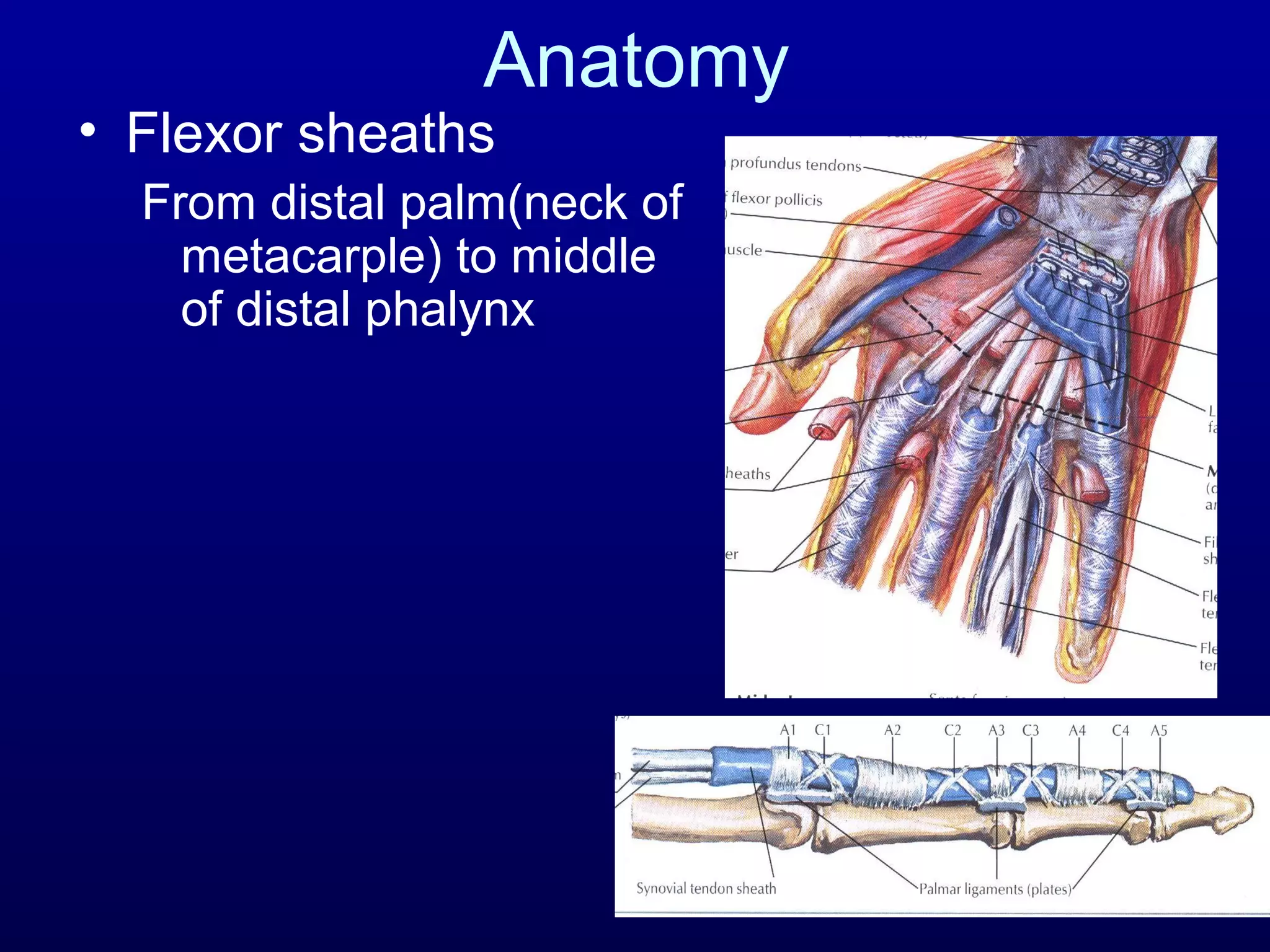
Flexor Tendon Injuries
Dr Sanjibani Sudha
FLEXOR TENDON INJURIES
• Anatomy
• Nutrition
• Healing
• Diagnosis
• Techniques
• Zone I
• Zone II injuries
• Partial lacerations
• Post op rehab
• Reconstruction ladder
 There are 12 flexor tendons in the hand and forearm
regions, they include finger, thumb flexor and wrist
flexor.
 Finger flexor are FDS,FDP
 Thumb flexor FPL
 Wrist flexor FCR,FCU,PL
Anatomy
• Extrinsic flexors
– Superficial group
• PT, FCR, FCU, PL
• Arise from medial
epicondyle, MCL,
coronoid process
Anatomy
• Extrinsic Flexors
– Intermediate group
• FDS
• Arises from medial
epicondyle, UCL,
coronoid process
• Usually have independent
musculotendinous origins
and act independantly
Anatomy
• Extrinsic flexors
– Deep group
• FPL – originates from
entire medial third of
volar radius
• FDP – originates on
proximal two thirds of the
ulna, often has common
musculotendinous origins
Anatomy
• Carpal
tunnel
– 9 tendons
– Median
nerve
Anatomy
• Flexor sheaths
From distal palm(neck of
metacarple) to middle
of distal phalynx
DIGITAL FLEXOR SHEATH
• Consist of SYNOVIAL SHEATH and PULLEYS(inter
woven condensed fibrous bands
• SYNOVIAL sheath is thin layer of continous smooth
paratenon, covering the inner surface of fibrous sheath.
• Provide smooth surface for tendon gliding
• Provide nutrition to tendon
• PULLEY SYSTEM is unique consists of annular pulley
and cruciate pulleys
• Cruciate pulleys are compressible , allowes
digital flexion to occur with condensation of fibro
osseous sheaths at inner part of flexed finger.
called as CONCERTINA effect
pulleys
• 5 annular pulleys and 3 cruciate pulleys
• Annular pulleys are heavier,condensed and rigid
• A1 A3 A5 originates from palmar plate of MCP PIP DIP
joints. A2 A4 from proximal and middle phalynx
• A2 is the broadest one,and encompasses the bifurcation
of fds
• Maintain the anatomical path of tendons clse to bone
and joints, optimising the mechanical efficiency of digital
flexion
• Thumb has 3 pulleys ;A1, oblique and A2
• A1 and oblique are fuctionaly imp
• A1 pulley located palmar to MCP jnt,
• Oblique pulley over proximal phalynx .
• A2 near the site of incertion of FPL
Anatomy
• Camper’s Chiasma
• FDS lies superficial to FDP upto bifurcation of
FDS at the level of MCP J. Then FDS tendon
becomes 2 slips coursing laterally and then
deeper to the FDP tendons .This FDS
bifurcation is in the A2 pulleys area . this part of
FDS also serves to constrain the FDS tendon.
• Deep to FDP tendon the FDS slips rejoin to form
CAMPERS chiasm and distally inserted to
proximal and middle part of middle phalynx
Tendon Morphology
• 70% collagen (Type I)
• Extracellular components
– Elastin
– Mucopolysaccharides (enhance water-binding
capability)
• Endotenon – around collagen bundles
• Epitenon – covers surface of tendon
• Paratenon – visceral/parietal adventitia
surrounding tendons in hand
• Synovial like fluid environment
Tendon Nutrition
1. Synovial fluid
:produced within tenosynovial
sheath
2. Blood supply
provide by vincular circulation
Vascular supply to flexor
tendon
Tendon Nutrition
• Vascular
– Longitudinal vessels
• Enter in palm
• Enter at proximal synovial fold
– Segmental branches from digital arteries
• Long and short vinculae
– Vessels at osseous insertions
• Synovial fluid diffusion
– Imbibition (pumping mechanism)
Tendon Nutrition
• Dorsal vascularity
• Avascular zones
– FDS (over proximal
phalanx
– FDP (over middle phalanx)
• Nutrition vital for rapid
healing, minimization of
adhesion and restoration
of gliding
 Nutrition of tendon
 Suspensory ligament of tendon
 Stabilization of tendon
FDP has 2 vincula; a fan like short vinculum and
a cord like long vinculum.
Short v is located at insertion of FDP tendon
Long v connects the FDP tendon through the short
v of FDS tendon on the floor of palmar surface of
phalynx
Flexor tendon healing
• 2 forms:
– Intrinsic healing:
– From prolifaration of tenocytes and extracellular
matrix from intrinsic cells. occurs without direct
blood flow to the tendon
– Intrinsic healing is innately weaker then extrinsic
healing
– If intrinsic capacity is disabled such as in
excessive trauma, or in case of post surgical
immobilisation extrinsic healing is favoured
• Extrinsic healing: occurs by proliferation of
fibroblasts from the peripheral epitendon;
adhesions occur because of extrinsic healing of
the tendon and limit tendon gliding within fibrous
synovial sheaths
Phases of Intrinsic healing
1.Inflammatory (0-5 days) : strength of the
repair is reliant on the strength of the
suture itself
2.Fibroblastic (5-28 days) : or so-called
collagen-producing phase
3.Remodelling (>28 days)
Tendon Adhesion
• Increased adhesion formation with:
– Traumatic/surgical injury
• Crush injuries
– Ischemia
• Disruption of vinculae
– Immobilization
– Gapping at repair site
– Excision/injury to flexor sheath components
• Debate over benefit of sheath repair
Tendon Adhesion
• Experimental attempts to minimize
adhesion formation
– Oral: steroids, antihistamines, NSAIDS
– Topical: beta-aminoproprionitrile,
hydrocyprolins, hyaluronic acid, collagen
solutions, fibrin
– Physical: silicone/cellophane wrapping,
polyethylene tubes, interposed sheath flaps
• Varying lab success but none proven
definitively or adopted into clinical practice
Diagnosis
• History
Diagnosis
• Physical exam
• Abnormal resting posture
• Absent FDP / FDS function
• Associated digital nerve and digital vessel
injury
• Discuss nature of injury and postoperative
course with patient
I Distal to
sublimis
II No man’s
land
III
Lumbrical
origin
IV carpal
tunnel
V proximal
to carpal tunnel
“No
Man’s
Land”
Bunnell
ZONES
• ZONE 1- distal to the insertion of
superficialis
• ZONE 2- no mans land b/w the distal
palmar crease and insertion of the
sublimis
• ZONE 3- lumbrical region b/w distal
palmar crease and the distal end of
transverse carpal ligament
• ZONE 4- zone covered by the flexor
retinaculum
• ZONE 5- proximal to the flexor
Zones of Injury
FDS Insertion
Flexor Sheath
(proximal)
TCL
(distal edge) Carpal Tunnel
RULES OF REPAIR
• All flexor tendons when severed should be repaired
irrespective of whatever is the zone.
• Ideally fix the fracture and repair the tendon and digital
primarily.
• When delayed repair is done may need a tendon graft
• Flexor tendon sheath should be repaired over the
tendon
• A2 and A4 pulleys of the flexor sheaths should be
preserved to prevent bowstringing and flexon
deformity
Timing of surgery
• primary –within 24 hrs
• Delayed primary -1 to 10 days
• Secondary – upto 4 wks
• Late secondary beyond 4 wks
• If wound is clean, primary repair, along
with care of neurovascular injury and
fracture.
Incisions
• Factors
– Avoid crossing joints
at 90 deg.
– Preference
– Existing lacerations
– Need to expose other
structures
Repair Techniques
• Ideal
– Gap resistant
– Strong enough to tolerate forces generated by
early controlled active motion protocols
• 10-50% decrease in repair strength from day 5-21
post repair in immobilized tendons
• This is effect is minimized (possibly eliminated)
through application of early motion stress
– Uncomplicated
– Minimal bulk
– Minimal interference with tendon vascularity
• Core sutures
• Epitenon sutures – reduce bulk and increase strength
• Core sutures –
- careful handling and alignment of tendons
- more dorsal suture placement  better outcome
- More sutures crossing the interaction site, more
strength
- lacerations involving more than 60 % cross-section of
the tendon should be repaired
- 4-0 braided sutures like capromid, polyester
Suture Configurations
Core Sutures
• Current literature supports several conclusions
regarding core sutures
– Strength proportional to number of strands
– Locking loops increase strength but may collapse and
lead to gapping
– Knots should be outside repair site
– Increased suture callibre = increases strength
– Braided 3-0 or 4-0 probably best suture material
– Dorsally placed suture stronger and biomechanically
advantageous
– Equal tension across all strands
Bunnel Stitch
Kessler Stitch
Masson-Allen Criss-cross
Modified kessler Tajima
Masson-Allen Criss-cross
Modified kessler Tajima
Sheath Repair
• Advantages
– Barrier to extrinsic adhesion formation
– More rapid return of synovial nutrition
• Disadvantages
– Technically difficult
– Increased foreign material at repair site
– May narrow sheathand restrict glide
• Presently, no clear cut advantage to
sheath repair has been established
Partial Lacerations
• Controversy in past as partial lacerations
were felt to predispose to entrapment,
triggering and rupture
• Repair if > 50%
• Some advocate repair of partial
lacerations > 60%
ZONE 1
• Repair primarily
• To distal stump or advance 1 cm
• More than 1 cm quadriga effect.
• Type 1: 7-10 days retracted in the palm. Repair
with a pull out wire technique.
• Type 2: few months retracted till PIP
• Type 3: retracted upto DIP.
• Old cases: tendon grafting, arthrodesis,
tenodesis
Tendon Advancement
– Previously advocated for zone 1 repairs, as
moving the repair site out of the sheath was
felt to decrease adhesion formation
– Disadvantages
• Shortening of flexor system
• Contracture
• Quadregia effect
• Little excursion distally, therefore adhesions near
insertion less of an issue
Pull out wire technique
ZONE 2
• Called bunnells zone or no mans land
zone.
• Notorious for bad results, adhesions are a
big problem. Area of pulleys
• Repair both tendons at the same time
• Tendons are sutured malrotated
• Suturing should be intratendonous
configuration.
ZONE 3
• Lumbrical zone
• Primarily sutured
• Do not suture the belly of the lumbricals as
it increases the tension of the muscles
• Lumbrical plus syndrome- paradoxical
extension on attempted flexion
Tendon Excursion
Summary
• Strong gap resistant repair
• 4 strand, locking epitendinous (or
equivalent), 3-0 suture needed for early
active motion
– 4-0 suture, modified Kessler, running
epitendinous suture adequate for more
conservative protocols
• No sheath repair
• Large grasping/locking loops
FDP Avulsions
• Commonly male athletes
• Forced extension at DIP during maximal
flexion (jersey finger)
• Often missed due to normal xray and
intact flexion at MP and PIP
– Opportunity for FDP reinsertion lost if
treatment delayed
FDP Avulsions
Leddy
and
Packer
FDP Avulsions
- Type 1: zig-zag exposure
- Tendon delivered
through pulley system
with catheter passed
retrograde
- Fixed to base of phalanx
with monofilament
suture through distal
phalanx and nail plate
and tied over button
- Fix within 7-10 days
before tendon
degeneration and
myostatic shortening
occurs
FDP Avulsions
- Type 2: small bony
fragment retracts to
A3 level
- Can fix up to 6 wks
post injury (less
shortening)
- May convert to type 1
if tendon slips through
A3 pulley and into
palm
- Use same technique
as for type 1
FDP Avulsions
- Type 3: large bony
fragment retracts to
A4 level
- Bony reduction and
fixation of fragment
Children
• Usually not able to reliably participate in
rehabilitation programs
• No benefit to early mobilization in patients
under 16 years
• Immobilization > 4 wks may lead to poorer
outcomes
Reconstruction
Ladder of tendon reconstruction
• Tenolysis
• Tendon transfer
• Primary tendon graft
• Two stage tendon graft
Tenolysis
• Indication
tendon adhesion
• Preop evaluation
discripency exists between passive and active
range of motion after hand therapy has platued
• Timing
– 3-6 mos. Post repair (minimum)
– Plateau with physiotherapy
• Anesthesia
– Local with sedation
• Allows patient participation
Tenolysis
• Technique
– Zig zag incisions
– Adhesions divided maintaining non-limiting
adhesions
– Pulleys reconstructed as needed
• If extensive or not possible convert to staged
reconstruction
– On post op- active range of motion should
begin immediately
Single Stage Tendon Grafting
Zone 2
Indications
 injuries resulting in segmental tendon loss.
 Delay in repair that obviates primary repair, such as
laceration that have been neglacted for 3 to 6 weeks and
show tendon degenerations
 Zone 2 injury where large section of tendon have been
damaged and surgeon belives delayed grafting is better
option
 Delay presentation of FDP avulsion injury associated
with significant tendon retraction
Supple joints with adequate passive ROM
Principles include
• Place only one graft in each finger
• Never sacrifice intact FDS
• Use a graft of smaller caliber
• Perform the junctions outside the tndon sheath
• Ensure adequate graft tension
• Before sx wound should be well healed with no extensive
scarring
• Joint should be free of contracture and maximum
passive range of motion shd have been attained
• Circulation is satisfactory
• At least one digital nerve in affected digit is intact
• Pts with joint contacture shd undergo hand therapy
regimen to achive maximum possible passive range of
motion
• In case of extensive scarring,pulley incompitance,joint
contacture single stage grafting is contraindicated.
Single Stage Tendon Grafting
Zone 2
• Technique
– 1 cm distal FDP stump left intact
– 1 cm of FDS insertion left intact (decreased
adhesion formation vs granulating insertion
site)
– Tenodesis of FDS tail to flexor sheath (10-20
deg of flexion) optional
• Hyperextension at PIP in absence of FDS tendon
occurs occasionally
Single Stage Tendon Grafting
Zone 2
• Technique
– Graft donors
• Palmaris longus
• Plantaris
• Long toe extensors
• (EIP)
• (EDM)
Single Stage Tendon Grafting
Zone 2
• Technique
– Graft passed through pulley system
• Atraumatic technique
– Distal fixation with tension set proximally or
proximal fixation first
– Multiple methods for fixation of graft ends
DISTAL JUNCTURE
• No gliding motion occures at distal juncture,making it as
paramount concern there shd be a durable and firm
union between the tendon and FDPstump or volar base
of distal phalynx
• If enough FDP stump is available graft can be sutured to
stump or woven through the stump
• If sufficient stump is not available BUNNEL TENDON TO
BONE PULL OUT technique is used
• Place a transverse drill hole across the base of distal
phalynx and the graft is threaded through the hole and
sutured to itself.in this case graft must be thin enough to
pass through bony tunnel
Single Stage Tendon Grafting
Zone 2
• Technique
– Distal
juncture
PROXIMAL JUNCTURE
• Is made into FDP tendon just distal to lumbrical origin
• Motor tendon is slit at its end and graft is threaded into
the slit
• pts graft length is estimated in an anesthetized pts by
relaxed position of finger with the wrist in neutral position
• Here each finger shd fall into semiflexion, slightly less
flexed then its ulnar neighbour.
Single Stage Tendon Grafting
Zone 2
• Technique
– proximal
juncture
Pulvertaft weave creates a
stronger repair vs end to end
techniques, and allows for
greater ease when setting
tension
Post op care
 Early motion exercise have been used
 Protected passive range of motion can be instituted after
2 to 3 days of sx if the juncture are strong enough to
withstand the tesile strength
 Post op splint- static dorsal bloking splint with wrist
positioned in neutral,MP in 45 degree flexion, Ipin
neutral. And worn for 6 weeks after sx

Secondary Reconstruction
Zone 1
• Zone 1 (functioning FDS)
– Eg. Late presentation of FDP avulsion
– DIP fusion
– Tendon graft
• Risks damaging FDS function through
injury/adhesions in a very functional finger
• ? Young patients, supple joints, need for active
DIP flexion
Secondary Reconstruction
Zones 3, 4 and 5
• Usually associated with 3 – 5 cm gap
– Interposition graft
– FDS to FDP transfer
– End to side profundus juncture
Two Stage Reconstruction
• Primary grafting likely to give poor result,
but salvage of functioning finger still
desirable
• Sub-optimal conditions
– Extensive soft tissue scarring
• Crush injuries
• Associated fractures, nerve injuries
– Loss of significant portion of pulley system
Two Stage Reconstruction
• Patient selection
– Motivated
– Absence of neurovascular injury
– Good passive joint motion
• Balance benefits of two additional
procedures in an already traumatized digit
with amputation/arthrodesis
Two Stage Reconstruction
• Stage 1
– Excision of tendon remnants
• Distal 1 cm of FDP left intact, remainder excised to
lumbrical level
• FDS tail preserved for potential pulley
reconstruction
– Incision proximal to wrist
• FDS removed/excised
• Hunter rod then placed through pulley system and
fixed distally (suture or plate and screw –
depending on implant)
Tenolysis
– Release of nongliding adhesions for salvage
in poorly functioning digits with previous
tendon injury
– Avoid in marginal digits
• May not tolerate additional vascular/neurologic
injury
– May need concomitant collateral ligament
release, capsulotomy
– Prepare patient for possible staged
reconstruction
Two Stage Reconstruction
• Stage 1
– Rod extends proximally to distal forearm in
plane between FDS and FDP
– Test glide
– Reconstruct pulleys as needed if implant
bowstrings
Two Stage Reconstruction
• Stage 1
– Postop
• Start passive motion at 7 days
• Continue x 3mos to allow pseodosheath to form
around implant
• Before stage 2 joints should be supple, and
wounds soft
Two Stage Reconstruction
• Stage 2 – implant removal and tendon
graft insertion
– Distal and proximal incisions opened
– Implant located proximally and motor selected
(FDP middle/ring/small, FDP index)
– Graft harvested, sutured to proximal implant
and delivered distally
• Fixed to distal phalanx with pull out wire over
button
Two Stage Reconstruction
• Stage 2 – implant removal and tendon
graft insertion
– Proximally sutured to motor with pulvertaft
weave
• FDS transfer from adjacent digit described
• Obviates need for graft
• Difficulty with length/tension
• Postop
• Early controlled motion x 3 wks, then slow
progression to active motion
Pulley Reconstruction
• Pulley loss
– Bowstringing = tendon taking shortest
distance between remaining pulleys
– Biomechanical disadvantage
• Excursion translates into less joint motion
– Adhesions/rupture at remaining pulleys due to
increased stress
– A2 and A4 needed (minimum)
• Most biomechanically important
• Some authors advocate reconstructing a 3 or 4
pulley system for optimal results
Pulley Reconstruction
• Most done in conjunction with a two stage
tendon reconstruction
• Can be done with single stage tendon
graft
• generally if extensive pulley reconstruction
is required it is better to do a two stage
procedure
Pulley Reconstruction
• Methods
– Superficialis tendon
• Insertion left intact
• Remnant sutured to original pulley rim, to
periosteum, or to bone through drill holes
– Tendon graft
• Sutured as above
• Passed through hole drilled in phalanx (risk of
fracture)
• Wrapped around phalanx (requires 6-8 cm of graft)
Pulley Reconstruction
• Methods
– Extensor retinaculum
• Excellent gliding surface
• Difficult to harvest the 8-6 cm required for fixation
around phalanx
– Artificial materials
• Dacron, PTFE, nylon silicone
• Due to abundant atogenous material and
disadvantages of artificial materials, this has not
become common clinical practice
• May be stronger in long term vs autogenous

Flexortendoninjuries1 140504003542-phpapp01

  • 1.
  • 2.
    FLEXOR TENDON INJURIES •Anatomy • Nutrition • Healing • Diagnosis • Techniques • Zone I • Zone II injuries • Partial lacerations • Post op rehab • Reconstruction ladder
  • 3.
     There are12 flexor tendons in the hand and forearm regions, they include finger, thumb flexor and wrist flexor.  Finger flexor are FDS,FDP  Thumb flexor FPL  Wrist flexor FCR,FCU,PL
  • 4.
    Anatomy • Extrinsic flexors –Superficial group • PT, FCR, FCU, PL • Arise from medial epicondyle, MCL, coronoid process
  • 5.
    Anatomy • Extrinsic Flexors –Intermediate group • FDS • Arises from medial epicondyle, UCL, coronoid process • Usually have independent musculotendinous origins and act independantly
  • 6.
    Anatomy • Extrinsic flexors –Deep group • FPL – originates from entire medial third of volar radius • FDP – originates on proximal two thirds of the ulna, often has common musculotendinous origins
  • 7.
    Anatomy • Carpal tunnel – 9tendons – Median nerve
  • 8.
    Anatomy • Flexor sheaths Fromdistal palm(neck of metacarple) to middle of distal phalynx
  • 9.
    DIGITAL FLEXOR SHEATH •Consist of SYNOVIAL SHEATH and PULLEYS(inter woven condensed fibrous bands • SYNOVIAL sheath is thin layer of continous smooth paratenon, covering the inner surface of fibrous sheath. • Provide smooth surface for tendon gliding • Provide nutrition to tendon • PULLEY SYSTEM is unique consists of annular pulley and cruciate pulleys
  • 10.
    • Cruciate pulleysare compressible , allowes digital flexion to occur with condensation of fibro osseous sheaths at inner part of flexed finger. called as CONCERTINA effect
  • 12.
    pulleys • 5 annularpulleys and 3 cruciate pulleys • Annular pulleys are heavier,condensed and rigid • A1 A3 A5 originates from palmar plate of MCP PIP DIP joints. A2 A4 from proximal and middle phalynx • A2 is the broadest one,and encompasses the bifurcation of fds • Maintain the anatomical path of tendons clse to bone and joints, optimising the mechanical efficiency of digital flexion
  • 14.
    • Thumb has3 pulleys ;A1, oblique and A2 • A1 and oblique are fuctionaly imp • A1 pulley located palmar to MCP jnt, • Oblique pulley over proximal phalynx . • A2 near the site of incertion of FPL
  • 15.
  • 16.
    • FDS liessuperficial to FDP upto bifurcation of FDS at the level of MCP J. Then FDS tendon becomes 2 slips coursing laterally and then deeper to the FDP tendons .This FDS bifurcation is in the A2 pulleys area . this part of FDS also serves to constrain the FDS tendon. • Deep to FDP tendon the FDS slips rejoin to form CAMPERS chiasm and distally inserted to proximal and middle part of middle phalynx
  • 17.
    Tendon Morphology • 70%collagen (Type I) • Extracellular components – Elastin – Mucopolysaccharides (enhance water-binding capability) • Endotenon – around collagen bundles • Epitenon – covers surface of tendon • Paratenon – visceral/parietal adventitia surrounding tendons in hand • Synovial like fluid environment
  • 18.
    Tendon Nutrition 1. Synovialfluid :produced within tenosynovial sheath 2. Blood supply provide by vincular circulation Vascular supply to flexor tendon
  • 19.
    Tendon Nutrition • Vascular –Longitudinal vessels • Enter in palm • Enter at proximal synovial fold – Segmental branches from digital arteries • Long and short vinculae – Vessels at osseous insertions • Synovial fluid diffusion – Imbibition (pumping mechanism)
  • 21.
    Tendon Nutrition • Dorsalvascularity • Avascular zones – FDS (over proximal phalanx – FDP (over middle phalanx) • Nutrition vital for rapid healing, minimization of adhesion and restoration of gliding
  • 22.
     Nutrition oftendon  Suspensory ligament of tendon  Stabilization of tendon
  • 23.
    FDP has 2vincula; a fan like short vinculum and a cord like long vinculum. Short v is located at insertion of FDP tendon Long v connects the FDP tendon through the short v of FDS tendon on the floor of palmar surface of phalynx
  • 24.
    Flexor tendon healing •2 forms: – Intrinsic healing: – From prolifaration of tenocytes and extracellular matrix from intrinsic cells. occurs without direct blood flow to the tendon – Intrinsic healing is innately weaker then extrinsic healing – If intrinsic capacity is disabled such as in excessive trauma, or in case of post surgical immobilisation extrinsic healing is favoured
  • 25.
    • Extrinsic healing:occurs by proliferation of fibroblasts from the peripheral epitendon; adhesions occur because of extrinsic healing of the tendon and limit tendon gliding within fibrous synovial sheaths
  • 26.
    Phases of Intrinsichealing 1.Inflammatory (0-5 days) : strength of the repair is reliant on the strength of the suture itself 2.Fibroblastic (5-28 days) : or so-called collagen-producing phase 3.Remodelling (>28 days)
  • 27.
    Tendon Adhesion • Increasedadhesion formation with: – Traumatic/surgical injury • Crush injuries – Ischemia • Disruption of vinculae – Immobilization – Gapping at repair site – Excision/injury to flexor sheath components • Debate over benefit of sheath repair
  • 28.
    Tendon Adhesion • Experimentalattempts to minimize adhesion formation – Oral: steroids, antihistamines, NSAIDS – Topical: beta-aminoproprionitrile, hydrocyprolins, hyaluronic acid, collagen solutions, fibrin – Physical: silicone/cellophane wrapping, polyethylene tubes, interposed sheath flaps • Varying lab success but none proven definitively or adopted into clinical practice
  • 29.
  • 30.
    Diagnosis • Physical exam •Abnormal resting posture • Absent FDP / FDS function • Associated digital nerve and digital vessel injury • Discuss nature of injury and postoperative course with patient
  • 31.
    I Distal to sublimis IINo man’s land III Lumbrical origin IV carpal tunnel V proximal to carpal tunnel “No Man’s Land” Bunnell
  • 32.
    ZONES • ZONE 1-distal to the insertion of superficialis • ZONE 2- no mans land b/w the distal palmar crease and insertion of the sublimis • ZONE 3- lumbrical region b/w distal palmar crease and the distal end of transverse carpal ligament • ZONE 4- zone covered by the flexor retinaculum • ZONE 5- proximal to the flexor
  • 33.
    Zones of Injury FDSInsertion Flexor Sheath (proximal) TCL (distal edge) Carpal Tunnel
  • 34.
    RULES OF REPAIR •All flexor tendons when severed should be repaired irrespective of whatever is the zone. • Ideally fix the fracture and repair the tendon and digital primarily. • When delayed repair is done may need a tendon graft • Flexor tendon sheath should be repaired over the tendon • A2 and A4 pulleys of the flexor sheaths should be preserved to prevent bowstringing and flexon deformity
  • 35.
    Timing of surgery •primary –within 24 hrs • Delayed primary -1 to 10 days • Secondary – upto 4 wks • Late secondary beyond 4 wks • If wound is clean, primary repair, along with care of neurovascular injury and fracture.
  • 36.
    Incisions • Factors – Avoidcrossing joints at 90 deg. – Preference – Existing lacerations – Need to expose other structures
  • 37.
    Repair Techniques • Ideal –Gap resistant – Strong enough to tolerate forces generated by early controlled active motion protocols • 10-50% decrease in repair strength from day 5-21 post repair in immobilized tendons • This is effect is minimized (possibly eliminated) through application of early motion stress – Uncomplicated – Minimal bulk – Minimal interference with tendon vascularity
  • 38.
    • Core sutures •Epitenon sutures – reduce bulk and increase strength • Core sutures – - careful handling and alignment of tendons - more dorsal suture placement  better outcome - More sutures crossing the interaction site, more strength - lacerations involving more than 60 % cross-section of the tendon should be repaired - 4-0 braided sutures like capromid, polyester Suture Configurations
  • 39.
    Core Sutures • Currentliterature supports several conclusions regarding core sutures – Strength proportional to number of strands – Locking loops increase strength but may collapse and lead to gapping – Knots should be outside repair site – Increased suture callibre = increases strength – Braided 3-0 or 4-0 probably best suture material – Dorsally placed suture stronger and biomechanically advantageous – Equal tension across all strands
  • 40.
  • 41.
  • 42.
  • 43.
    Sheath Repair • Advantages –Barrier to extrinsic adhesion formation – More rapid return of synovial nutrition • Disadvantages – Technically difficult – Increased foreign material at repair site – May narrow sheathand restrict glide • Presently, no clear cut advantage to sheath repair has been established
  • 44.
    Partial Lacerations • Controversyin past as partial lacerations were felt to predispose to entrapment, triggering and rupture • Repair if > 50% • Some advocate repair of partial lacerations > 60%
  • 45.
    ZONE 1 • Repairprimarily • To distal stump or advance 1 cm • More than 1 cm quadriga effect. • Type 1: 7-10 days retracted in the palm. Repair with a pull out wire technique. • Type 2: few months retracted till PIP • Type 3: retracted upto DIP. • Old cases: tendon grafting, arthrodesis, tenodesis
  • 46.
    Tendon Advancement – Previouslyadvocated for zone 1 repairs, as moving the repair site out of the sheath was felt to decrease adhesion formation – Disadvantages • Shortening of flexor system • Contracture • Quadregia effect • Little excursion distally, therefore adhesions near insertion less of an issue
  • 47.
    Pull out wiretechnique
  • 48.
    ZONE 2 • Calledbunnells zone or no mans land zone. • Notorious for bad results, adhesions are a big problem. Area of pulleys • Repair both tendons at the same time • Tendons are sutured malrotated • Suturing should be intratendonous configuration.
  • 49.
    ZONE 3 • Lumbricalzone • Primarily sutured • Do not suture the belly of the lumbricals as it increases the tension of the muscles • Lumbrical plus syndrome- paradoxical extension on attempted flexion
  • 50.
  • 51.
    Summary • Strong gapresistant repair • 4 strand, locking epitendinous (or equivalent), 3-0 suture needed for early active motion – 4-0 suture, modified Kessler, running epitendinous suture adequate for more conservative protocols • No sheath repair • Large grasping/locking loops
  • 52.
    FDP Avulsions • Commonlymale athletes • Forced extension at DIP during maximal flexion (jersey finger) • Often missed due to normal xray and intact flexion at MP and PIP – Opportunity for FDP reinsertion lost if treatment delayed
  • 53.
  • 54.
    FDP Avulsions - Type1: zig-zag exposure - Tendon delivered through pulley system with catheter passed retrograde - Fixed to base of phalanx with monofilament suture through distal phalanx and nail plate and tied over button - Fix within 7-10 days before tendon degeneration and myostatic shortening occurs
  • 55.
    FDP Avulsions - Type2: small bony fragment retracts to A3 level - Can fix up to 6 wks post injury (less shortening) - May convert to type 1 if tendon slips through A3 pulley and into palm - Use same technique as for type 1
  • 56.
    FDP Avulsions - Type3: large bony fragment retracts to A4 level - Bony reduction and fixation of fragment
  • 57.
    Children • Usually notable to reliably participate in rehabilitation programs • No benefit to early mobilization in patients under 16 years • Immobilization > 4 wks may lead to poorer outcomes
  • 58.
  • 59.
    Ladder of tendonreconstruction • Tenolysis • Tendon transfer • Primary tendon graft • Two stage tendon graft
  • 60.
    Tenolysis • Indication tendon adhesion •Preop evaluation discripency exists between passive and active range of motion after hand therapy has platued • Timing – 3-6 mos. Post repair (minimum) – Plateau with physiotherapy • Anesthesia – Local with sedation • Allows patient participation
  • 61.
    Tenolysis • Technique – Zigzag incisions – Adhesions divided maintaining non-limiting adhesions – Pulleys reconstructed as needed • If extensive or not possible convert to staged reconstruction – On post op- active range of motion should begin immediately
  • 62.
    Single Stage TendonGrafting Zone 2 Indications  injuries resulting in segmental tendon loss.  Delay in repair that obviates primary repair, such as laceration that have been neglacted for 3 to 6 weeks and show tendon degenerations  Zone 2 injury where large section of tendon have been damaged and surgeon belives delayed grafting is better option  Delay presentation of FDP avulsion injury associated with significant tendon retraction Supple joints with adequate passive ROM
  • 63.
    Principles include • Placeonly one graft in each finger • Never sacrifice intact FDS • Use a graft of smaller caliber • Perform the junctions outside the tndon sheath • Ensure adequate graft tension
  • 64.
    • Before sxwound should be well healed with no extensive scarring • Joint should be free of contracture and maximum passive range of motion shd have been attained • Circulation is satisfactory • At least one digital nerve in affected digit is intact • Pts with joint contacture shd undergo hand therapy regimen to achive maximum possible passive range of motion • In case of extensive scarring,pulley incompitance,joint contacture single stage grafting is contraindicated.
  • 65.
    Single Stage TendonGrafting Zone 2 • Technique – 1 cm distal FDP stump left intact – 1 cm of FDS insertion left intact (decreased adhesion formation vs granulating insertion site) – Tenodesis of FDS tail to flexor sheath (10-20 deg of flexion) optional • Hyperextension at PIP in absence of FDS tendon occurs occasionally
  • 66.
    Single Stage TendonGrafting Zone 2 • Technique – Graft donors • Palmaris longus • Plantaris • Long toe extensors • (EIP) • (EDM)
  • 67.
    Single Stage TendonGrafting Zone 2 • Technique – Graft passed through pulley system • Atraumatic technique – Distal fixation with tension set proximally or proximal fixation first – Multiple methods for fixation of graft ends
  • 68.
    DISTAL JUNCTURE • Nogliding motion occures at distal juncture,making it as paramount concern there shd be a durable and firm union between the tendon and FDPstump or volar base of distal phalynx • If enough FDP stump is available graft can be sutured to stump or woven through the stump • If sufficient stump is not available BUNNEL TENDON TO BONE PULL OUT technique is used • Place a transverse drill hole across the base of distal phalynx and the graft is threaded through the hole and sutured to itself.in this case graft must be thin enough to pass through bony tunnel
  • 69.
    Single Stage TendonGrafting Zone 2 • Technique – Distal juncture
  • 70.
    PROXIMAL JUNCTURE • Ismade into FDP tendon just distal to lumbrical origin • Motor tendon is slit at its end and graft is threaded into the slit • pts graft length is estimated in an anesthetized pts by relaxed position of finger with the wrist in neutral position • Here each finger shd fall into semiflexion, slightly less flexed then its ulnar neighbour.
  • 71.
    Single Stage TendonGrafting Zone 2 • Technique – proximal juncture Pulvertaft weave creates a stronger repair vs end to end techniques, and allows for greater ease when setting tension
  • 72.
    Post op care Early motion exercise have been used  Protected passive range of motion can be instituted after 2 to 3 days of sx if the juncture are strong enough to withstand the tesile strength  Post op splint- static dorsal bloking splint with wrist positioned in neutral,MP in 45 degree flexion, Ipin neutral. And worn for 6 weeks after sx 
  • 73.
    Secondary Reconstruction Zone 1 •Zone 1 (functioning FDS) – Eg. Late presentation of FDP avulsion – DIP fusion – Tendon graft • Risks damaging FDS function through injury/adhesions in a very functional finger • ? Young patients, supple joints, need for active DIP flexion
  • 74.
    Secondary Reconstruction Zones 3,4 and 5 • Usually associated with 3 – 5 cm gap – Interposition graft – FDS to FDP transfer – End to side profundus juncture
  • 75.
    Two Stage Reconstruction •Primary grafting likely to give poor result, but salvage of functioning finger still desirable • Sub-optimal conditions – Extensive soft tissue scarring • Crush injuries • Associated fractures, nerve injuries – Loss of significant portion of pulley system
  • 76.
    Two Stage Reconstruction •Patient selection – Motivated – Absence of neurovascular injury – Good passive joint motion • Balance benefits of two additional procedures in an already traumatized digit with amputation/arthrodesis
  • 77.
    Two Stage Reconstruction •Stage 1 – Excision of tendon remnants • Distal 1 cm of FDP left intact, remainder excised to lumbrical level • FDS tail preserved for potential pulley reconstruction – Incision proximal to wrist • FDS removed/excised • Hunter rod then placed through pulley system and fixed distally (suture or plate and screw – depending on implant)
  • 78.
    Tenolysis – Release ofnongliding adhesions for salvage in poorly functioning digits with previous tendon injury – Avoid in marginal digits • May not tolerate additional vascular/neurologic injury – May need concomitant collateral ligament release, capsulotomy – Prepare patient for possible staged reconstruction
  • 79.
    Two Stage Reconstruction •Stage 1 – Rod extends proximally to distal forearm in plane between FDS and FDP – Test glide – Reconstruct pulleys as needed if implant bowstrings
  • 80.
    Two Stage Reconstruction •Stage 1 – Postop • Start passive motion at 7 days • Continue x 3mos to allow pseodosheath to form around implant • Before stage 2 joints should be supple, and wounds soft
  • 81.
    Two Stage Reconstruction •Stage 2 – implant removal and tendon graft insertion – Distal and proximal incisions opened – Implant located proximally and motor selected (FDP middle/ring/small, FDP index) – Graft harvested, sutured to proximal implant and delivered distally • Fixed to distal phalanx with pull out wire over button
  • 82.
    Two Stage Reconstruction •Stage 2 – implant removal and tendon graft insertion – Proximally sutured to motor with pulvertaft weave • FDS transfer from adjacent digit described • Obviates need for graft • Difficulty with length/tension • Postop • Early controlled motion x 3 wks, then slow progression to active motion
  • 83.
    Pulley Reconstruction • Pulleyloss – Bowstringing = tendon taking shortest distance between remaining pulleys – Biomechanical disadvantage • Excursion translates into less joint motion – Adhesions/rupture at remaining pulleys due to increased stress – A2 and A4 needed (minimum) • Most biomechanically important • Some authors advocate reconstructing a 3 or 4 pulley system for optimal results
  • 84.
    Pulley Reconstruction • Mostdone in conjunction with a two stage tendon reconstruction • Can be done with single stage tendon graft • generally if extensive pulley reconstruction is required it is better to do a two stage procedure
  • 85.
    Pulley Reconstruction • Methods –Superficialis tendon • Insertion left intact • Remnant sutured to original pulley rim, to periosteum, or to bone through drill holes – Tendon graft • Sutured as above • Passed through hole drilled in phalanx (risk of fracture) • Wrapped around phalanx (requires 6-8 cm of graft)
  • 86.
  • 87.
    Pulley Reconstruction • Methods –Extensor retinaculum • Excellent gliding surface • Difficult to harvest the 8-6 cm required for fixation around phalanx – Artificial materials • Dacron, PTFE, nylon silicone • Due to abundant atogenous material and disadvantages of artificial materials, this has not become common clinical practice • May be stronger in long term vs autogenous

Editor's Notes

  • #8 At the wrist level ten structures pass through the carpal tunnel 4xfds, 4xfdp, fpl and median nerve Bordered by:hamate, triquetrum and pisiform and ulnarly Scaphoid and trapezium radially Roof = flexor retinaculum Consists of three components – the the deep forearm facsia, the transverse carpal ligament (which traverses from the scaphoid tuberosity and trapezial beak radially to the hook of the hamate and pisiform ulnary) and the distal aponeurosis btw the thenar and hypothenar muscles
  • #9 Pulleys A2 and a4 (most biomechanically important) arise from periosteum of proximal aspect of proximal phalanx, and middile aspect of middle phalanx (respectively) A1 a3 and a5 (joint pulleys) arise from volar plates of mp, pip and dip joints respectively Intervening cruciate pulleys (c1 c2 and c3) are thin and collapse to allow annular pulley approximation during flexion
  • #16 In the palm FDS is superficial to FDP In the proximal sheath fds divides to allow fdp to pass through and go on to insert at the base of the distal phalanx Fds the reunites at camper’s chiasm before dividing into two slips of insertion on the base of the middle phalanx
  • #34 Zone 1: distal to FDS insertion Zone 2: FDS insertion proximal beginning of flexor sheath Previously “no man’s land” Zone 3: distal edge of TCL to flexor sheath origin Zone 4: carpal tunnel Zone 5: distal portion of forearm
  • #37 Generally a combination of zig zag or mid lateral incisions are used Depend on Direction of initial laceration Need to expose other injured structures Surgical preference Little advantage to limiting exposure Avoid crossing jt creases at rigth angles
  • #54 Type 1: zig-zag exposure Tendon delivered through pulley system with catheter passed retrograde Fixed to base of phalanx with monofilament suture through distal phalanx and nail plate and tied over button Fix within 7-10 days before tendon degeneration and myostatic shortening occurs Type 2: small bony fragment retracts to A3 level Can fix up to 6 wks post injury (less shortening) May convert to type 1 if tendon slips through A3 pulley and into palm Use same technique as for type 1 Type 3: large bony fragment retracts to A4 level Bony reduction and fixation of fragment
  • #55 Type 1: zig-zag exposure Tendon delivered through pulley system with catheter passed retrograde Fixed to base of phalanx with monofilament suture through distal phalanx and nail plate and tied over button Fix within 7-10 days before tendon degeneration and myostatic shortening occurs Type 2: small bony fragment retracts to A3 level Can fix up to 6 wks post injury (less shortening) May convert to type 1 if tendon slips through A3 pulley and into palm Use same technique as for type 1 Type 3: large bony fragment retracts to A4 level Bony reduction and fixation of fragment
  • #56 Type 1: zig-zag exposure Tendon delivered through pulley system with catheter passed retrograde Fixed to base of phalanx with monofilament suture through distal phalanx and nail plate and tied over button Fix within 7-10 days before tendon degeneration and myostatic shortening occurs Type 2: small bony fragment retracts to A3 level Can fix up to 6 wks post injury (less shortening) May convert to type 1 if tendon slips through A3 pulley and into palm Use same technique as for type 1 Type 3: large bony fragment retracts to A4 level Bony reduction and fixation of fragment
  • #57 Type 1: zig-zag exposure Tendon delivered through pulley system with catheter passed retrograde Fixed to base of phalanx with monofilament suture through distal phalanx and nail plate and tied over button Fix within 7-10 days before tendon degeneration and myostatic shortening occurs Type 2: small bony fragment retracts to A3 level Can fix up to 6 wks post injury (less shortening) May convert to type 1 if tendon slips through A3 pulley and into palm Use same technique as for type 1 Type 3: large bony fragment retracts to A4 level Bony reduction and fixation of fragment