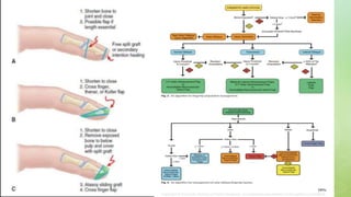
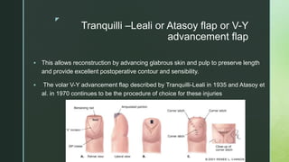
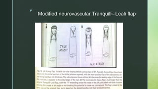
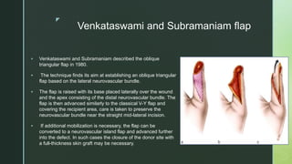
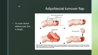
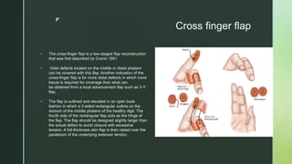
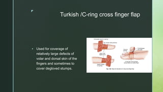
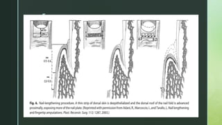
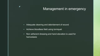
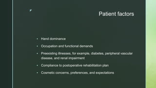
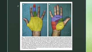
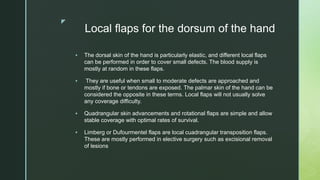
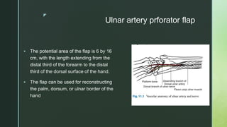
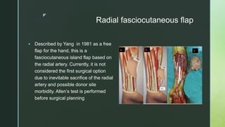
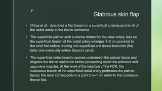
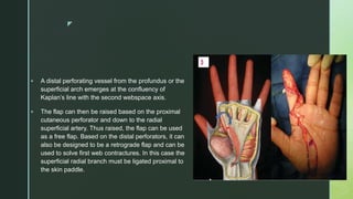
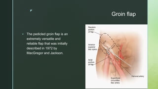
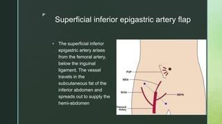
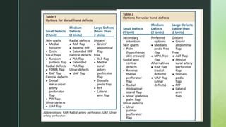
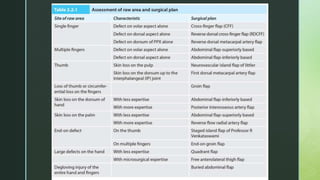
z
FINGER TIP AND
SOFT TISSUE
RECONSTRUCTION
OF HAND
Dr. Aditi Sharma
M.Ch. Resident
Department of Plastic Surgery
z
Finger tip injury- Introduction
 Finger tip injury is any soft tissue and /or nail or bony injury distal
to insertion of long flexor and extensor tendon of a finger or
thumb.
 Can occur at any age but common in working class adults and
children.
 Common causes in adults are laceration and crush or avulsion
whereas in children commonly caused by jamming or crushing
by doors.
z
Anatomy
z
 The digital arteries and nerves
trifurcate and arborize near the DIP
 They send branches to nail bed,nail
fold and pulp
 Pulp Vascularization.
 1, Pulp anastomotic arch; 2, palmar
digital artery; 3, flexor
tendon; 4, pulp-destined terminal
branch; 5, central artery of the pulp.
z
 Each digital nerve trifurcates near the distal interphalangeal
joint, sending branches to the perionychium, fingertip, and volar
pad. The digital nerves lie volar to the digital arteries near the
fingertip
 The fingertip is the organ of touch and feel and is abundantly
supplied with sensory receptors, including Pacinian and
Meissner corpuscles and Merkel cell neurite complexes.
z
Classification of fingertip amputation
z
PNB classification
z
Types based on angle of injury
z
z
Composite Tip Grafts or Cap technique
 Reapplication of composite tip grafts, amputated parts containing bone
fat or nail bed, can be considered for children younger than age 6 years.
As composite tip grafts must initially survive by plasmatic imbibition until
neovascularization, revascularization is not reliable for adults and tip
grafts should not be reapplied for adults.
 The distal phalanx fracture is reduced and secured with internal fixation
using a K-wire. The surrounding skin is approximated with absorbable
chromic suture to avoid the hassle of suture removal in a child. Soft
splinting is done for 2 weeks.
z
Free (Nonvascularized) Pulp Grafts
From the Toe
 In 1959 Mac Cash proposed the use of pulp grafts cut on a toe to the exact size of the defect and carefully sutured at the
recipient site by eliminating dead space. The risk of loss with such a composite graft seems to condemn the method.
 The idea of using the pulp of a toe to rebuild a mechanically satisfactory fingertip is worth considering however.
 Thin grafts (“dressing-graft”): some pulp or digital distal soft tissue defects will require a complex reconstruction by
microsurgical transfer; these free transfers are not performed in an emergency setting but preferably secondarily or in a
“deferred” emergency. A thin graft applied to the defect protects the future recipient site, improves the comfort of the
dressing and prevents excessive granulation that would complicate the subsequent reconstruction procedure.
 Palmar oblique amputations in area 1: these come under the scope of healing by secondary intention; however, when
this soft tissue defect is relatively large, healing time can reach 6–7 weeks. So when job requirements demand it, in the
case of a nondominant pulp, a thick or full-thickness skin graft can be used to shorten the healing time
z
Lateral advancement flap of Kutler
 Used for transverse or
dorsal oblique
amputations
z
Tranquilli –Leali or Atasoy flap or V-Y
advancement flap
 This allows reconstruction by advancing glabrous skin and pulp to preserve length
and provide excellent postoperative contour and sensibility.
 The volar V-Y advancement flap described by Tranquilli-Leali in 1935 and Atasoy et
al. in 1970 continues to be the procedure of choice for these injuries
z
Modified neurovascular Tranquilli–Leali flap
z
Venkataswami and Subramaniam flap
 Venkataswami and Subramaniam described the oblique
triangular flap in 1980.
 The technique finds its aim at establishing an oblique triangular
flap based on the lateral neurovascular bundle.
 The flap is raised with its base placed laterally over the wound
and the apex consisting of the distal neurovascular bundle. The
flap is then advanced similarly to the classical V-Y flap and
covering the recipient area, care is taken to preserve the
neurovascular bundle near the straight mid-lateral incision.
 If additional mobilization is necessary, the flap can be
converted to a neurovascular island flap and advanced further
into the defect. In such cases the closure of the donor site with
a full-thickness skin graft may be necessary.
z
Homodigital artery flap
 This flap is similar to the reverse digital artery flap, as it is an island
flap harvested from the same injured finger in one stage.
 This flap is harvested based on dorsal branches of the proper
digital artery and venous supply within a 5-mm wide bridge of fat of
the dorsal branch of the proper digital artery at the distal finger
crease.
 This flap can be harvested with dorsal digital nerve branches to be
a sensate flap.
 The donor site usually requires a skin graft.
 The arc of motion of this flap makes it difficult to reach the fingertip.
z
z
Reverse homodigital arterial flap
 described by Lai in 1989
 The flap is harvested from the lateral aspect of the proximal phalanx of
the same finger, preferably the nonopposition side.
 It is an axial flap based on collateral flow through the contralateral
digital artery, thus it is only applicable to defects distal to the crossover
communication from the contralateral digital artery
 .
z
•The pedicle is harvested with a cuff of
soft tissue to include the digital artery
venae comitante. The digital nerve can
be preserved.
•The pedicle is harvested to 5 mm
proximal to the distal interphalangeal
joint to capture crossover vessels from
the contralateral digital artery. If doubt
exists concerning reverse blood supply
to the flap, the proximal digital artery can
be temporarily clamped to evaluate
retrograde flow to the skin island.
• The donor site usually requires a skin
graft
z
Hueston Flap and Souquet Flap
 Hueston described a lateral palmar
advancement flap to cover the tip of the
amputated finger. Souquet described a
similar flap.
 A transverse back cut is made across the
volar aspect of the finger, and the flap is
elevated off the flexor tendon sheath. The
flap is then elevated and rotated into the
defect. There will generally be a triangular
defect proximally, and this must be closed
with a skin graft.
z
 The difference between the 2 flaps is
that the Hueston’s flap includes only
one neurovascular bundle at the base
of the flap, whereas Souquet’s flap
includes both neurovascular bundles.
 In the Hueston flap the longitudinal
incision is made volar to the
neurovascular bundle, and in the
Souquet flap the incision is made
dorsal to the bundle.Both flaps are
technically rotation advancement flaps
z
Adipofascial turnover flap
 To cover dorsal
defects upto 3cm
in length
z
Cross finger flap
 The cross-finger flap is a two-staged flap reconstruction
that was first described by Cronin 1951
 Volar defects located on the middle or distal phalanx
can be covered with this flap. Another indication of the
cross-finger flap is for more distal defects in which more
tissue is required for coverage than what can
be obtained from a local advancement flap such as V-Y
flap.
 The flap is outlined and elevated in an open book
fashion in which a 3-sided rectangular outline on the
dorsum of the middle phalanx of the healthy digit. The
fourth side of the rectangular flap acts as the hinge of
the flap. The flap should be designed slightly larger than
the actual defect to avoid closure with excessive
tension. A full-thickness skin flap is then raised over the
paratenon of the underlying extensor tendon.
z
 Care must be taken to avoid injury to the paratenon so that the
secondary defect can be closed easily with a skin graft.
 After flap elevation, the flap is rotated 180 degrees around its hinge
and secured over the palmar defect of the adjacent injured finger.
 A full-thickness skin graft is then harvested mostly from the
muscular forearm to close the secondary defect.
 A K-wire can be placed to stabilize the fingers together to avoid
tension over the flap. The fingers are immobilized for approximately
2-3 weeks after which a secondary procedure is performed to divide
the skin bridge.
z
z
Reverse Cross finger flap
z
Turkish /C-ring cross finger flap
 Used for coverage of
relatively large defects of
volar and dorsal skin of the
fingers and sometimes to
cover degloved stumps.
z
Thenar flap
 random-pattern flap which is best suited to repair volar oblique distal fingertip
amputations, with subtotal or total pulp loss but no major bony or nail
involvement
 The thenar flap is superior to the cross-finger flap as it provides more
appropriate soft tissue bulk and obviates the need for adjacent finger
involvement and subsequent disfiguring donor defect
 It provides excellent reconstitution of three-dimensional bulk, contour of finger
pulp, and fingertip projection due to the significant amount of subcutaneous
tissue available, reducing the risk of claw deformity.
 This two-stage technique is simple to perform with excellent tissue color and
texture match, excellent recovery of functional sensibility, and low-donor site
morbidity due to its inconspicuous flap design
z
z
Visor flap
 The visor flap is a bipedicled dorsal finger skin
flap that can be used to cover fingertip wounds
after digital amputations proximal to the nail bed
 Blood supply through this flap is provided
through dorsal branches of the volar digital
arteries and this is more reliable distal to the
proximal phalanx. In elevating the visor flap it is
critical to preserve the extensor paratenon to
allow this donor site to be skin grafted.
z
Nail lengthening
 The extent of perionychial injury is a significant consideration when treating dorsal
oblique injuries.
 Some authors suggest that if less than half of the original length remains, the nail bed
should be ablated.
 Others state that the nail bed should be ablated if less than 5 mm remains. Most
surgeons agree that if the injury is proximal to the lunula, the nail should be ablated and
revision amputation performed.
 Nail-lengthening procedures have been described for injuries that sacrifice all but the
most proximal sterile matrix. With these techniques, the nail can reliably be relatively
lengthened 2 to 3 mm
z
z
Soft tissue reconstruction of hand
z
Goals of management
 Minimization of pain
 Preserving sensation and length
 Reducing the time of impairement
 Providing a cosmetically enhanced finger tip
 Good functional outcome
z
Management in emergency
 Adequate cleaning and debridement of wound
 Achieve bloodless field using torniquet
 Non adherent dressing and hand elevation is used for
hemostasis
z
Assessment of injury
 Site of the defect
 Size of the defect
 Shape of the defect
 Floor of the defect
 Edges of the defect
 Distal deficit:
 Associated tendon injury or loss
 Associated bone fracture with or without loss
 Associated nerve injury or loss
z
Patient factors
 Hand dominance
 Occupation and functional demands
 Preexisting illnesses, for example, diabetes, peripheral vascular
disease, and renal impairment
 Compliance to postoperative rehabilitation plan
 Cosmetic concerns, preferences, and expectations
z
Qualities of an ideal skin cover for a
defect on the hand
 Must be durable skin
 Must be cosmetically pleasing
 Must be sensate
 Must be pain-free.
z
z
Local flaps for the dorsum of the hand
 The dorsal skin of the hand is particularly elastic, and different local flaps
can be performed in order to cover small defects. The blood supply is
mostly at random in these flaps.
 They are useful when small to moderate defects are approached and
mostly if bone or tendons are exposed. The palmar skin of the hand can be
considered the opposite in these terms. Local flaps will not usually solve
any coverage difficulty.
 Quadrangular skin advancements and rotational flaps are simple and allow
stable coverage with optimal rates of survival.
 Limberg or Dufourmentel flaps are local cuadrangular transposition flaps.
These are mostly performed in elective surgery such as excisional removal
of lesions
z
 the wound margins should be excised back to healthy skin prior
to flap transfer.
 recreate the original defect in secondary reconstruction and all
scarred skin should be excised so that the flap will be sutured to
healthy margins.
z
Posterior interosseous fasciocutaneous flap
 Described by Zancolli and Angrigiani in 1988 for the dorsal coverage of the hand, this flap is
based on the existence of an anastomosis between the posterior interosseous artery and the
dorsal branch of the anterior interosseous artery at the dorsal aspect of the wrist.
 The posterior interosseous artery will be ligated, and the blood flow will course retrogradely
from the anterior interosseous artery to the posterior interosseous pedicle
 The design of the posterior interosseous flap starts with the marking of the cutaneous island.
A line is drawn between the lateral humeral epicondyle and the distal radioulnar joint. The
island must be outlined in between the proximal and distal thirds of the forearm.
 The main posterior interosseous cutaneous branch emerges 9 centimetres distal to the lateral
epicondyle in the same line; this can also be easily identified with a Doppler ultrasound.
z
 The vascular anastomosis between the two interosseous
arteries can be found 2 cm proximal to the radiocarpal joint
at the proximal border of the pronator quadratus.
 The interosseous posterior artery is found at the septum
between the extensor carpi ulnaris (ECU) and the extensor
digiti minimi (EDM). This septum and the anastomosis are
easily identified distally, which is the reason why many
surgeons prefer to first identify the anastomosis and the
posterior interosseous artery and eventually raise the flap.
The posterior interosseous artery is closely related with the
posterior interosseous nerve. This condition might
represent a challenge in the hands of an inexperienced
surgeon.
 Donor site may be close directly or with a split skin graft
depending on the width of the cutaneous island.
z
Dorsoulnar fasciocutaneous flap
 This is a flap that is based on the distal
branch of the ulnar artery which emerges
2–5 cm proximal to the pisiform bone.
 This branch courses between the flexor
carpi ulnaris (FCU) and the ECU and then
reaches the cutaneous skin crossing
between the ulnar nerve and the FCU
distally.
 It then divides into descending and
ascending branches. The descending
branch anastomoses with skin perforators
of the dorsal arch
z
Ulnar artery prforator flap
 The potential area of the flap is 6 by 16
cm, with the length extending from the
distal third of the forearm to the distal
third of the dorsal surface of the hand.
 The flap can be used for reconstructing
the palm, dorsum, or ulnar border of the
hand
z
Radial fasciocutaneous flap
 Described by Yang in 1981 as a free
flap for the hand, this is a
fasciocutaneous island flap based on
the radial artery. Currently, it is not
considered the first surgical option
due to inevitable sacrifice of the radial
artery and possible donor site
morbidity. Allen’s test is performed
before surgical planning
z
Glabrous skin flap
 Orbay et al. described a flap based on a superficial cutaneous branch of
the radial artery at the thenar eminence
 The superficial palmar arch is mainly formed by the ulnar artery, less so
the superficial branch of the radial artery emerges 1–2 cm proximal to
the wrist fold before dividing into superficial and dorsal branches (the
latter one eventually enters Guyon’s canal).
 The superficial radial branch courses underneath the palmar fascia and
irrigates the thenar eminence before proceeding under the adductor and
opponens muscles. At the level of the insertion of the FCR, the
cutaneous branch of the superficial radial artery perforates the palmar
fascia; this level corresponds to a point 0.5–1 cm radial to the cutaneous
thenar fold.
z
 A distal perforating vessel from the profundus or the
superficial arch emerges at the confluency of
Kaplan’s line with the second webspace axis.
 The flap can then be raised based on the proximal
cutaneous perforator and down to the radial
superficial artery. Thus raised, the flap can be used
as a free flap. Based on the distal perforators, it can
also be designed to be a retrograde flap and can be
used to solve first web contractures. In this case the
superficial radial branch must be ligated proximal to
the skin paddle.
z
Groin flap
 The pedicled groin flap is an
extremely versatile and
reliable flap that was initially
described in 1972 by
MacGregor and Jackson.
z
Superficial inferior epigastric artery flap
 The superficial inferior
epigastric artery arises
from the femoral artery,
below the inguinal
ligament. The vessel
travels in the
subcutaneous fat of the
inferior abdomen and
spreads out to supply the
hemi-abdomen
z
z

Fingertip injury.pptx

  • 1.
    z FINGER TIP AND SOFTTISSUE RECONSTRUCTION OF HAND Dr. Aditi Sharma M.Ch. Resident Department of Plastic Surgery
  • 2.
    z Finger tip injury-Introduction  Finger tip injury is any soft tissue and /or nail or bony injury distal to insertion of long flexor and extensor tendon of a finger or thumb.  Can occur at any age but common in working class adults and children.  Common causes in adults are laceration and crush or avulsion whereas in children commonly caused by jamming or crushing by doors.
  • 3.
  • 4.
    z  The digitalarteries and nerves trifurcate and arborize near the DIP  They send branches to nail bed,nail fold and pulp  Pulp Vascularization.  1, Pulp anastomotic arch; 2, palmar digital artery; 3, flexor tendon; 4, pulp-destined terminal branch; 5, central artery of the pulp.
  • 5.
    z  Each digitalnerve trifurcates near the distal interphalangeal joint, sending branches to the perionychium, fingertip, and volar pad. The digital nerves lie volar to the digital arteries near the fingertip  The fingertip is the organ of touch and feel and is abundantly supplied with sensory receptors, including Pacinian and Meissner corpuscles and Merkel cell neurite complexes.
  • 6.
  • 7.
  • 8.
    z Types based onangle of injury
  • 9.
  • 10.
    z Composite Tip Graftsor Cap technique  Reapplication of composite tip grafts, amputated parts containing bone fat or nail bed, can be considered for children younger than age 6 years. As composite tip grafts must initially survive by plasmatic imbibition until neovascularization, revascularization is not reliable for adults and tip grafts should not be reapplied for adults.  The distal phalanx fracture is reduced and secured with internal fixation using a K-wire. The surrounding skin is approximated with absorbable chromic suture to avoid the hassle of suture removal in a child. Soft splinting is done for 2 weeks.
  • 11.
    z Free (Nonvascularized) PulpGrafts From the Toe  In 1959 Mac Cash proposed the use of pulp grafts cut on a toe to the exact size of the defect and carefully sutured at the recipient site by eliminating dead space. The risk of loss with such a composite graft seems to condemn the method.  The idea of using the pulp of a toe to rebuild a mechanically satisfactory fingertip is worth considering however.  Thin grafts (“dressing-graft”): some pulp or digital distal soft tissue defects will require a complex reconstruction by microsurgical transfer; these free transfers are not performed in an emergency setting but preferably secondarily or in a “deferred” emergency. A thin graft applied to the defect protects the future recipient site, improves the comfort of the dressing and prevents excessive granulation that would complicate the subsequent reconstruction procedure.  Palmar oblique amputations in area 1: these come under the scope of healing by secondary intention; however, when this soft tissue defect is relatively large, healing time can reach 6–7 weeks. So when job requirements demand it, in the case of a nondominant pulp, a thick or full-thickness skin graft can be used to shorten the healing time
  • 12.
    z Lateral advancement flapof Kutler  Used for transverse or dorsal oblique amputations
  • 13.
    z Tranquilli –Leali orAtasoy flap or V-Y advancement flap  This allows reconstruction by advancing glabrous skin and pulp to preserve length and provide excellent postoperative contour and sensibility.  The volar V-Y advancement flap described by Tranquilli-Leali in 1935 and Atasoy et al. in 1970 continues to be the procedure of choice for these injuries
  • 14.
  • 15.
    z Venkataswami and Subramaniamflap  Venkataswami and Subramaniam described the oblique triangular flap in 1980.  The technique finds its aim at establishing an oblique triangular flap based on the lateral neurovascular bundle.  The flap is raised with its base placed laterally over the wound and the apex consisting of the distal neurovascular bundle. The flap is then advanced similarly to the classical V-Y flap and covering the recipient area, care is taken to preserve the neurovascular bundle near the straight mid-lateral incision.  If additional mobilization is necessary, the flap can be converted to a neurovascular island flap and advanced further into the defect. In such cases the closure of the donor site with a full-thickness skin graft may be necessary.
  • 16.
    z Homodigital artery flap This flap is similar to the reverse digital artery flap, as it is an island flap harvested from the same injured finger in one stage.  This flap is harvested based on dorsal branches of the proper digital artery and venous supply within a 5-mm wide bridge of fat of the dorsal branch of the proper digital artery at the distal finger crease.  This flap can be harvested with dorsal digital nerve branches to be a sensate flap.  The donor site usually requires a skin graft.  The arc of motion of this flap makes it difficult to reach the fingertip.
  • 17.
  • 18.
    z Reverse homodigital arterialflap  described by Lai in 1989  The flap is harvested from the lateral aspect of the proximal phalanx of the same finger, preferably the nonopposition side.  It is an axial flap based on collateral flow through the contralateral digital artery, thus it is only applicable to defects distal to the crossover communication from the contralateral digital artery  .
  • 19.
    z •The pedicle isharvested with a cuff of soft tissue to include the digital artery venae comitante. The digital nerve can be preserved. •The pedicle is harvested to 5 mm proximal to the distal interphalangeal joint to capture crossover vessels from the contralateral digital artery. If doubt exists concerning reverse blood supply to the flap, the proximal digital artery can be temporarily clamped to evaluate retrograde flow to the skin island. • The donor site usually requires a skin graft
  • 20.
    z Hueston Flap andSouquet Flap  Hueston described a lateral palmar advancement flap to cover the tip of the amputated finger. Souquet described a similar flap.  A transverse back cut is made across the volar aspect of the finger, and the flap is elevated off the flexor tendon sheath. The flap is then elevated and rotated into the defect. There will generally be a triangular defect proximally, and this must be closed with a skin graft.
  • 21.
    z  The differencebetween the 2 flaps is that the Hueston’s flap includes only one neurovascular bundle at the base of the flap, whereas Souquet’s flap includes both neurovascular bundles.  In the Hueston flap the longitudinal incision is made volar to the neurovascular bundle, and in the Souquet flap the incision is made dorsal to the bundle.Both flaps are technically rotation advancement flaps
  • 22.
    z Adipofascial turnover flap To cover dorsal defects upto 3cm in length
  • 23.
    z Cross finger flap The cross-finger flap is a two-staged flap reconstruction that was first described by Cronin 1951  Volar defects located on the middle or distal phalanx can be covered with this flap. Another indication of the cross-finger flap is for more distal defects in which more tissue is required for coverage than what can be obtained from a local advancement flap such as V-Y flap.  The flap is outlined and elevated in an open book fashion in which a 3-sided rectangular outline on the dorsum of the middle phalanx of the healthy digit. The fourth side of the rectangular flap acts as the hinge of the flap. The flap should be designed slightly larger than the actual defect to avoid closure with excessive tension. A full-thickness skin flap is then raised over the paratenon of the underlying extensor tendon.
  • 24.
    z  Care mustbe taken to avoid injury to the paratenon so that the secondary defect can be closed easily with a skin graft.  After flap elevation, the flap is rotated 180 degrees around its hinge and secured over the palmar defect of the adjacent injured finger.  A full-thickness skin graft is then harvested mostly from the muscular forearm to close the secondary defect.  A K-wire can be placed to stabilize the fingers together to avoid tension over the flap. The fingers are immobilized for approximately 2-3 weeks after which a secondary procedure is performed to divide the skin bridge.
  • 25.
  • 26.
  • 27.
    z Turkish /C-ring crossfinger flap  Used for coverage of relatively large defects of volar and dorsal skin of the fingers and sometimes to cover degloved stumps.
  • 28.
    z Thenar flap  random-patternflap which is best suited to repair volar oblique distal fingertip amputations, with subtotal or total pulp loss but no major bony or nail involvement  The thenar flap is superior to the cross-finger flap as it provides more appropriate soft tissue bulk and obviates the need for adjacent finger involvement and subsequent disfiguring donor defect  It provides excellent reconstitution of three-dimensional bulk, contour of finger pulp, and fingertip projection due to the significant amount of subcutaneous tissue available, reducing the risk of claw deformity.  This two-stage technique is simple to perform with excellent tissue color and texture match, excellent recovery of functional sensibility, and low-donor site morbidity due to its inconspicuous flap design
  • 29.
  • 30.
    z Visor flap  Thevisor flap is a bipedicled dorsal finger skin flap that can be used to cover fingertip wounds after digital amputations proximal to the nail bed  Blood supply through this flap is provided through dorsal branches of the volar digital arteries and this is more reliable distal to the proximal phalanx. In elevating the visor flap it is critical to preserve the extensor paratenon to allow this donor site to be skin grafted.
  • 31.
    z Nail lengthening  Theextent of perionychial injury is a significant consideration when treating dorsal oblique injuries.  Some authors suggest that if less than half of the original length remains, the nail bed should be ablated.  Others state that the nail bed should be ablated if less than 5 mm remains. Most surgeons agree that if the injury is proximal to the lunula, the nail should be ablated and revision amputation performed.  Nail-lengthening procedures have been described for injuries that sacrifice all but the most proximal sterile matrix. With these techniques, the nail can reliably be relatively lengthened 2 to 3 mm
  • 32.
  • 33.
  • 34.
    z Goals of management Minimization of pain  Preserving sensation and length  Reducing the time of impairement  Providing a cosmetically enhanced finger tip  Good functional outcome
  • 35.
    z Management in emergency Adequate cleaning and debridement of wound  Achieve bloodless field using torniquet  Non adherent dressing and hand elevation is used for hemostasis
  • 36.
    z Assessment of injury Site of the defect  Size of the defect  Shape of the defect  Floor of the defect  Edges of the defect  Distal deficit:  Associated tendon injury or loss  Associated bone fracture with or without loss  Associated nerve injury or loss
  • 37.
    z Patient factors  Handdominance  Occupation and functional demands  Preexisting illnesses, for example, diabetes, peripheral vascular disease, and renal impairment  Compliance to postoperative rehabilitation plan  Cosmetic concerns, preferences, and expectations
  • 38.
    z Qualities of anideal skin cover for a defect on the hand  Must be durable skin  Must be cosmetically pleasing  Must be sensate  Must be pain-free.
  • 39.
  • 40.
    z Local flaps forthe dorsum of the hand  The dorsal skin of the hand is particularly elastic, and different local flaps can be performed in order to cover small defects. The blood supply is mostly at random in these flaps.  They are useful when small to moderate defects are approached and mostly if bone or tendons are exposed. The palmar skin of the hand can be considered the opposite in these terms. Local flaps will not usually solve any coverage difficulty.  Quadrangular skin advancements and rotational flaps are simple and allow stable coverage with optimal rates of survival.  Limberg or Dufourmentel flaps are local cuadrangular transposition flaps. These are mostly performed in elective surgery such as excisional removal of lesions
  • 41.
    z  the woundmargins should be excised back to healthy skin prior to flap transfer.  recreate the original defect in secondary reconstruction and all scarred skin should be excised so that the flap will be sutured to healthy margins.
  • 42.
    z Posterior interosseous fasciocutaneousflap  Described by Zancolli and Angrigiani in 1988 for the dorsal coverage of the hand, this flap is based on the existence of an anastomosis between the posterior interosseous artery and the dorsal branch of the anterior interosseous artery at the dorsal aspect of the wrist.  The posterior interosseous artery will be ligated, and the blood flow will course retrogradely from the anterior interosseous artery to the posterior interosseous pedicle  The design of the posterior interosseous flap starts with the marking of the cutaneous island. A line is drawn between the lateral humeral epicondyle and the distal radioulnar joint. The island must be outlined in between the proximal and distal thirds of the forearm.  The main posterior interosseous cutaneous branch emerges 9 centimetres distal to the lateral epicondyle in the same line; this can also be easily identified with a Doppler ultrasound.
  • 43.
    z  The vascularanastomosis between the two interosseous arteries can be found 2 cm proximal to the radiocarpal joint at the proximal border of the pronator quadratus.  The interosseous posterior artery is found at the septum between the extensor carpi ulnaris (ECU) and the extensor digiti minimi (EDM). This septum and the anastomosis are easily identified distally, which is the reason why many surgeons prefer to first identify the anastomosis and the posterior interosseous artery and eventually raise the flap. The posterior interosseous artery is closely related with the posterior interosseous nerve. This condition might represent a challenge in the hands of an inexperienced surgeon.  Donor site may be close directly or with a split skin graft depending on the width of the cutaneous island.
  • 44.
    z Dorsoulnar fasciocutaneous flap This is a flap that is based on the distal branch of the ulnar artery which emerges 2–5 cm proximal to the pisiform bone.  This branch courses between the flexor carpi ulnaris (FCU) and the ECU and then reaches the cutaneous skin crossing between the ulnar nerve and the FCU distally.  It then divides into descending and ascending branches. The descending branch anastomoses with skin perforators of the dorsal arch
  • 45.
    z Ulnar artery prforatorflap  The potential area of the flap is 6 by 16 cm, with the length extending from the distal third of the forearm to the distal third of the dorsal surface of the hand.  The flap can be used for reconstructing the palm, dorsum, or ulnar border of the hand
  • 46.
    z Radial fasciocutaneous flap Described by Yang in 1981 as a free flap for the hand, this is a fasciocutaneous island flap based on the radial artery. Currently, it is not considered the first surgical option due to inevitable sacrifice of the radial artery and possible donor site morbidity. Allen’s test is performed before surgical planning
  • 47.
    z Glabrous skin flap Orbay et al. described a flap based on a superficial cutaneous branch of the radial artery at the thenar eminence  The superficial palmar arch is mainly formed by the ulnar artery, less so the superficial branch of the radial artery emerges 1–2 cm proximal to the wrist fold before dividing into superficial and dorsal branches (the latter one eventually enters Guyon’s canal).  The superficial radial branch courses underneath the palmar fascia and irrigates the thenar eminence before proceeding under the adductor and opponens muscles. At the level of the insertion of the FCR, the cutaneous branch of the superficial radial artery perforates the palmar fascia; this level corresponds to a point 0.5–1 cm radial to the cutaneous thenar fold.
  • 48.
    z  A distalperforating vessel from the profundus or the superficial arch emerges at the confluency of Kaplan’s line with the second webspace axis.  The flap can then be raised based on the proximal cutaneous perforator and down to the radial superficial artery. Thus raised, the flap can be used as a free flap. Based on the distal perforators, it can also be designed to be a retrograde flap and can be used to solve first web contractures. In this case the superficial radial branch must be ligated proximal to the skin paddle.
  • 49.
    z Groin flap  Thepedicled groin flap is an extremely versatile and reliable flap that was initially described in 1972 by MacGregor and Jackson.
  • 50.
    z Superficial inferior epigastricartery flap  The superficial inferior epigastric artery arises from the femoral artery, below the inguinal ligament. The vessel travels in the subcutaneous fat of the inferior abdomen and spreads out to supply the hemi-abdomen
  • 51.
  • 52.