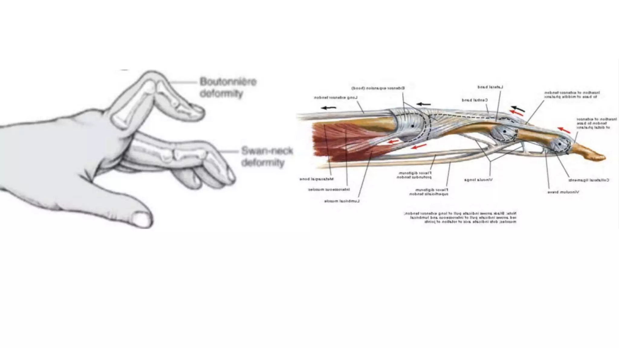
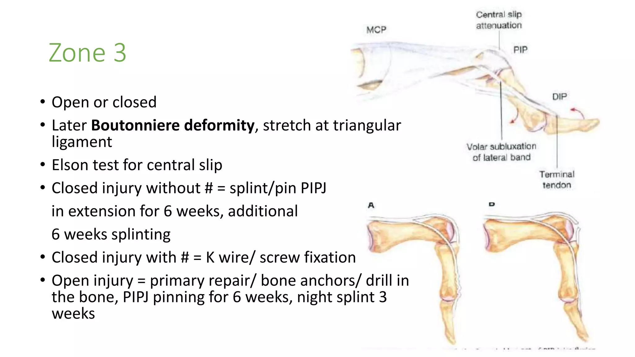
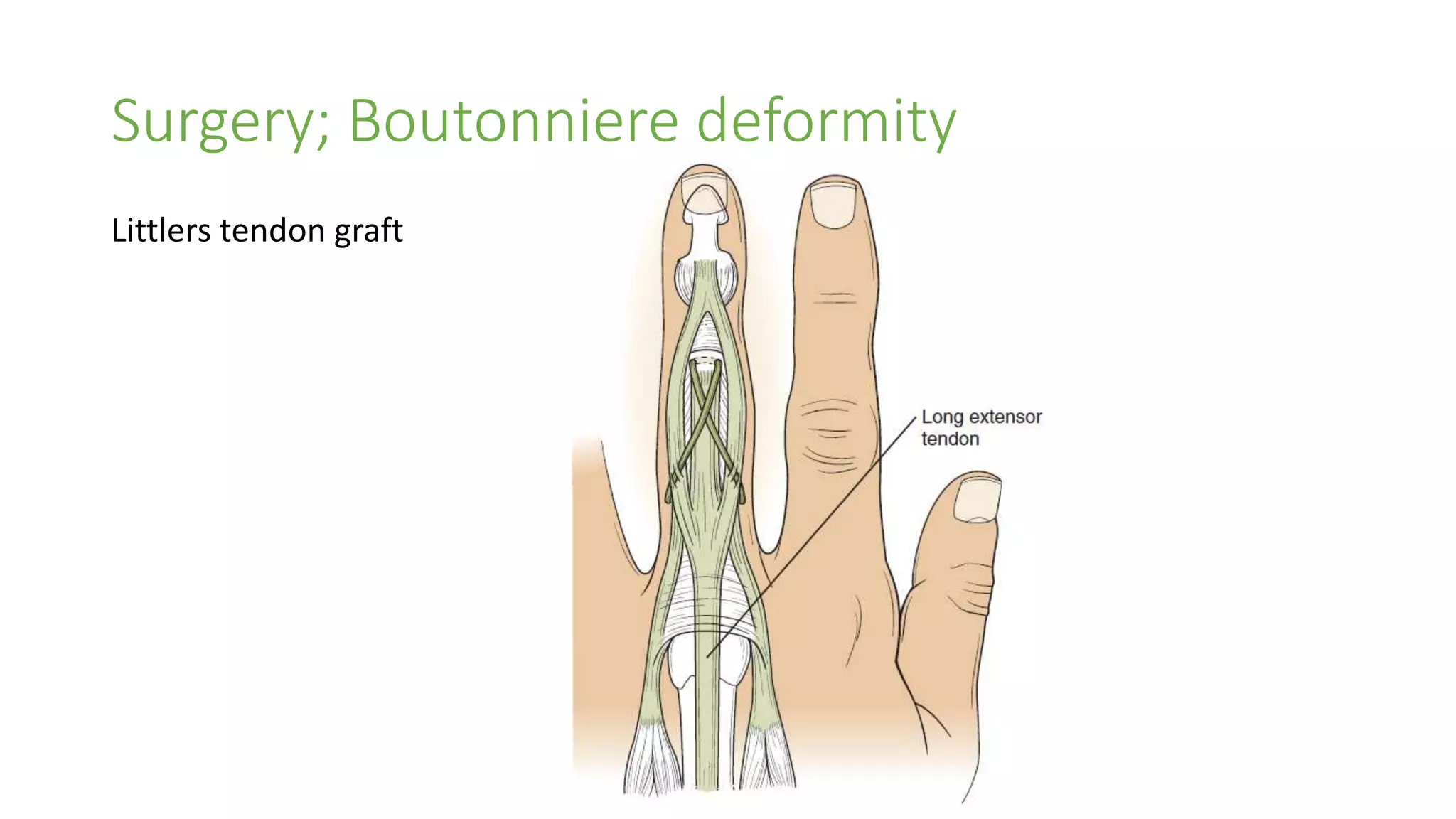
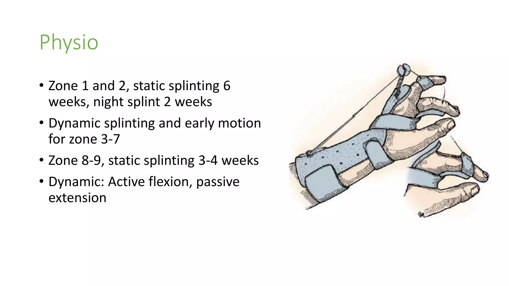
Extensor Tendon Injury
Dr Mujtuba Pervez Khan
Resident Plastic Surgery
DUHS/CHK
Zone 1 injury (mallet finger)
• Terminal extensor tendon
• Sudden resisted flexion, baseball
finger
• Proximal and dorsal migration of
lateral bands -> swan neck
deformity
Mallet finger type 1 (closed)
• 6 wks splint, 2 wks night splint, splint weaning and active flexion
• If extensor lag at 8 wks, resume splinting till resolves
Mallet finger type 2 (open)
• Running tendon repair / Tenodermal suture
• DIPJ axial or oblique K wire fixation for 6 weeks
• Splint for 4 weeks
• *irrigation of DIPJ
Mallet finger type 3 (loss of skin/tendon)
• Primary repair if possible
• DIPJ fixation in hyperextension
• Flap coverage
• Stage reconstruction
Mallet finger type 4 (#)
• K wire fixation in children
• In adults, operative and conservative
treatment
• 4C volar sublaxation (hyper extension
injury) -> operative
Chronic Mallet finger
• Several month after injury ,SND
• 1. Skin imbrication, K wire
fixation for 6 weeks
• 2. Fowler tenotomy
• 3. Spiral ORL reconstruction
Zone 2
• Injury to conjoined lateral bands and triangular ligaments
• One band is sufficient for DIPJ extension
• Running sutures and splint for 6 weeks
• Chronic injuries, treat like mallet finger, ORL recon
Zone 3
• Open or closed
• Later Boutonniere deformity, stretch at triangular
ligament
• Elson test for central slip
• Closed injury without # = splint/pin PIPJ
in extension for 6 weeks, additional
6 weeks splinting
• Closed injury with # = K wire/ screw fixation
• Open injury = primary repair/ bone anchors/ drill in
the bone, PIPJ pinning for 6 weeks, night splint 3
weeks
Boutonniere deformity
• DIPJ hyperextension, PIPJ flexion (central slip rupture)
• Volar migration of lateral bands
• Arthritis, burns, trauma, synovitis
Splint (PIPJ extension) for 2-3
months, if fail  Surgery
Joint fusion
Surgery; Boutonniere deformity
Fowler tenotomy
Surgery; Boutonniere deformity
Littlers tendon graft
Surgery; Boutonniere deformity
Curtis staged reconstruction
Stage 1 and 2 = Intrinsic release
and transverse retinacular
ligament is freed and transected if
necessary
Stage 3 = Fowler tenotomy if
extensor lag present
Stage 4 = Repair of central slip
Zone 4
• Lateral bands are shielded by convexity of prox phalanx
• Repair of tendon and splint PIPJ, DIPJ free for 6 weeks
Zone 5
• Fight bite
• Wound CS, broad spectrum ABX, Joint irrigation, primary repair
• Isolated sagittal band injuries, radial bands, mostly closed, due to resisted extension of the finger
• Mistaken for trigger finger due to snapping
Zone 6
• Juncturae tendons can hide the injury
• Core suture and epitendinous repair
Zone 7
• Treatment same as zone 6
• Ext retinaculum prevents bowstringing, excise portion for exposure
and prevents friction
• Preserve 50% of length
• EPL, repair over the retinculum
Zone 8
• Most distal belly of EIP, EPL, EPB, APL
• 4cm proximal to wrist are musculotendinous junction
• Tendon to tendon repair is stronger
Zone 9
• Muscle bellies,
• Repairs are weaker
• Figure of 8 repair
• Nerve injury
Physio
• Zone 1 and 2, static splinting 6
weeks, night splint 2 weeks
• Dynamic splinting and early motion
for zone 3-7
• Zone 8-9, static splinting 3-4 weeks
• Dynamic: Active flexion, passive
extension
Swan neck Deformity
• Tendon imbalance -> PIPJ hyperextension,
DIPJ and MCPJ flexion
• PIPJ
1. FDS rupture
2. PIPJ synovitis (attenuation of volar plate
or TRL)
• MCPJ
3. MCPJ synovitis (weak insertion of EDC,
force at PIPJ), weak volar plate 
sublaxation ->
4. Intrinsic tightness (RA -> myofibrosis)
• DIPJ = 5. Mallet finger (force shifts to PIPJ)
• Wrist = 6. Carpal collapse (lengthening of
flexors and extensors)
• Injuries can be overlapping
Swan neck Deformity
• TYPE 1. Full PIPJ flexibility – no intrinsic tightness
conservative, DIPJ fusion (if due to mallet finger), flexor
tenodesis (slip to A1), ORL reconstruction by ulnar lateral
band repair to distal phalanx)
• TYPE 2. PIPJ flexible – intrinsic tightness, MCPJ sublaxation –
MCPJ relocation, intrinsic release, relocation of MCPJ, distal
intrinsic release
• TYPE 3. Limited PIPJ mobility, joint surface normal
release of lateral bands (shift volar),
• TYPE 4. Stiff PIPJ, intra articular joint destruction
joint fusion/arthroplasty
thanks

Extensor tendon injury

  • 1.
    Extensor Tendon Injury DrMujtuba Pervez Khan Resident Plastic Surgery DUHS/CHK
  • 8.
    Zone 1 injury(mallet finger) • Terminal extensor tendon • Sudden resisted flexion, baseball finger • Proximal and dorsal migration of lateral bands -> swan neck deformity
  • 9.
    Mallet finger type1 (closed) • 6 wks splint, 2 wks night splint, splint weaning and active flexion • If extensor lag at 8 wks, resume splinting till resolves
  • 10.
    Mallet finger type2 (open) • Running tendon repair / Tenodermal suture • DIPJ axial or oblique K wire fixation for 6 weeks • Splint for 4 weeks • *irrigation of DIPJ
  • 11.
    Mallet finger type3 (loss of skin/tendon) • Primary repair if possible • DIPJ fixation in hyperextension • Flap coverage • Stage reconstruction
  • 12.
    Mallet finger type4 (#) • K wire fixation in children • In adults, operative and conservative treatment • 4C volar sublaxation (hyper extension injury) -> operative
  • 13.
    Chronic Mallet finger •Several month after injury ,SND • 1. Skin imbrication, K wire fixation for 6 weeks • 2. Fowler tenotomy • 3. Spiral ORL reconstruction
  • 14.
    Zone 2 • Injuryto conjoined lateral bands and triangular ligaments • One band is sufficient for DIPJ extension • Running sutures and splint for 6 weeks • Chronic injuries, treat like mallet finger, ORL recon
  • 15.
    Zone 3 • Openor closed • Later Boutonniere deformity, stretch at triangular ligament • Elson test for central slip • Closed injury without # = splint/pin PIPJ in extension for 6 weeks, additional 6 weeks splinting • Closed injury with # = K wire/ screw fixation • Open injury = primary repair/ bone anchors/ drill in the bone, PIPJ pinning for 6 weeks, night splint 3 weeks
  • 16.
    Boutonniere deformity • DIPJhyperextension, PIPJ flexion (central slip rupture) • Volar migration of lateral bands • Arthritis, burns, trauma, synovitis Splint (PIPJ extension) for 2-3 months, if fail  Surgery Joint fusion
  • 17.
  • 18.
  • 19.
    Surgery; Boutonniere deformity Curtisstaged reconstruction Stage 1 and 2 = Intrinsic release and transverse retinacular ligament is freed and transected if necessary Stage 3 = Fowler tenotomy if extensor lag present Stage 4 = Repair of central slip
  • 20.
    Zone 4 • Lateralbands are shielded by convexity of prox phalanx • Repair of tendon and splint PIPJ, DIPJ free for 6 weeks
  • 21.
    Zone 5 • Fightbite • Wound CS, broad spectrum ABX, Joint irrigation, primary repair • Isolated sagittal band injuries, radial bands, mostly closed, due to resisted extension of the finger • Mistaken for trigger finger due to snapping
  • 22.
    Zone 6 • Juncturaetendons can hide the injury • Core suture and epitendinous repair
  • 23.
    Zone 7 • Treatmentsame as zone 6 • Ext retinaculum prevents bowstringing, excise portion for exposure and prevents friction • Preserve 50% of length • EPL, repair over the retinculum
  • 24.
    Zone 8 • Mostdistal belly of EIP, EPL, EPB, APL • 4cm proximal to wrist are musculotendinous junction • Tendon to tendon repair is stronger
  • 25.
    Zone 9 • Musclebellies, • Repairs are weaker • Figure of 8 repair • Nerve injury
  • 26.
    Physio • Zone 1and 2, static splinting 6 weeks, night splint 2 weeks • Dynamic splinting and early motion for zone 3-7 • Zone 8-9, static splinting 3-4 weeks • Dynamic: Active flexion, passive extension
  • 27.
    Swan neck Deformity •Tendon imbalance -> PIPJ hyperextension, DIPJ and MCPJ flexion • PIPJ 1. FDS rupture 2. PIPJ synovitis (attenuation of volar plate or TRL) • MCPJ 3. MCPJ synovitis (weak insertion of EDC, force at PIPJ), weak volar plate  sublaxation -> 4. Intrinsic tightness (RA -> myofibrosis) • DIPJ = 5. Mallet finger (force shifts to PIPJ) • Wrist = 6. Carpal collapse (lengthening of flexors and extensors) • Injuries can be overlapping
  • 28.
    Swan neck Deformity •TYPE 1. Full PIPJ flexibility – no intrinsic tightness conservative, DIPJ fusion (if due to mallet finger), flexor tenodesis (slip to A1), ORL reconstruction by ulnar lateral band repair to distal phalanx) • TYPE 2. PIPJ flexible – intrinsic tightness, MCPJ sublaxation – MCPJ relocation, intrinsic release, relocation of MCPJ, distal intrinsic release • TYPE 3. Limited PIPJ mobility, joint surface normal release of lateral bands (shift volar), • TYPE 4. Stiff PIPJ, intra articular joint destruction joint fusion/arthroplasty
  • 30.