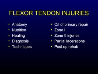
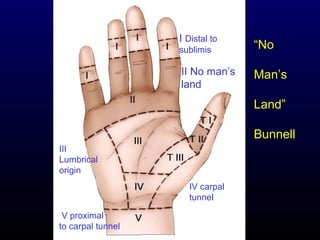
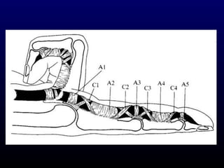
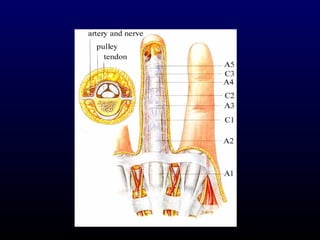
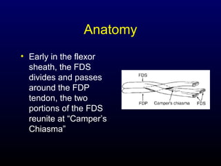
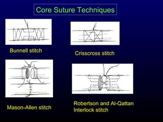
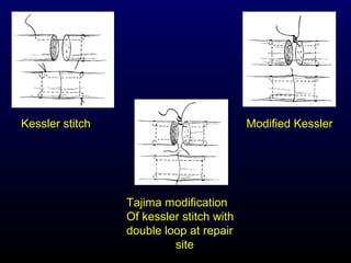
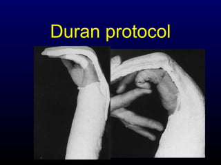
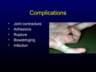
FLEXOR TENDON
INJURIES
Abdulaziz Al-Ahaideb
FLEXOR TENDON INJURIES
•
•
•
•
•

Anatomy
Nutrition
Healing
Diagnosis
Techniques

•
•
•
•
•

C/I of primary repair
Zone I
Zone II injuries
Partial lacerations
Post op rehab
I Distal to
sublimis

II No man’s
land

ā€œNo
Man’s
Landā€
Bunnell

III
Lumbrical
origin
IV carpal
tunnel
V proximal
to carpal tunnel
Anatomy
• Early in the flexor
sheath, the FDS
divides and passes
around the FDP
tendon, the two
portions of the FDS
reunite at ā€œCamper’s
Chiasmaā€
Anatomy
• FDS tendons usually arise from single
muscle bundles and act independently
• There is often a common muscle origin
for several FDP tendons with the result
that there is simultaneous flexion of
multiple digits
Blood Supply
• From segmental branches of the paired
digital arteries which enter the tendon
through:
– long and short vincula
– at the osseous insertions

• Synovial fluid diffusion
Excursions of FDP & FDS
Up to 9 cm of flexor tendon
excursion may be required
to produce composite wrist
and digital flexion, while only
2.5 cm of excursion is required
for full digital flexion with the wrist
stabilized in neutral position
Flexor tendon healing
• 2 forms:
– Intrinsic healing: occurs without direct
blood flow to the tendon
– Extrinsic healing: occurs by proliferation
of fibroblasts from the peripheral
epitendon; adhesions occur because of
extrinsic healing of the tendon and limit
tendon gliding within fibrous synovial
sheaths
Phases of Intrinsic healing
1.Inflammatory (0-5 days) : strength of the
repair is reliant on the strength of the
suture itself
2.Fibroblastic (5-28 days) : or so-called
collagen-producing phase
3.Remodelling (>28 days)
Loss of strength after repair
4500
4000
3500
3000
2500
2000
1500
1000
500
0

Bunnell
Kessler

0

5

10
Days

15

20
Adhesions formation
• Healing that is largely based on intrinsic
cellular activity will result in fewer , less dense
adhesions
• Factors that influence the formation of
adhesions:
– Trauma to the tendon and its sheath from the
initial injury and reparative surgery
– Ischemia
– Tendon immobilization
– Gapping at the repair site
– Sheath resection
Diagnosis
• Alteration in the normal resting posture
of the fingers
• Functional tests of FDS and FDP
• Lacerations on the palmar aspect of the
fingers almost always injure the FD
before FDS
• A careful sensory evaluation is
mandatory
Preoperative planning
• Severed flexor
tendon ends will
retract well
away from the
laceration site ,
esp. when the
digit is in flexion
at the time of
injury
Flexor tendon repair
Contraindications of primary
repair
• When there are severe multiple tissue
injuries to the involved fingers or palm
• When the wounds are badly
contaminated by potentially infecting
materials
• When there has been significant skin
loss over the flexor system
• Inability of patient to cooperate with
rehabilitation
Cont’

Contraindications of
primary repair

• Concomitant fractures or neurovascular
injuries are NOT necessarily C/I to
primary or delayed primary repair
Facts
• Flexor tendon repair is not a surgical
emergency. It is proved that equal or
better results can be achieved by
delayed primary repair.
• Better to repair both FDP & FDS
tendons rather than FDP alone
Surgical Incisions
• Incisions should not compromise the
viability of the skin flaps and when
healed, will not create contractures or
cosmetically unsightly scars
• Zigzag (Bruner) or midaxial incisions
Ideal repair
•
•
•
•
•
•

Easy placement of the sutures in the tendon
Secure suture knots
Smooth juncture of tendon ends
Minimal gapping at the repair site
Minimal interference with the vascularity
Strong enough to stand early motion stress
What can we provide?
• Minimal dissection and handling
• Tendon apposition without gapping
• Early protected mobilization
Core Suture Techniques

Bunnell stitch

Mason-Allen stitch

Crisscross stitch

Robertson and Al-Qattan
Interlock stitch
Kessler stitch

Modified Kessler

Tajima modification
Of kessler stitch with
double loop at repair
site
Sheath repair
• Conflicting lab and clinical studies
• Advantages:
– barrier to the formation of extrinsic
adhesions
– quicker return of synovial nutrition
– better tendon-sheath biomechanics
Sheath repair
• Disadvantages:
– technically difficult
– may narrow and restrict tendon gliding
Suture Materials
• Core Non-absorbable 4/0 suture
• Different configurations
• 6/0 monofilament running epitenon
suture.
• As noted by Singer MD et al. 1998, 3-0
prolene or mersilene suture may be
suture of choice
• The strength of the a tendon repair is
proportional to the number of the suture
strands that cross the repair site
• The number of the suture knots in the
repair site should be minimized
• Repairs are stronger when the core
sutures are placed dorsally
Zone I injuries
• Direct repair: if laceration is more than
1 cm from FDP insertion
• Tendon advancement: if the laceration
is less then 1 cm from insertion. In this
case, a pull thru technique is used.
Direct repair
• There is usually little difficulty in finding the
proximal tendon end, which is retained in the
finger by its vinculum and can usually be
located in the proximal phalanx or at the level
of the PIP
• When exposing the distal stump, the entire
A4 annular pulley should be preserved
• The proximal tendon is retrieved and passed
underneath A4 pulley
Tendon advancement
• When the distal stump is insufficient to
hold a suture, the proximal FDP stump
may be reattached by first elevating an
osteoperiosteal flap from the base of
the distal phalanx and then drilling an
oblique hole beneath the flap, directed
so as to penetrate the dorsal cortex just
beneath the proximal fingernail.
• A doublearmed (straight needles) 3-0
suture is placed in the proximal tendon
stump and passed through the bone
hole. Tie the suture over felt and a
button
• Another alternative is to use suture
anchors (but they are weaker than the
button as noticed by Silva et al 1998)

• When possible, the tendon attachment
should be supplemented by sutures
through the adjacent sheath or
periosteum.
Zone II Injuries
No man’s land
• Dissection proceeds with identification
and protection of the digital nerves and
arteries
• It is necessary to open either the C1
(between A2 and A3) or C2 (between
A3 and A4) cruciate-synovial sheath
• Always restore the normal relation
between the two tendons
Proximal Tendon Retrieval
1. Try to milk the tendon with the wrist
flexed
2. Morris and Martin (J. Hand Surg. Br. 1993; 18: 33-34) :
single skin hook is carefully inserted into
sheath, then the hook is then turned
toward the tendons and when it is
secured to the tendon, withdrawal of the
hook should retrieve both tendons
Proximal Tendon Retrieval
3. Sourmelis and McGrouther (J. Hand Surg. Br.
1987; 12:109-111) :

a small catheter is passed into the sheath
and is delivered proximally into a small
wound in the palm, just proximal to the
A1 pulley, the catheter is sutured to both
tendons 2 cm proximal to A1 pulley,
which is then pulled distally to deliver the
tendons into the synovial window
Partial laceration
• Partially severed tendon should not be
repaired if at least 40% of the tendon
remains intact
• J Hand Surg 2000;25A:1118-1121 :
– over a 5-year-period 15 patients with zone II partial flexor
tendon lacerations that were larger than half the width of the
tendon were treated conservatively without tendon suturing
– if present, the cause of triggering was determined and
eliminated by trimming any beveled tendon edge
– early protected mobilization was started the first day after
injury using a dorsal splint
– the results were excellent in 93% of cases and good in the
remaining 7%
Postoperative Management
Different Methods
1. Active Extention-Rubber Band Flexion
Method: e.g. Kleinert , and BrookeArmy
2. Controlled Passive Motion Methods:
e.g. Duran’s protocol
3. Controlled Active Motion Methods
Kleinert Protocol
• Combines dorsal extension block with
rubber-band traction proximal to wrist
• Originally, included a nylon loop placed
thru the nail, and around the nail is
placed a rubber band
• This passively flexes fingers, & the
patient actively extends within the limits
of the splint
Kleinert Protocol
Duran protocol
• At surgery, a dorsal extension-block
splint is applied with the wrist at 20-30°
of flexion, the MCP joints at 50-60° of
flexion, and the IP joints straight
Duran protocol
Complications
•
•
•
•
•

Joint contracture
Adhesions
Rupture
Bowstringing
Infection
Summary
• Meticulous technique
– Minimal handling
– Appropriate suture configuration
– Minimal resection of tendon sheath

• Postoperative mobilization
• Supervision!
THANK YOU
Complications &
Late reconstruction of
Flexor tendon injury
Nov 3/04

Flexor tendon injuries

  • 1.
  • 3.
  • 4.
    I Distal to sublimis IINo man’s land ā€œNo Man’s Landā€ Bunnell III Lumbrical origin IV carpal tunnel V proximal to carpal tunnel
  • 7.
    Anatomy • Early inthe flexor sheath, the FDS divides and passes around the FDP tendon, the two portions of the FDS reunite at ā€œCamper’s Chiasmaā€
  • 8.
    Anatomy • FDS tendonsusually arise from single muscle bundles and act independently • There is often a common muscle origin for several FDP tendons with the result that there is simultaneous flexion of multiple digits
  • 9.
    Blood Supply • Fromsegmental branches of the paired digital arteries which enter the tendon through: – long and short vincula – at the osseous insertions • Synovial fluid diffusion
  • 10.
    Excursions of FDP& FDS Up to 9 cm of flexor tendon excursion may be required to produce composite wrist and digital flexion, while only 2.5 cm of excursion is required for full digital flexion with the wrist stabilized in neutral position
  • 11.
    Flexor tendon healing •2 forms: – Intrinsic healing: occurs without direct blood flow to the tendon – Extrinsic healing: occurs by proliferation of fibroblasts from the peripheral epitendon; adhesions occur because of extrinsic healing of the tendon and limit tendon gliding within fibrous synovial sheaths
  • 12.
    Phases of Intrinsichealing 1.Inflammatory (0-5 days) : strength of the repair is reliant on the strength of the suture itself 2.Fibroblastic (5-28 days) : or so-called collagen-producing phase 3.Remodelling (>28 days)
  • 13.
    Loss of strengthafter repair 4500 4000 3500 3000 2500 2000 1500 1000 500 0 Bunnell Kessler 0 5 10 Days 15 20
  • 14.
    Adhesions formation • Healingthat is largely based on intrinsic cellular activity will result in fewer , less dense adhesions • Factors that influence the formation of adhesions: – Trauma to the tendon and its sheath from the initial injury and reparative surgery – Ischemia – Tendon immobilization – Gapping at the repair site – Sheath resection
  • 15.
    Diagnosis • Alteration inthe normal resting posture of the fingers • Functional tests of FDS and FDP • Lacerations on the palmar aspect of the fingers almost always injure the FD before FDS • A careful sensory evaluation is mandatory
  • 16.
    Preoperative planning • Severedflexor tendon ends will retract well away from the laceration site , esp. when the digit is in flexion at the time of injury
  • 17.
  • 18.
    Contraindications of primary repair •When there are severe multiple tissue injuries to the involved fingers or palm • When the wounds are badly contaminated by potentially infecting materials • When there has been significant skin loss over the flexor system • Inability of patient to cooperate with rehabilitation
  • 19.
    Cont’ Contraindications of primary repair •Concomitant fractures or neurovascular injuries are NOT necessarily C/I to primary or delayed primary repair
  • 20.
    Facts • Flexor tendonrepair is not a surgical emergency. It is proved that equal or better results can be achieved by delayed primary repair. • Better to repair both FDP & FDS tendons rather than FDP alone
  • 21.
    Surgical Incisions • Incisionsshould not compromise the viability of the skin flaps and when healed, will not create contractures or cosmetically unsightly scars • Zigzag (Bruner) or midaxial incisions
  • 22.
    Ideal repair • • • • • • Easy placementof the sutures in the tendon Secure suture knots Smooth juncture of tendon ends Minimal gapping at the repair site Minimal interference with the vascularity Strong enough to stand early motion stress
  • 23.
    What can weprovide? • Minimal dissection and handling • Tendon apposition without gapping • Early protected mobilization
  • 24.
    Core Suture Techniques Bunnellstitch Mason-Allen stitch Crisscross stitch Robertson and Al-Qattan Interlock stitch
  • 25.
    Kessler stitch Modified Kessler Tajimamodification Of kessler stitch with double loop at repair site
  • 26.
    Sheath repair • Conflictinglab and clinical studies • Advantages: – barrier to the formation of extrinsic adhesions – quicker return of synovial nutrition – better tendon-sheath biomechanics
  • 27.
    Sheath repair • Disadvantages: –technically difficult – may narrow and restrict tendon gliding
  • 28.
    Suture Materials • CoreNon-absorbable 4/0 suture • Different configurations • 6/0 monofilament running epitenon suture. • As noted by Singer MD et al. 1998, 3-0 prolene or mersilene suture may be suture of choice
  • 29.
    • The strengthof the a tendon repair is proportional to the number of the suture strands that cross the repair site • The number of the suture knots in the repair site should be minimized • Repairs are stronger when the core sutures are placed dorsally
  • 30.
    Zone I injuries •Direct repair: if laceration is more than 1 cm from FDP insertion • Tendon advancement: if the laceration is less then 1 cm from insertion. In this case, a pull thru technique is used.
  • 31.
    Direct repair • Thereis usually little difficulty in finding the proximal tendon end, which is retained in the finger by its vinculum and can usually be located in the proximal phalanx or at the level of the PIP • When exposing the distal stump, the entire A4 annular pulley should be preserved • The proximal tendon is retrieved and passed underneath A4 pulley
  • 32.
    Tendon advancement • Whenthe distal stump is insufficient to hold a suture, the proximal FDP stump may be reattached by first elevating an osteoperiosteal flap from the base of the distal phalanx and then drilling an oblique hole beneath the flap, directed so as to penetrate the dorsal cortex just beneath the proximal fingernail.
  • 33.
    • A doublearmed(straight needles) 3-0 suture is placed in the proximal tendon stump and passed through the bone hole. Tie the suture over felt and a button • Another alternative is to use suture anchors (but they are weaker than the button as noticed by Silva et al 1998) • When possible, the tendon attachment should be supplemented by sutures through the adjacent sheath or periosteum.
  • 34.
    Zone II Injuries Noman’s land
  • 35.
    • Dissection proceedswith identification and protection of the digital nerves and arteries • It is necessary to open either the C1 (between A2 and A3) or C2 (between A3 and A4) cruciate-synovial sheath • Always restore the normal relation between the two tendons
  • 36.
    Proximal Tendon Retrieval 1.Try to milk the tendon with the wrist flexed 2. Morris and Martin (J. Hand Surg. Br. 1993; 18: 33-34) : single skin hook is carefully inserted into sheath, then the hook is then turned toward the tendons and when it is secured to the tendon, withdrawal of the hook should retrieve both tendons
  • 37.
    Proximal Tendon Retrieval 3.Sourmelis and McGrouther (J. Hand Surg. Br. 1987; 12:109-111) : a small catheter is passed into the sheath and is delivered proximally into a small wound in the palm, just proximal to the A1 pulley, the catheter is sutured to both tendons 2 cm proximal to A1 pulley, which is then pulled distally to deliver the tendons into the synovial window
  • 39.
    Partial laceration • Partiallysevered tendon should not be repaired if at least 40% of the tendon remains intact • J Hand Surg 2000;25A:1118-1121 : – over a 5-year-period 15 patients with zone II partial flexor tendon lacerations that were larger than half the width of the tendon were treated conservatively without tendon suturing – if present, the cause of triggering was determined and eliminated by trimming any beveled tendon edge – early protected mobilization was started the first day after injury using a dorsal splint – the results were excellent in 93% of cases and good in the remaining 7%
  • 40.
  • 41.
    Different Methods 1. ActiveExtention-Rubber Band Flexion Method: e.g. Kleinert , and BrookeArmy 2. Controlled Passive Motion Methods: e.g. Duran’s protocol 3. Controlled Active Motion Methods
  • 42.
    Kleinert Protocol • Combinesdorsal extension block with rubber-band traction proximal to wrist • Originally, included a nylon loop placed thru the nail, and around the nail is placed a rubber band • This passively flexes fingers, & the patient actively extends within the limits of the splint
  • 43.
  • 44.
    Duran protocol • Atsurgery, a dorsal extension-block splint is applied with the wrist at 20-30° of flexion, the MCP joints at 50-60° of flexion, and the IP joints straight
  • 45.
  • 46.
  • 47.
    Summary • Meticulous technique –Minimal handling – Appropriate suture configuration – Minimal resection of tendon sheath • Postoperative mobilization • Supervision!
  • 48.
    THANK YOU Complications & Latereconstruction of Flexor tendon injury Nov 3/04