Dr.MD.Monsur Rahman,PT
MPT-Musculoskeletal Disorders
Maharishi Markandeshwar Institute Of Physiotherapy And Rehabilitation, Maharishi Markandeshwar (Deemed to be University), Mullana - Ambala,133-207 (Haryana)
Bicipital tendonitis is inflammation of long head of the biceps tendon under the bicipital groove.
In early stage, tendon becomes red and swollen, as tendonitis develops the tendon sheath can thicken.
In late stage, often become dark red in color due to inflammation.
Hand rehabilitation following flexor tendon injuriesAbey P Rajan
hand rehabilitation following flexor tendon injuries include introduction, clinical anatomy, tendon nutrition, tendon healing, post op. management, special cases, summary
Myofascial release refers to the manual
technique for stretching the fascia and
releasing bonds between fascia and
Lintegument, musles,and bones, with the goal of
eliminating pain, increasing range of motion
and balancing the body.
This is the presentation which was delivered to third year Bachelor of Physiotherapy students at Kathmandu University School of Medical Sciences (KUSMS), Dhulikhel, Nepal. Different schools of thoughts in manual therapy are the part of curriculum for the undergraduate students at KUSMS.
Introduction:-
Hoffa's pad syndrome also known as Infrapatellar fat syndrome is an intrinsic disease of Hoffa's fat pad and a problem of knee joint which causes the pain at the front of knee joint so this pain is known as anterior knee pain.
Hoffa's fat pad contains pluripotent cells that can differentiate into osteoblasts and chondrocytes.
Hoffa's pad becomes inflamed or Damaged by The crushing of the pad between the femur and tibia during extension, causes inflammation of Hoffa's fat pad.
Nerve supply of fat pad is excellent( It receives branches of the femoral , common peroneal and saphenous nerves) so if it occurs any injury ,causes a sharp anterior knee pain.
The infrapatellar fat pad is a pad of adipose tissue underneath or deep to the patella tendon and the top of the fat pad attaches to the lower part of patella (knee cap).
hoffa's pad is a shock absorber ,when there is a direct force on the patella can result in pinching of the fat pad between femur and tibial plateau. The tibial plateau is the proximal tibial surface on which the femur rests.
HFP is surrounded anteriorly by the patellar tendon and the joint capsule, superiorly by the inferior pole of the patella, inferiorly by the proximal tibia and the deep infrapatellar bursa, and posteriorly by the joint synovium .
The main function of the HFP is to reduce friction between the patella, the patellar tendon, and the deep skeletal structures. In addition, it prevents pinching of the synovial membrane and it facilitates the vascularization of adjacent structures.
Causes:-
cause is usually due to single or repetitive traumatic episodes.
when you extend your knee the fat pad act as a cushion and reduces friction between outer patella facets and quadriceps tendons .
when you flex your knee ,upper part of fat pad becomes tensioned, it moves backwards in the knee.
it develops gradually over time if you repeatedly move your knee.
This is when your knee is forced to move forward from its completely straight normal position.
You may have always been able to over straighten your knee, which is called knee hyperextension or genu recurvatum .
hyperextension sports such as basketball, volleyball or high jumping may also cause inflammation of Hoffa's pad.
Hoffa's disease is more frequent in young women and the symptoms are anterior knee pain when upstairs and downstairs.
Sign and Symptoms:-
Complaints of anterior knee pain occurs when playing hyperextension sports such as basketball ,volleyball or high jumping .effusion and inflammation may be occurs and decreases the ROM of joint , stair negotiation .
Symptoms may worsen if the knee is overly straightened or bent for too long a period. Complications may include an inability to fully straighten the knee.
Diagnosis:-
Hoffa's syndrome completely diagnosed by MRI .we have requirement of an experienced orthopaedics to diagnose it.
primary Assessment have to check the Active and Passive Range Of Motion(AROM/PROM) of Hip joint and Knee joint.
Tendinopathy of wrist and hand ppt presentation by Dr Dinesh Chandra Sharma DNB Orthopaedics, Dr Hardas singh orthopaedic hospital and superspeciality research centre, Amritsar
TMJ is a ginglymo-diarthroidal joint that is freely mobile with superior and inferior joint spaces separated by articular disc.
The type of imaging technique depends upon the clinical problems associated, so either imaging of hard tissue (OSSEOUS) or soft tissue is desired.
Certain protocols are to be taken care before the imaging procedure:
the amount of diagnostic information available from particular imaging modality.
The cost of examination
The radiation dose
Claw Hand,Definition,Causes,Types,Symptoms and ManagementDr.Md.Monsur Rahman
Dr.Md.Monsur Rahman, Bachelor of Physiotherapy (BPT), Master of Physiotherapy (MPT) in Musculoskeletal Disorders, ABC-Spine in Osteopathic Approach,
Maharishi Markandeshwar (Deemed to be University), Ambala -Haryana.
1.Anatomy
a.Course
b.Motor distribution
c.Sensory distribution
2.Common sites affected
3.Level of median nerve injury
4.Clinical feature with various test performed
5.Various syndromes related to median nerve
6.Treatment
7.Summary
Sacroiliac(SI) Joint Dysfunction,Evaluation and Treatment Dr.Md.Monsur Rahman
Dr.MD.Monsur Rahman,PT
MPT-Musculoskeletal Disorders
Maharishi Markandeshwar Institute Of Physiotherapy And Rehabilitation, Maharishi Markandeshwar (Deemed to be University), Mullana - Ambala,133-207 (Haryana)
Understanding your spine and how it works can help you better understand some of the problems that occur from aging or injury.
Many demands are placed on your spine. It holds up your head, shoulders, and upper body. It gives you support to stand up straight, and gives you flexibility to bend and twist. It also protects your spinal cord.
Dr.MD.Monsur Rahman,PT
MPT-Musculoskeletal Disorders
Maharishi Markandeshwar Institute Of Physiotherapy And Rehabilitation, Maharishi Markandeshwar (Deemed to be University), Mullana - Ambala,133-207 (Haryana)
Dr.MD.Monsur Rahman,PT
MPT-Musculoskeletal Disorders
Maharishi Markandeshwar Institute Of Physiotherapy And Rehabilitation, Maharishi Markandeshwar (Deemed to be University), Mullana - Ambala,133-207 (Haryana)
HOT NEW PRODUCT! BIG SALES FAST SHIPPING NOW FROM CHINA!! EU KU DB BK substit...GL Anaacs
Contact us if you are interested:
Email / Skype : kefaya1771@gmail.com
Threema: PXHY5PDH
New BATCH Ku !!! MUCH IN DEMAND FAST SALE EVERY BATCH HAPPY GOOD EFFECT BIG BATCH !
Contact me on Threema or skype to start big business!!
Hot-sale products:
NEW HOT EUTYLONE WHITE CRYSTAL!!
5cl-adba precursor (semi finished )
5cl-adba raw materials
ADBB precursor (semi finished )
ADBB raw materials
APVP powder
5fadb/4f-adb
Jwh018 / Jwh210
Eutylone crystal
Protonitazene (hydrochloride) CAS: 119276-01-6
Flubrotizolam CAS: 57801-95-3
Metonitazene CAS: 14680-51-4
Payment terms: Western Union,MoneyGram,Bitcoin or USDT.
Deliver Time: Usually 7-15days
Shipping method: FedEx, TNT, DHL,UPS etc.Our deliveries are 100% safe, fast, reliable and discreet.
Samples will be sent for your evaluation!If you are interested in, please contact me, let's talk details.
We specializes in exporting high quality Research chemical, medical intermediate, Pharmaceutical chemicals and so on. Products are exported to USA, Canada, France, Korea, Japan,Russia, Southeast Asia and other countries.
TEST BANK for Operations Management, 14th Edition by William J. Stevenson, Ve...kevinkariuki227
TEST BANK for Operations Management, 14th Edition by William J. Stevenson, Verified Chapters 1 - 19, Complete Newest Version.pdf
Acute scrotum is a general term referring to an emergency condition affecting the contents or the wall of the scrotum.
There are a number of conditions that present acutely, predominantly with pain and/or swelling
A careful and detailed history and examination, and in some cases, investigations allow differentiation between these diagnoses. A prompt diagnosis is essential as the patient may require urgent surgical intervention
Testicular torsion refers to twisting of the spermatic cord, causing ischaemia of the testicle.
Testicular torsion results from inadequate fixation of the testis to the tunica vaginalis producing ischemia from reduced arterial inflow and venous outflow obstruction.
The prevalence of testicular torsion in adult patients hospitalized with acute scrotal pain is approximately 25 to 50 percent
New Directions in Targeted Therapeutic Approaches for Older Adults With Mantl...i3 Health
i3 Health is pleased to make the speaker slides from this activity available for use as a non-accredited self-study or teaching resource.
This slide deck presented by Dr. Kami Maddocks, Professor-Clinical in the Division of Hematology and
Associate Division Director for Ambulatory Operations
The Ohio State University Comprehensive Cancer Center, will provide insight into new directions in targeted therapeutic approaches for older adults with mantle cell lymphoma.
STATEMENT OF NEED
Mantle cell lymphoma (MCL) is a rare, aggressive B-cell non-Hodgkin lymphoma (NHL) accounting for 5% to 7% of all lymphomas. Its prognosis ranges from indolent disease that does not require treatment for years to very aggressive disease, which is associated with poor survival (Silkenstedt et al, 2021). Typically, MCL is diagnosed at advanced stage and in older patients who cannot tolerate intensive therapy (NCCN, 2022). Although recent advances have slightly increased remission rates, recurrence and relapse remain very common, leading to a median overall survival between 3 and 6 years (LLS, 2021). Though there are several effective options, progress is still needed towards establishing an accepted frontline approach for MCL (Castellino et al, 2022). Treatment selection and management of MCL are complicated by the heterogeneity of prognosis, advanced age and comorbidities of patients, and lack of an established standard approach for treatment, making it vital that clinicians be familiar with the latest research and advances in this area. In this activity chaired by Michael Wang, MD, Professor in the Department of Lymphoma & Myeloma at MD Anderson Cancer Center, expert faculty will discuss prognostic factors informing treatment, the promising results of recent trials in new therapeutic approaches, and the implications of treatment resistance in therapeutic selection for MCL.
Target Audience
Hematology/oncology fellows, attending faculty, and other health care professionals involved in the treatment of patients with mantle cell lymphoma (MCL).
Learning Objectives
1.) Identify clinical and biological prognostic factors that can guide treatment decision making for older adults with MCL
2.) Evaluate emerging data on targeted therapeutic approaches for treatment-naive and relapsed/refractory MCL and their applicability to older adults
3.) Assess mechanisms of resistance to targeted therapies for MCL and their implications for treatment selection
These lecture slides, by Dr Sidra Arshad, offer a quick overview of physiological basis of a normal electrocardiogram.
Learning objectives:
1. Define an electrocardiogram (ECG) and electrocardiography
2. Describe how dipoles generated by the heart produce the waveforms of the ECG
3. Describe the components of a normal electrocardiogram of a typical bipolar leads (limb II)
4. Differentiate between intervals and segments
5. Enlist some common indications for obtaining an ECG
Study Resources:
1. Chapter 11, Guyton and Hall Textbook of Medical Physiology, 14th edition
2. Chapter 9, Human Physiology - From Cells to Systems, Lauralee Sherwood, 9th edition
3. Chapter 29, Ganong’s Review of Medical Physiology, 26th edition
4. Electrocardiogram, StatPearls - https://www.ncbi.nlm.nih.gov/books/NBK549803/
5. ECG in Medical Practice by ABM Abdullah, 4th edition
6. ECG Basics, http://www.nataliescasebook.com/tag/e-c-g-basics
Report Back from SGO 2024: What’s the Latest in Cervical Cancer?bkling
Are you curious about what’s new in cervical cancer research or unsure what the findings mean? Join Dr. Emily Ko, a gynecologic oncologist at Penn Medicine, to learn about the latest updates from the Society of Gynecologic Oncology (SGO) 2024 Annual Meeting on Women’s Cancer. Dr. Ko will discuss what the research presented at the conference means for you and answer your questions about the new developments.
The prostate is an exocrine gland of the male mammalian reproductive system
It is a walnut-sized gland that forms part of the male reproductive system and is located in front of the rectum and just below the urinary bladder
Function is to store and secrete a clear, slightly alkaline fluid that constitutes 10-30% of the volume of the seminal fluid that along with the spermatozoa, constitutes semen
A healthy human prostate measures (4cm-vertical, by 3cm-horizontal, 2cm ant-post ).
It surrounds the urethra just below the urinary bladder. It has anterior, median, posterior and two lateral lobes
It’s work is regulated by androgens which are responsible for male sex characteristics
Generalised disease of the prostate due to hormonal derangement which leads to non malignant enlargement of the gland (increase in the number of epithelial cells and stromal tissue)to cause compression of the urethra leading to symptoms (LUTS
Prix Galien International 2024 Forum ProgramLevi Shapiro
June 20, 2024, Prix Galien International and Jerusalem Ethics Forum in ROME. Detailed agenda including panels:
- ADVANCES IN CARDIOLOGY: A NEW PARADIGM IS COMING
- WOMEN’S HEALTH: FERTILITY PRESERVATION
- WHAT’S NEW IN THE TREATMENT OF INFECTIOUS,
ONCOLOGICAL AND INFLAMMATORY SKIN DISEASES?
- ARTIFICIAL INTELLIGENCE AND ETHICS
- GENE THERAPY
- BEYOND BORDERS: GLOBAL INITIATIVES FOR DEMOCRATIZING LIFE SCIENCE TECHNOLOGIES AND PROMOTING ACCESS TO HEALTHCARE
- ETHICAL CHALLENGES IN LIFE SCIENCES
- Prix Galien International Awards Ceremony
Lung Cancer: Artificial Intelligence, Synergetics, Complex System Analysis, S...Oleg Kshivets
RESULTS: Overall life span (LS) was 2252.1±1742.5 days and cumulative 5-year survival (5YS) reached 73.2%, 10 years – 64.8%, 20 years – 42.5%. 513 LCP lived more than 5 years (LS=3124.6±1525.6 days), 148 LCP – more than 10 years (LS=5054.4±1504.1 days).199 LCP died because of LC (LS=562.7±374.5 days). 5YS of LCP after bi/lobectomies was significantly superior in comparison with LCP after pneumonectomies (78.1% vs.63.7%, P=0.00001 by log-rank test). AT significantly improved 5YS (66.3% vs. 34.8%) (P=0.00000 by log-rank test) only for LCP with N1-2. Cox modeling displayed that 5YS of LCP significantly depended on: phase transition (PT) early-invasive LC in terms of synergetics, PT N0—N12, cell ratio factors (ratio between cancer cells- CC and blood cells subpopulations), G1-3, histology, glucose, AT, blood cell circuit, prothrombin index, heparin tolerance, recalcification time (P=0.000-0.038). Neural networks, genetic algorithm selection and bootstrap simulation revealed relationships between 5YS and PT early-invasive LC (rank=1), PT N0—N12 (rank=2), thrombocytes/CC (3), erythrocytes/CC (4), eosinophils/CC (5), healthy cells/CC (6), lymphocytes/CC (7), segmented neutrophils/CC (8), stick neutrophils/CC (9), monocytes/CC (10); leucocytes/CC (11). Correct prediction of 5YS was 100% by neural networks computing (area under ROC curve=1.0; error=0.0).
CONCLUSIONS: 5YS of LCP after radical procedures significantly depended on: 1) PT early-invasive cancer; 2) PT N0--N12; 3) cell ratio factors; 4) blood cell circuit; 5) biochemical factors; 6) hemostasis system; 7) AT; 8) LC characteristics; 9) LC cell dynamics; 10) surgery type: lobectomy/pneumonectomy; 11) anthropometric data. Optimal diagnosis and treatment strategies for LC are: 1) screening and early detection of LC; 2) availability of experienced thoracic surgeons because of complexity of radical procedures; 3) aggressive en block surgery and adequate lymph node dissection for completeness; 4) precise prediction; 5) adjuvant chemoimmunoradiotherapy for LCP with unfavorable prognosis.
Title: Sense of Taste
Presenter: Dr. Faiza, Assistant Professor of Physiology
Qualifications:
MBBS (Best Graduate, AIMC Lahore)
FCPS Physiology
ICMT, CHPE, DHPE (STMU)
MPH (GC University, Faisalabad)
MBA (Virtual University of Pakistan)
Learning Objectives:
Describe the structure and function of taste buds.
Describe the relationship between the taste threshold and taste index of common substances.
Explain the chemical basis and signal transduction of taste perception for each type of primary taste sensation.
Recognize different abnormalities of taste perception and their causes.
Key Topics:
Significance of Taste Sensation:
Differentiation between pleasant and harmful food
Influence on behavior
Selection of food based on metabolic needs
Receptors of Taste:
Taste buds on the tongue
Influence of sense of smell, texture of food, and pain stimulation (e.g., by pepper)
Primary and Secondary Taste Sensations:
Primary taste sensations: Sweet, Sour, Salty, Bitter, Umami
Chemical basis and signal transduction mechanisms for each taste
Taste Threshold and Index:
Taste threshold values for Sweet (sucrose), Salty (NaCl), Sour (HCl), and Bitter (Quinine)
Taste index relationship: Inversely proportional to taste threshold
Taste Blindness:
Inability to taste certain substances, particularly thiourea compounds
Example: Phenylthiocarbamide
Structure and Function of Taste Buds:
Composition: Epithelial cells, Sustentacular/Supporting cells, Taste cells, Basal cells
Features: Taste pores, Taste hairs/microvilli, and Taste nerve fibers
Location of Taste Buds:
Found in papillae of the tongue (Fungiform, Circumvallate, Foliate)
Also present on the palate, tonsillar pillars, epiglottis, and proximal esophagus
Mechanism of Taste Stimulation:
Interaction of taste substances with receptors on microvilli
Signal transduction pathways for Umami, Sweet, Bitter, Sour, and Salty tastes
Taste Sensitivity and Adaptation:
Decrease in sensitivity with age
Rapid adaptation of taste sensation
Role of Saliva in Taste:
Dissolution of tastants to reach receptors
Washing away the stimulus
Taste Preferences and Aversions:
Mechanisms behind taste preference and aversion
Influence of receptors and neural pathways
Impact of Sensory Nerve Damage:
Degeneration of taste buds if the sensory nerve fiber is cut
Abnormalities of Taste Detection:
Conditions: Ageusia, Hypogeusia, Dysgeusia (parageusia)
Causes: Nerve damage, neurological disorders, infections, poor oral hygiene, adverse drug effects, deficiencies, aging, tobacco use, altered neurotransmitter levels
Neurotransmitters and Taste Threshold:
Effects of serotonin (5-HT) and norepinephrine (NE) on taste sensitivity
Supertasters:
25% of the population with heightened sensitivity to taste, especially bitterness
Increased number of fungiform papillae
3. Intrinsic muscles of the hand
1.(a)three muscles of thinner eminence
Abductor pollicis brevis .
Flexor pollicis brevis.
Apponens pollicis brevis .
(b) one adductor of thumb
Adductor pollicis
2. Four hypothenar muscles
Palmaris brevis.
Abductor digiti minimi.
Flexor digiti minimi.
Opponense digiti minimi.
3. Four lumbricals.
4. Four palmar interossei.
5. Four dorsal interossei.
4.
5. Blood Supply(BS):
i. Hand and digits has dual
(BS) with contributions
from the radial and ulnar
arteries.
i. Proximal portions of the hand
(BS) come from the deep and
superficial arches on the
palmar and dorsal side.
ii. BS of the fingers is distributed
by the digital arteries that
arises from the superficial
palmer arch.
6. Introduction
• Common in all age groups.
• Hand is the most important functional unit of the upper limb.
(Motor or sensory)
• Stiffness of fingers
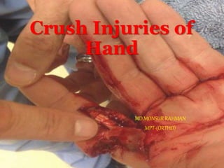
8. CRUSH INJURIES
Compressive type of force to the tissues.
Most Common Causes:
-Machine Injuries In The Industries
-RTA
-Agricultural Injuries
-Fall of heavy objects in building
collapse, during earthquakes etc.
9. Crush injuries result in:
Damage to the overlying soft tissue envelope
Laceration
Fracture
Bleeding
Loss of vascular integrity
The neurovascular structures and the bony ligamentous structures.
10. Danger signs in evaluation of crush
injuries
• Volar swelling, especially in the palm and digits
• Loss of active range of motion
• Pain on passive motion of the digits or hand
• Altered neurovascular status
• Increased swelling, even with elevation above heart level
• Profuse bleeding from an open crush injury.
12. Skin and Subcutaneous Tissue
Lacerations and contusions.
Foreign material --- embedded in the wounds.
Alternatively, the skin may look largely intact.
13. Muscles
Overstretching and tearing of the muscle bleeding and
swelling within the muscle itself.
A disruption of muscle-tendon connections may result in
loss of function.
14. Tendons
The stretching forces may create small, partial tears.
During the healing process, scar tissue forms to heal such
tears and may cause the tendons to adhere to surrounding
tissues, resulting in loss of joint motion and hand function.
15. Nerves
Usually, nerves are not torn by a crush injury.
Conduction disrupted
It may take weeks to even months to determine whether the
loss of nerve activity is permanent.
16. Blood Vessels
Direct compression or shearing forces, which may injure
the inner layer
If the injured vessel is an artery --- Ischemia
If the injured vessels are veins--- restriction of venous
outflow
17. Bone and Joints
Joint capsules and surrounding ligaments may rupture
In children, the growth plates of the bones may be
disrupted.
Disruption of growth plates interferes with subsequent bone
growth, and the bone may not grow to its proper length.
18. The Anatomic Zones
• Five flexor zones:
zone 1
From the insertion of the profundus tendon at the distal phalanx to
just distal to the insertion of the FDS.
19.
20. Flexor digitorum Profundus Tendon
Injury
• Disruption of the FDP tendon, also known as jersey finger
• In an athlete’s finger
- football or rugby.
• The injury causes forced extension of the DIP joint during
active flexion. (finger lies in slight extension relative to other
fingers in resting position)
• pain and swelling
21.
22.
23. Treatment
• Type I injuries (partial rupture of the tendon) can be treated without
surgery with rest, ice and elevation.
A finger splint is often used to hold the digit in place until healed.
• Type II (full tendon rupture) and
• Type III (rupture with bone chip attached)
24. TENODESIS
• Flexor tenodesis to prevent hyperextension & to stabilize the
distal joint
• Often, surgical pins are inserted into the injured digit to stabilize
the bone and tendon in their proper alignment.
• Securely re-attach the distal FDP tendon to the middle phalanx
with appropriate tension.
25. Collateral Ligament Injuries
• Forced ulnar or radial deviation at any of the IP joints can cause
partial or complete collateral ligament tears.
• The PIP joint usually is involved in collateral ligament injuries,
which are commonly classified as “jammed fingers.”
• pain located only at the affected ligament.
26. Treatment
• If the joints are stable and no large fracture fragments are present, the
injury can be treated with buddy taping (i.e., taping the injured finger,
above and below the joint, to an adjacent finger)
Am Fam Physician. 2006 Mar 1;73(5):810-816. News & Publications
Journals afp Vol. 73/No. 5(March 1, 2006)
28. Zone II
• Zone II is often referred to as "Bunnell's no man's land," the critical
area of pulleys between the insertion of the FDS and the distal
palmar crease.
• Both flexor tendons interweave in a complex manner, therefore even
minimum swelling can cause adhesions with pulleys & thereby
impair the free motion of the tendon.
29.
30. Trigger Finger
• Trigger finger, or flexor tenosynovitis, is a condition in which the
tendons that flex the fingers become swollen and inflamed. This results
in pain at the base of one or more of the fingers
• Inability of FDS &FDP tendons to slide smoothly under the A1 pulley
31.
32. TREATMENT
• Corticosteroids with local anesthetic into the flexor sheath.
TISSUE RELEASE
• A small (less than 2 cm) incision is made in the skin, and
the tight portion of the flexor tendon sheath is released.
• After the surgery, a sterile bandage is applied to the site of
surgery.
• This bandage is removed after a few days,
• And full use of the finger may then begin to prevent new
adhesions (scar).
(Full recovery is expected for surgery. By Jonathan Cluett, M.D., Journal About.com Guide
Updated March 29, 2007)
33. Zone-III
Extends from the distal edge of the carpal ligament to the proximal
edge of the A1 pulley, which is the entrance of the tendon sheath.
The distal palmar crease superficially marks the termination of zone
III and the beginning of zone II.
34.
35.
36. Dupuytren’s Contracture
• This condition is due to inflammation of involving the ulnar side of
the palmar aponeurosis. Localized thickening and shortening of the
palmar fascia.
• The fascia is thickened to form nodules and it contracts so that the
affected fingers are drawn into flexion.
39. Skin Graft Method
• A skin graft may be needed if the skin surface has contracted so
much that the finger cannot relax and the palm cannot be stretched
out flat.
• Surgeons graft skin from the wrist, elbow, or groin. The skin is
grafted into the area near the incision to give the finger extra
mobility for movement.
40. Zone IV
Includes the carpal tunnel and its contents (i.e., the 9 digital
flexors and the median nerve).
41. Carpal Tunnel Syndrome
• Cause of CTS - The tendons in the wrist swell and put compression
on the median nerve,
• Hand numbness, pain and tingling in the distribution of median
nerve.
42.
43. Treatment
• During surgery, an incision is made in the palm.
• The roof of the carpal tunnel is divided to increase the size of the
carpal tunnel and decrease pressure on the median nerve.
44. Extensor Zones
• zone I - mallet finger
DIP jt. (finger) and IP jt. In thumb.
• zone II - middle phalanx (finger)
proximal phalanx (thumb)
• zone III - apex PIP joint (finger)
MCP jt. (thumb)
• zone IV- proximal phalanx (finger)
metacarpal (thumb)
• zone V - over apex MCP joint:
• zone VI - dorsum of hand
• Zone VII - dorsal retinaculum
• Zone VIII- distal forearm
45.
46. Mallet Finger
• The trauma results in the avulsion
of the extensor tendon from the
point of attachment to the distal
phalanx
47. • A segment of the distal phalanx,
which comprises the distal
portion of DIP joint, may break
off along with the tendon.
• If not treated, mallet finger leaves
a deformity with the DIP in
permanent flexion.
48. Treatment
• Most mallet finger injuries can be closed-reduction and fixed by
percutaneous placement of K-wires.
• The longitudinal K-wire is blocking the DIP joint from flexion
to protect the repair.
49. A/P Radiographic view of
finger.
• The smaller oblique K-wire is
placed through the bone fragment,
fixing it to the distal fragment.
50. Swan Neck Deformity
• Finger with a hyper-extended PIP joint and a flexed DIP joint.
• The extensor tendon gets out of balance, which allows the DIP
joint to get pulled downward into flexion.
51. The Journal of hand surgery J Hand
Surg Am. 2010 Aug 13;: 20709465
Distraction arthrolysis using an external fixator followed by flexor
tenolysis- useful treatment for patient with pip joint extension
contracture and tendon adhesions after severe crush injury.
52. • On the day of attaching the external fixator, moderate
distraction was applied to the joint and the gap was widened
to approximately 2 mm.
• Pip joint was gradually widened for 10 days until a gap of
about 5 mm was attained.
• Passive range of motion was performed for about 1week until
swelling of the affected digit subsided. Then, flexor tenolysis
was performed.
53. Boutonniere Deformity
• Buttonhole deformity
• The middle finger joint is bent in a fixed position inward and the
outermost finger joint is bent excessively outward (away from the
palm)
• Most common causes are Rheumatoid Arthritis and trauma
54. Treatment
Gutter splinting will help stretch and straighten the PIP joint .
Best results occur when the PIP joint is limber, rather than
stuck in a bent position.
55. De Quervain’s Tenosynovitis
• Injury occurs because of inflammation around the tendon sheath of
the APL and EPB in the first dorsal compartment .
• Fibrous sheath (APL & EPB) tendon becomes fibrosed and thickened.
• Lateral aspect of lower end of the radius where the tendons lie in
shallow bony groove.
56. • A splint can be used - one that immobilizes the wrist, and also involves the
thumb.
• Corticosteroid injections into the tendon sheath. Surgical release may be
required in chronic cases.
57. Splint used for conservative treatment (left) & bandage used
following surgery (right).
58. SURGERY
• An incision is made over the first dorsal compartment and the dorsal
carpal ligament is cut to expose the tendons.
• The tendons APL and EPB are identified and motion is checked. The
wound is then closed and a compressive dressing with a plaster splint is
applied.
59. PHYSIOTHERAPY
• Visits will include heat treatments, soft tissue massage, and vigorous
stretching.
• Active and assisted active finger exercises may restore the hand
functions.
• Passive Stretching to correct the residual tightness of the soft tissues.
60. Prognosis
Crush injuries can be severe and devastating to the individual.
Long-term impairment and disability may occur, and disability is
sometimes permanent.