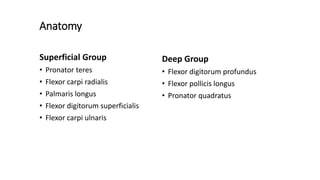
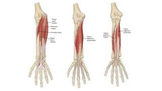
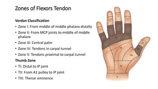
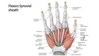
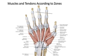
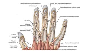
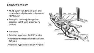
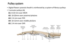
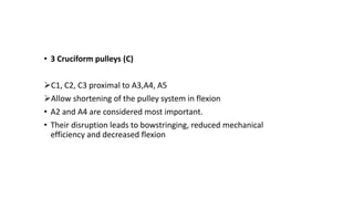
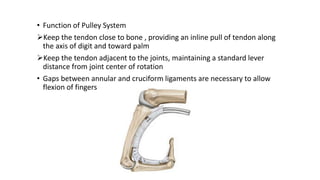
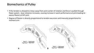
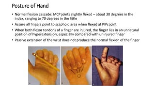
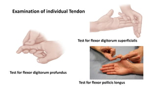
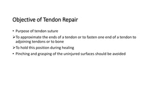
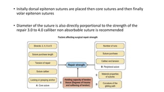
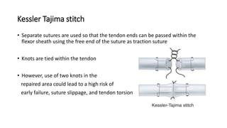
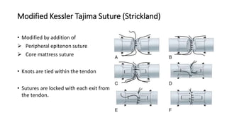
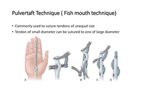
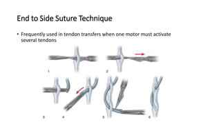
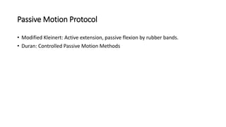
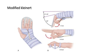
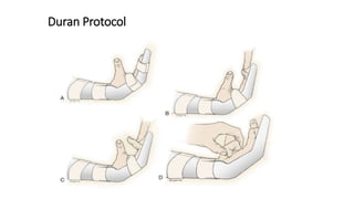
SlideShare a Scribd company logo
Flexors Tendon Injuries
Moderator : Assoc Prof Dr Suresh Uprety
Presenter : Dr Ajay Shah (Resident)
Department of Orthopedics and Trauma Surgery
IOM,TUTH
Anatomy
Superficial Group
• Pronator teres
• Flexor carpi radialis
• Palmaris longus
• Flexor digitorum superficialis
• Flexor carpi ulnaris
Deep Group
• Flexor digitorum profundus
• Flexor pollicis longus
• Pronator quadratus
Zones of Flexors Tendon
Verdan Classification
• Zone I: From middle of middle phalanx distally
• Zone II: From MCP joints to middle of middle
phalanx
• Zone III: Central palm
• Zone IV: Tendons in carpal tunnel
• Zone V: Tendons proximal to carpal tunnel
Thumb Zone
• TI: Distal to IP joint
• TII: From A1 pulley to IP joint
• TIII: Thenar eminence
Flexors Synovial
sheath
Muscles and Tendons According to Zones
Carpal Tunnel
Neurovascular Distribution According to Zones
Camper’s chiasm
• At A1 pulley FDS tendon splits and
rotates laterally then dorsally around
FDP tendon
• Two splits tendon join together
proximal to PIP joint at camper’s
chiasm
• Functions
Provides a pathway for FDP tendon
Increases the stability and balance of
PIP joint
Prevents hyperextension of PIP joint
No Man’s Land
• Historic derivation of “no man's land”
dates back to 14th century
• It was used to describe an area outside
London used for executions
• Sterling Bunnel used this term in hand
surgery who most probably derived it
with his experience in the World War 1
• He described it as no man's land because of perennial anatomical
characteristics
• This zone has a fibro osseous digital canal where both the tendons interweave
in a complex manner
• Multiple pulleys increase its complexity because minimal swelling of the
epitenon can impair free motion of the tendon
• Any infection, fibrosis, scarring, overcrowding can lead to dense adhesions and
hence compromising the results
Pulley system
• Digital flexors synovial sheath is reinforced by a system of fibrous pulleys
• 5 annular pulleys (A)
A1: 8-10 mm over MCPJ
A2: 18-20mm over proximal phalanx
A3: 2-4 mm over PIPJ
A4: 10-12mm over middle phalanx
A5: 2-4 mm over DIPJ
• 3 Cruciform pulleys (C)
C1, C2, C3 proximal to A3,A4, A5
Allow shortening of the pulley system in flexion
• A2 and A4 are considered most important.
• Their disruption leads to bowstringing, reduced mechanical
efficiency and decreased flexion
• Function of Pulley System
Keep the tendon close to bone , providing an inline pull of tendon along
the axis of digit and toward palm
Keep the tendon adjacent to the joints, maintaining a standard lever
distance from joint center of rotation
• Gaps between annular and cruciform ligaments are necessary to allow
flexion of fingers
Biomechanics of Pulley
• If the tendon is allowed to move away from joint center of rotation and force is pulled through
flexor system , lever distance increases, causing increase in work performance at joint leading to
excess flexion at DIP joint
• Degree of flexion is directly proportional to tendon excursion and inversely proportional to
moment arm
Tendon Nutrition
Tendon nutrition 2 basic sources:
• Synovial fluid produced within the tenosynovial sheath
• Blood supply provided through longitudinal vessels in the paratenon,
intraosseous vessels at the tendon insertion, and vincular circulation
Blood supply
• Suture placement in volar half of tendon
• Intratendinous placement of crisscross sutures
disturb the microcirculation
Tendon healing
Tendon healing is believed to occur through the activity of extrinsic and intrinsic
mechanisms
• Extrinsic: Through the activity of peripheral fibroblasts derived from tendon
sheath and seems dominant mechanism contributing to the formation of scar and
adhesions.
• Intrinsic: Through the activity of the fibroblasts derived from the tendon
Phases of Tendon healing
• Following laceration, tendon healing proceeds through 3 sequential but
overlapping phases
Examination
• General condition of patient
• Possibility of other injuries
• Neurologic examination
Assessment of light touch and static two point
discrimination
• Vascular examination
CRT of volar digital pulp and nail bed is assessed
When necessary, digital Allen test can be done
Posture of Hand
• Normal flexion cascade: MCP joints slightly flexed – about 30 degrees in the
index, ranging to 70 degrees in the little
• Assure all fingers point to scaphoid area when flexed at PIPs joint
• When both flexor tendons of a finger are injured, the finger lies in an unnatural
position of hyperextension, especially compared with uninjured finger
• Passive extension of the wrist does not produce the normal flexion of the finger
Test for flexor digitorum profundus
Test for flexor digitorum superficialis
Test for flexor pollicis longus
Examination of individual Tendon
Objective of Tendon Repair
• Purpose of tendon suture
To approximate the ends of a tendon or to fasten one end of a tendon to
adjoining tendons or to bone
To hold this position during healing
• Pinching and grasping of the uninjured surfaces should be avoided
Principles of Tendon Repair
• All flexor tendons should be repaired at whatever level they are transected.
• Whenever possible the repair should be done primarily. When delayed a tendon
graft may be required
• A2 and A4 annular pulley areas of the flexor sheath should be preserved;
otherwise, tendon bowstringing and flexion deformity of the finger can develop
and excursion of the tendon becomes impaired.
• Tendon laceration of 60 % or more is treated as complete transection
• Initally dorsal epitenon sutures are placed then core sutures and then finally
volar epitenon sutures
• Diameter of the suture is also directly porportional to the strength of the
repair 3.0 to 4.0 calliber non absorbable suture is recommended
• Repair is supplemented with a running circumferential epitenon suture
technique.
Advantages:
smooth repair and improves tendon gliding
minimize the adhesion formation
less gap formation
• Suture technique must withstand gap formation of 3 mm at the repair site during
the initial 3 weeks following repair.
Strickland stressed six characteristics of an ideal tendon repair
• Easy placement of sutures in the tendon,
• Secure suture knots,
• Smooth juncture of tendon ends,
• Minimal gapping at the repair site,
• Minimal interference with tendon vascularity, and
• Sufficient strength throughout healing to permit application of early
motion stress to the tendon.
Timing of Flexor Tendon Repair
• Primary tendon repair
within the first 12 hours of injury
can be extended to within 24 hours of injury in rare situations
• Delayed primary repair: within 24 hours to approximately 10 days.
• Secondary after 10 to 14 days
• Late secondary repair after 4 weeks
Ideal properties of suture material
• Non reactive
• Hold securely when knotted
• Small caliber
• High tensile strength
• Easy to handle
Common material: Ethibond , Nylon, Prolene
Suture Configurations
Tendon repair types can be divided into three groups
• Group 1: Simple suture
• Group 2 : Bunnell suture
• Group 3: Pulvertaft technique (fish-mouth weave)
An abundance of research has shown that four-strand, six-strand, and
eight-strand core sutures
• Create stronger repairs
• Reduce the possibility of gap formation
• Permit greater active forces applied to the repaired tendons
• Allowing earlier active motion
End to End Suture Techniques
Kessler technique
• Modification of the Mason-Allen suture.
• Effective for tendon repair in the fingers and palm
• Knots being left exposed on the tendon surface.
Modified Kessler technique
• Modifications:
Only one knot inside the repair site
Knot is left in the cut surface of the tendon
• Easier to use a monofilament suture like a
4.0 Prolene to reapproximate tendon edges
Kessler Tajima stitch
• Separate sutures are used so that the tendon ends can be passed within the
flexor sheath using the free end of the suture as traction suture
• Knots are tied within the tendon
• However, use of two knots in the
repaired area could lead to a high risk of
early failure, suture slippage, and tendon torsion
Modified Kessler Tajima Suture (Strickland)
• Modified by addition of
 Peripheral epitenon suture
 Core mattress suture
• Knots are tied within the tendon
• Sutures are locked with each exit from
the tendon.
Pulvertaft Technique ( Fish mouth technique)
• Commonly used to suture tendons of unequal size
• Tendon of small diameter can be sutured to one of large diameter
End to Side Suture Technique
• Frequently used in tendon transfers when one motor must activate
several tendons
Tendon to Bone Attachment
• Frequently requires a pull-out technique (usually distal phalanx)
• For tendon to bone repairs ,core suture techniques used most often are
Kessler
Bunnell crisscross suture
Suture Anchor Tendon Attachment
• As effective as pull-out wire or suture but without
the potential complications with fingernail that
can occur with pull-out technique
• Two suture anchors are placed in the distal
phalanx from distal-volar to proximal-dorsal
• To purchase in the thickest portion of the distal
phalanx to provide greatest pullout strength
Surgical Incision
• Incisions should not compromise viability of skin flaps
• Should not create contractures or cosmetically unsightly scars
• Less exposure needed distally than proximally
• Zigzag (Brunner),mid radial ,mid ulnar or midline oblique incisions
Zone I Repair
• Contains only the FDP tendon
• Direct repair (primary tenorraphy): laceration >1 cm from FDP insertion
• Proximal tendon retrieved by feeding tube and passed underneath A4
pulley
Tendon advancement (<1cm stump)
• Pull-out suture
Free ends of sutures are passed
through or around distal phalanx, tied
over dorsum of fingernail with button.
Removed after 6 weeks
• Internal suture methods
Suture anchors or other methods to
affix the tendon directly to the bone
When the diagnosis of interruption of FDP tendon is delayed, and the tendon has
retracted into the palm, its vinculum has been disrupted and a decision must be
made regarding repair depending upon types
• Type I: FDP tendon is avulsed from its
insertion and retracts into palm
• Type II: Avulsed from its insertion but
stump remains within digital sheath
• Type III: Avulsed bony fragment attached
to tendon stump, remains within flexor
sheath
Modified Leddy and Packer classification for FDP avulsion
Type IV avulsions were first described by Robins and Dobyns in 1974. Inclusion
of type V avulsions in the classification was proposed by Al-Qattan in 2001
• Type 1, if it is within 7 to 10 days of the injury, the tendon should be threaded
back into the finger and reattached with a pull-out wire into distal phalanx
• Type 2, despite the passage of a few months, these tendons can be reattached as
well because circulation is thought to be maintained
• Type 3, usually fracture repair with k-wire or miniature screw fixation
• Type 4, fracture fixation followed by advancement of tendon to distal phalanx
• Type 5, FDP tendon with avulsed bone fragment is reduced using a pull-out
technique, and a K-wire is placed for avulsed fragment
Zone II Repair
Primary repair at this level frequently fails because of adhesion in the area of
pulley
Technical concerns during the repair procedure includes
• Management of lacerations of FDP and FDS tendons
• Appropriate orientation of the FDP with FDS slips
• Attachment of the FDS slips in the thin flat area
• Management of the flexor sheath, including annular thickening (pulleys)
• Postoperative management,
• Timing and technique for tenolysis
• Care should be taken when FDS has been injured in area just proximal to PIP joint
• Care also should be taken to deliver FDP tendon through the split portion of FDS
when profundus tendon has retracted proximally
• Method of repair however is controversial but following are the different options
of treatment:
Repair of the FDP tendon only with debridement of the FDS stump
Repair of both tendons or
Repair of FDP with repair of one slip of FDS tendon
Most hand surgeons prefer to repair the FDP and one slip of FDS , reasonably a
good option as the repair of both slips of FDS may produce overcrowding within the
sheath and pulleys and compromise the result
Coats RW, 2nd, Echevarría-Oré JC, Mass DP. Acute flexor tendon repairs in zone II. Hand Clin. 2005;21:173–9
• Core suture with two or more strands, locking components, and buried knots is
usually preferred
• A running, circumferential 5-0 or 6-0 suture is used to complete a smooth repair
and to minimize adhesion formation to the sheath
• Tenolysis may be required in an estimated 18% to 25% of patients after flexor
tendon repair
• Three months is considered to be earliest time for flexor tenolysis, assuming no
improvement in motion in previous 1 to 2 months
• Function in the finger can be improved by 50% by tenolysis
Strickland technique for flexor tendon injury in Zone II
Zone III Repair
• If both tendons are lacerated, both are repaired, end to end with circumferential
re-enforcing sutures
• Muscles bellies of lumbricals and tendon are frequently affected so additional
incision often needed to expose further area
• If conditions permit, primary repair of sharply severed nerve is crucial
• If wound condition preclude tendon and nerve repair, the ends of the tendon and
nerve are sutured to adjacent fascia to prevent undue retraction
• Lumbricals muscle bellies usually are not sutured, causes lumbrical plus finger
Zone IV Repair
• Associated with median ,ulnar nerve or ulnar/radial vessel injury
• If the laceration occurs beneath the transverse carpal ligament, partial or complete release
of the transverse carpal ligament may be required
• Preserve, if possible, a portion of the transverse carpal ligament to avoid bowstringing
postoperatively
• If it cannot be preserved, release it in a Z-lengthening configuration
• Best suture configuration is intratendinous one with a locking core
• Immobilize the wrist in about 45 degrees of flexion, 50 to 60 degrees of flexion at MCP
joints and the IP joints in full extension
Zone V Repair
• In zone V, multiple tendons, nerves, and vessels frequently are injured so its important to identify
the tendons accurately
• Properly match the tendon ends by careful attention to their location and level in the wound,
their relation to neighboring structures
• Blood clots within the tenosynovium usually serve as clues in locating severed tendons
• Repairs done in the distal forearm do not absolutely require an intratendinous repair, double
right-angled or mattress suture may be used
• Immobilize the wrist in about 45 degrees of flexion, with fingers in 50 to 60 degrees of flexion at
MCP joints and the IP joints in full extension
Postoperative Rehabilitation
Immobilization
Indication for immobilization after flexor tendon surgery is usually not required,
exceptions
• Children or adults unable to cooperate with hand therapy
• Unstable bone repair
• Concerns about the effect of tension on microneurovascular repairs.
• Unwilling to adhere to strict early mobilisation protocols
Passive Motion Protocol
• Modified Kleinert: Active extension, passive flexion by rubber bands.
• Duran: Controlled Passive Motion Methods
Modified kleinert
Duran Protocol
Flexor Tendons Reconstruction
When FDS and FDP tendons have been divided in the critical area of the pulleys,
flexor tendon grafting may be indicated if
• Injuries resulting in segmental tendon loss
• Neglected >3 to 6 weeks with tendon degeneration and scar within the tendon
sheath i.e. chronic flexor tendon injuries
• If a gap cannot be closed because of myocontracture
• Large section of tendon has been damaged in Zone II injury
• Delayed presentation of FDP avulsion injuries associated with significant tendon
retraction
Requirements before tendon graft
• Skin is pliable;
• Any wounds are well healed
• Tissues through which tendon is expected to glide must be relatively free
of scar
• Edema has subsided
• Joints allow a full passive range of motion
• Sensation in the finger is normal or at least one digital nerve is intact
Boyes preoperative classification helps to identify the severity of injury, with
higher grades portending worse outcomes
Single-Stage Tendon Graft
• Considered when the flexor tendon sheath is relatively intact and unscarred and
the pulley system is competent
• Attempt can be made to dilate collapsed pulleys with urethral dilators
• However, if unsuccessful, a two-stage procedure with pulley reconstruction
should be undertaken
Age is a strong prognostic factor. Results are best in patients 10 to 30 years old, and
worst results in very young and in patients older than 50 years
Bora FW Jr: Profundus tendon grafting with unimpaired sublimis function in children. Clin Orthop Relat Res
1970;71 (71):118-123.
Two-Stage Tendon Graft
Indications
• Crushing injuries associated with fracture or skin damage
• Damaged pulley system
• Excessive scarring of the tendon bed
• Failure of previous operations
• Stiff joints
First stage
Excision of tendon and scar from the flexor tendon bed
Preserving or reconstructing the flexor pulley system
• Dacron-impregnated silicone rod is inserted to maintain the tunnel in the area of
excised tendons until passive motion and sensitivity have been restored to the digit
Second stage
• Done when the finger is soft, supple, and well healed with mobile joints
• Earliest time for the second stage is about 8 weeks, but usually 3 months is
required, depending on the patient’s needs and the surgeon’s judgment
• Consists of
Rod removal
Tendon graft insertion
Pulley Reconstruction
Goal
• To enable maximum excursion and
strength with controlled gliding of flexor
tendons
• To minimize friction
Two main techniques are
• Weaving the pulley graft through
remaining pulley rim
• Creating a new pulley loop around bone
Graft Choices
• Palmaris longus tendon
• Plantaris tendon
• Extensor Digitorum longus to 2nd 3rd or 4th toes
• Flexor digitorum longus to 2nd toe
• Flexor digitorum superficialis
Intrasynovial grafts (FDS and toe flexors) have better
morphologic and functional characteristics than that of
extrasynovial grafts. Extrasynovial grafts have been
thought to lead to more adhesions
Flexor tendon grafting to the hand: An assessment of the intrasynovial
donor tendon. A preliminary single-cohort study. J Hand Surg Am 2000;
25(4):721-730.
Complications
Short term:
• Infection
• Injury to neurovascular structures or pulley system
• Abnormal scarring
Long term:
• Adhesion
• Rupture
• Joint contracture
• Triggering
Rarer complications
• Lumbrical plus finger
• Quadrigia effect
Adhesions
• Most common complication despite early motion protocols
• Higher risk with Zone II injuries
• Treatment
Physical therapy
Tenolysis when patients progressive gain in digital motion has plateaued,
usually 3- 6 months after repair
Adhesion formation with restriction of tendon excursion and the need for
tenolysis has reported rates from 12% to 47%
LaSalle WB, Strickland JW: An evaluation of the two-stage flexor tendon reconstruction technique. J
Hand Surg Am 1983;8(3):263-267.
Tendon rupture
• Noted by the patient as “popping” in the hand
• 7-10 days postop when tensile strength is weakest
• MRI may help in diagnosis
• Treatment
If < 1cm of scar is present, resect the scar and perform primary repair
If > 1cm of scar is present, perform tendon graft
• if the sheath is intact and allows passage of a pediatric urethral catheter or vascular
dilator, perform primary tendon grafting
• if the sheath is collapsed, place Hunter rod and perform staged graft
References
• Campbells Operative Orthopaedics,13th edition
• Green Operative Hand Surgery, 7th edition
• Apleys system of Orthopedics,9th edition
• Articles

More Related Content

What's hot

Tendon injuries of hand
Tendon injuries of handTendon injuries of hand
Tendon injuries of hand
Vivek Mathew Philip
 
Flexor tendon injury, management and rehabilitation
Flexor tendon injury, management and rehabilitationFlexor tendon injury, management and rehabilitation
Flexor tendon injury, management and rehabilitation
Khadijah Nordin
 
Tendon transfer- principles and techniques
Tendon transfer- principles and techniquesTendon transfer- principles and techniques
Tendon transfer- principles and techniques
Dr Sourabh Shankar Chakraborty
 
Tendon transfer for radial nerve palsy
Tendon transfer for radial nerve palsyTendon transfer for radial nerve palsy
Tendon transfer for radial nerve palsy
Mohammed Aljodah
 
Hand rehabilitation after flexor tendon repair
Hand rehabilitation after flexor tendon repairHand rehabilitation after flexor tendon repair
Hand rehabilitation after flexor tendon repair
Mohammed Aljodah
 
RADIAL NERVE PALSY AND TENDON TRANSFER
RADIAL NERVE PALSY AND TENDON TRANSFERRADIAL NERVE PALSY AND TENDON TRANSFER
RADIAL NERVE PALSY AND TENDON TRANSFER
Benthungo Tungoe
 
Median nerve injuries and mangement
Median nerve injuries and mangementMedian nerve injuries and mangement
Median nerve injuries and mangement
sanyal1981
 
Clinical testing ulnar nerve
Clinical testing ulnar nerveClinical testing ulnar nerve
Clinical testing ulnar nerve
Roopchand Ps
 
Flexor tendon injuries.m
Flexor tendon injuries.mFlexor tendon injuries.m
Flexor tendon injuries.mMansoor Khan
 
Hand rehabilitation following flexor tendon injuries
Hand rehabilitation following flexor tendon injuriesHand rehabilitation following flexor tendon injuries
Hand rehabilitation following flexor tendon injuries
Abey P Rajan
 
Finger tip injuries
Finger tip injuriesFinger tip injuries
Finger tip injuries
Sushil Sharma
 
Radial nerve-palsy-tendon-transfers
Radial nerve-palsy-tendon-transfersRadial nerve-palsy-tendon-transfers
Radial nerve-palsy-tendon-transfersdrpouriamoradi
 
management of claw hand
management of claw handmanagement of claw hand
management of claw hand
Prashanth Kumar
 
Finger tip injury
Finger tip injuryFinger tip injury
Finger tip injury
Abdulla Kamal
 
Intramedullary nailing seminar by dr ashwani panchal
Intramedullary nailing seminar by dr ashwani panchalIntramedullary nailing seminar by dr ashwani panchal
Intramedullary nailing seminar by dr ashwani panchal
Dr ashwani panchal
 
Hand deformity in rheumatoid arthritis
Hand deformity in rheumatoid arthritisHand deformity in rheumatoid arthritis
Hand deformity in rheumatoid arthritis
sushilonlines
 
4.position of safe immobilisation
4.position of safe immobilisation4.position of safe immobilisation
4.position of safe immobilisation
Ngoc Quang
 
Principles of management of volkmann’s contracture
Principles of management of volkmann’s contracturePrinciples of management of volkmann’s contracture
Principles of management of volkmann’s contracture
Soliudeen Arojuraye
 
MEDIAN NERVE PALSY AND TENDON TRANSFERS
MEDIAN NERVE PALSY AND TENDON TRANSFERSMEDIAN NERVE PALSY AND TENDON TRANSFERS
MEDIAN NERVE PALSY AND TENDON TRANSFERS
Benthungo Tungoe
 
Common hand injuries
Common hand injuriesCommon hand injuries
Common hand injuries
tbf413
 

What's hot (20)

Tendon injuries of hand
Tendon injuries of handTendon injuries of hand
Tendon injuries of hand
 
Flexor tendon injury, management and rehabilitation
Flexor tendon injury, management and rehabilitationFlexor tendon injury, management and rehabilitation
Flexor tendon injury, management and rehabilitation
 
Tendon transfer- principles and techniques
Tendon transfer- principles and techniquesTendon transfer- principles and techniques
Tendon transfer- principles and techniques
 
Tendon transfer for radial nerve palsy
Tendon transfer for radial nerve palsyTendon transfer for radial nerve palsy
Tendon transfer for radial nerve palsy
 
Hand rehabilitation after flexor tendon repair
Hand rehabilitation after flexor tendon repairHand rehabilitation after flexor tendon repair
Hand rehabilitation after flexor tendon repair
 
RADIAL NERVE PALSY AND TENDON TRANSFER
RADIAL NERVE PALSY AND TENDON TRANSFERRADIAL NERVE PALSY AND TENDON TRANSFER
RADIAL NERVE PALSY AND TENDON TRANSFER
 
Median nerve injuries and mangement
Median nerve injuries and mangementMedian nerve injuries and mangement
Median nerve injuries and mangement
 
Clinical testing ulnar nerve
Clinical testing ulnar nerveClinical testing ulnar nerve
Clinical testing ulnar nerve
 
Flexor tendon injuries.m
Flexor tendon injuries.mFlexor tendon injuries.m
Flexor tendon injuries.m
 
Hand rehabilitation following flexor tendon injuries
Hand rehabilitation following flexor tendon injuriesHand rehabilitation following flexor tendon injuries
Hand rehabilitation following flexor tendon injuries
 
Finger tip injuries
Finger tip injuriesFinger tip injuries
Finger tip injuries
 
Radial nerve-palsy-tendon-transfers
Radial nerve-palsy-tendon-transfersRadial nerve-palsy-tendon-transfers
Radial nerve-palsy-tendon-transfers
 
management of claw hand
management of claw handmanagement of claw hand
management of claw hand
 
Finger tip injury
Finger tip injuryFinger tip injury
Finger tip injury
 
Intramedullary nailing seminar by dr ashwani panchal
Intramedullary nailing seminar by dr ashwani panchalIntramedullary nailing seminar by dr ashwani panchal
Intramedullary nailing seminar by dr ashwani panchal
 
Hand deformity in rheumatoid arthritis
Hand deformity in rheumatoid arthritisHand deformity in rheumatoid arthritis
Hand deformity in rheumatoid arthritis
 
4.position of safe immobilisation
4.position of safe immobilisation4.position of safe immobilisation
4.position of safe immobilisation
 
Principles of management of volkmann’s contracture
Principles of management of volkmann’s contracturePrinciples of management of volkmann’s contracture
Principles of management of volkmann’s contracture
 
MEDIAN NERVE PALSY AND TENDON TRANSFERS
MEDIAN NERVE PALSY AND TENDON TRANSFERSMEDIAN NERVE PALSY AND TENDON TRANSFERS
MEDIAN NERVE PALSY AND TENDON TRANSFERS
 
Common hand injuries
Common hand injuriesCommon hand injuries
Common hand injuries
 

Similar to Flexor tendon injuries slideshare

Acute Extensor tendon injuries diagnosis and management.pptx
Acute Extensor tendon injuries diagnosis and management.pptxAcute Extensor tendon injuries diagnosis and management.pptx
Acute Extensor tendon injuries diagnosis and management.pptx
Rohie3
 
Acute Tendon injury of the Hand and its Repair.pptx
Acute Tendon injury of the Hand and its Repair.pptxAcute Tendon injury of the Hand and its Repair.pptx
Acute Tendon injury of the Hand and its Repair.pptx
Okpako Isaac
 
Tendon repair
Tendon repairTendon repair
Tendon repair
MonitoshPaul
 
Flexor tendon injury
Flexor tendon injuryFlexor tendon injury
Flexor tendon injury
Dr Mujtuba Pervez Khan
 
Tendon injuries of hand by Dr Saumya Agarwal
Tendon injuries of hand by Dr Saumya AgarwalTendon injuries of hand by Dr Saumya Agarwal
Fracture Fixation Techniques.pptx
Fracture Fixation Techniques.pptxFracture Fixation Techniques.pptx
Fracture Fixation Techniques.pptx
Rekha Pathak
 
Flexor tendon repair
Flexor tendon repairFlexor tendon repair
Flexor tendon repair
Punith Vasanthan
 
Flexor Tendon injury.pptx
Flexor Tendon injury.pptxFlexor Tendon injury.pptx
Flexor Tendon injury.pptx
Athishjaims1
 
Tendon injuries of hand
Tendon injuries of handTendon injuries of hand
Tendon injuries of hand
rohit raj
 
Distal femur fracture
Distal femur fractureDistal femur fracture
Distal femur fracture
Dr Sharanprasad Hongal
 
distalfemur-170720141254.pdf
distalfemur-170720141254.pdfdistalfemur-170720141254.pdf
distalfemur-170720141254.pdf
DrShubhamNagdev
 
Squint surgeries
Squint surgeriesSquint surgeries
Squint surgeries
Mahrukh Khan
 
MRI shoulder and knee- Anatomy, Scan Planning & Its Techniques
MRI shoulder and knee- Anatomy, Scan Planning & Its Techniques  MRI shoulder and knee- Anatomy, Scan Planning & Its Techniques
MRI shoulder and knee- Anatomy, Scan Planning & Its Techniques
Nitish Virmani
 
Flexortendoninjuries1 140504003542-phpapp01
Flexortendoninjuries1 140504003542-phpapp01Flexortendoninjuries1 140504003542-phpapp01
Flexortendoninjuries1 140504003542-phpapp01
ssanjibanisudha
 
Total elbow arthroplasty
Total elbow arthroplastyTotal elbow arthroplasty
Total elbow arthroplasty
Anil Kumar Prakash
 
Pollicization
PollicizationPollicization
Principles of knee arthoscopy
Principles of knee arthoscopyPrinciples of knee arthoscopy
Principles of knee arthoscopy
Kaushal Kafle
 
Rejul journal club- tendoscopy
Rejul   journal club- tendoscopyRejul   journal club- tendoscopy
Rejul journal club- tendoscopy
Rejul Raj
 
Flexion tendon injuries of hand
Flexion tendon injuries of handFlexion tendon injuries of hand
Flexion tendon injuries of hand
darshann77
 

Similar to Flexor tendon injuries slideshare (20)

Acute Extensor tendon injuries diagnosis and management.pptx
Acute Extensor tendon injuries diagnosis and management.pptxAcute Extensor tendon injuries diagnosis and management.pptx
Acute Extensor tendon injuries diagnosis and management.pptx
 
Acute Tendon injury of the Hand and its Repair.pptx
Acute Tendon injury of the Hand and its Repair.pptxAcute Tendon injury of the Hand and its Repair.pptx
Acute Tendon injury of the Hand and its Repair.pptx
 
Tendon repair
Tendon repairTendon repair
Tendon repair
 
Flexor tendon injuries
Flexor tendon injuriesFlexor tendon injuries
Flexor tendon injuries
 
Flexor tendon injury
Flexor tendon injuryFlexor tendon injury
Flexor tendon injury
 
Tendon injuries of hand by Dr Saumya Agarwal
Tendon injuries of hand by Dr Saumya AgarwalTendon injuries of hand by Dr Saumya Agarwal
Tendon injuries of hand by Dr Saumya Agarwal
 
Fracture Fixation Techniques.pptx
Fracture Fixation Techniques.pptxFracture Fixation Techniques.pptx
Fracture Fixation Techniques.pptx
 
Flexor tendon repair
Flexor tendon repairFlexor tendon repair
Flexor tendon repair
 
Flexor Tendon injury.pptx
Flexor Tendon injury.pptxFlexor Tendon injury.pptx
Flexor Tendon injury.pptx
 
Tendon injuries of hand
Tendon injuries of handTendon injuries of hand
Tendon injuries of hand
 
Distal femur fracture
Distal femur fractureDistal femur fracture
Distal femur fracture
 
distalfemur-170720141254.pdf
distalfemur-170720141254.pdfdistalfemur-170720141254.pdf
distalfemur-170720141254.pdf
 
Squint surgeries
Squint surgeriesSquint surgeries
Squint surgeries
 
MRI shoulder and knee- Anatomy, Scan Planning & Its Techniques
MRI shoulder and knee- Anatomy, Scan Planning & Its Techniques  MRI shoulder and knee- Anatomy, Scan Planning & Its Techniques
MRI shoulder and knee- Anatomy, Scan Planning & Its Techniques
 
Flexortendoninjuries1 140504003542-phpapp01
Flexortendoninjuries1 140504003542-phpapp01Flexortendoninjuries1 140504003542-phpapp01
Flexortendoninjuries1 140504003542-phpapp01
 
Total elbow arthroplasty
Total elbow arthroplastyTotal elbow arthroplasty
Total elbow arthroplasty
 
Pollicization
PollicizationPollicization
Pollicization
 
Principles of knee arthoscopy
Principles of knee arthoscopyPrinciples of knee arthoscopy
Principles of knee arthoscopy
 
Rejul journal club- tendoscopy
Rejul   journal club- tendoscopyRejul   journal club- tendoscopy
Rejul journal club- tendoscopy
 
Flexion tendon injuries of hand
Flexion tendon injuries of handFlexion tendon injuries of hand
Flexion tendon injuries of hand
 

Recently uploaded

Are There Any Natural Remedies To Treat Syphilis.pdf
Are There Any Natural Remedies To Treat Syphilis.pdfAre There Any Natural Remedies To Treat Syphilis.pdf
Are There Any Natural Remedies To Treat Syphilis.pdf
Little Cross Family Clinic
 
HOT NEW PRODUCT! BIG SALES FAST SHIPPING NOW FROM CHINA!! EU KU DB BK substit...
HOT NEW PRODUCT! BIG SALES FAST SHIPPING NOW FROM CHINA!! EU KU DB BK substit...HOT NEW PRODUCT! BIG SALES FAST SHIPPING NOW FROM CHINA!! EU KU DB BK substit...
HOT NEW PRODUCT! BIG SALES FAST SHIPPING NOW FROM CHINA!! EU KU DB BK substit...
GL Anaacs
 
Superficial & Deep Fascia of the NECK.pptx
Superficial & Deep Fascia of the NECK.pptxSuperficial & Deep Fascia of the NECK.pptx
Superficial & Deep Fascia of the NECK.pptx
Dr. Rabia Inam Gandapore
 
Physiology of Chemical Sensation of smell.pdf
Physiology of Chemical Sensation of smell.pdfPhysiology of Chemical Sensation of smell.pdf
Physiology of Chemical Sensation of smell.pdf
MedicoseAcademics
 
Physiology of Special Chemical Sensation of Taste
Physiology of Special Chemical Sensation of TastePhysiology of Special Chemical Sensation of Taste
Physiology of Special Chemical Sensation of Taste
MedicoseAcademics
 
Lung Cancer: Artificial Intelligence, Synergetics, Complex System Analysis, S...
Lung Cancer: Artificial Intelligence, Synergetics, Complex System Analysis, S...Lung Cancer: Artificial Intelligence, Synergetics, Complex System Analysis, S...
Lung Cancer: Artificial Intelligence, Synergetics, Complex System Analysis, S...
Oleg Kshivets
 
KDIGO 2024 guidelines for diabetologists
KDIGO 2024 guidelines for diabetologistsKDIGO 2024 guidelines for diabetologists
KDIGO 2024 guidelines for diabetologists
د.محمود نجيب
 
24 Upakrama.pptx class ppt useful in all
24 Upakrama.pptx class ppt useful in all24 Upakrama.pptx class ppt useful in all
24 Upakrama.pptx class ppt useful in all
DrSathishMS1
 
ARTHROLOGY PPT NCISM SYLLABUS AYURVEDA STUDENTS
ARTHROLOGY PPT NCISM SYLLABUS AYURVEDA STUDENTSARTHROLOGY PPT NCISM SYLLABUS AYURVEDA STUDENTS
ARTHROLOGY PPT NCISM SYLLABUS AYURVEDA STUDENTS
Dr. Vinay Pareek
 
Factory Supply Best Quality Pmk Oil CAS 28578–16–7 PMK Powder in Stock
Factory Supply Best Quality Pmk Oil CAS 28578–16–7 PMK Powder in StockFactory Supply Best Quality Pmk Oil CAS 28578–16–7 PMK Powder in Stock
Factory Supply Best Quality Pmk Oil CAS 28578–16–7 PMK Powder in Stock
rebeccabio
 
Ozempic: Preoperative Management of Patients on GLP-1 Receptor Agonists
Ozempic: Preoperative Management of Patients on GLP-1 Receptor Agonists  Ozempic: Preoperative Management of Patients on GLP-1 Receptor Agonists
Ozempic: Preoperative Management of Patients on GLP-1 Receptor Agonists
Saeid Safari
 
Surgical Site Infections, pathophysiology, and prevention.pptx
Surgical Site Infections, pathophysiology, and prevention.pptxSurgical Site Infections, pathophysiology, and prevention.pptx
Surgical Site Infections, pathophysiology, and prevention.pptx
jval Landero
 
ARTIFICIAL INTELLIGENCE IN HEALTHCARE.pdf
ARTIFICIAL INTELLIGENCE IN  HEALTHCARE.pdfARTIFICIAL INTELLIGENCE IN  HEALTHCARE.pdf
ARTIFICIAL INTELLIGENCE IN HEALTHCARE.pdf
Anujkumaranit
 
Report Back from SGO 2024: What’s the Latest in Cervical Cancer?
Report Back from SGO 2024: What’s the Latest in Cervical Cancer?Report Back from SGO 2024: What’s the Latest in Cervical Cancer?
Report Back from SGO 2024: What’s the Latest in Cervical Cancer?
bkling
 
The hemodynamic and autonomic determinants of elevated blood pressure in obes...
The hemodynamic and autonomic determinants of elevated blood pressure in obes...The hemodynamic and autonomic determinants of elevated blood pressure in obes...
The hemodynamic and autonomic determinants of elevated blood pressure in obes...
Catherine Liao
 
Non-respiratory Functions of the Lungs.pdf
Non-respiratory Functions of the Lungs.pdfNon-respiratory Functions of the Lungs.pdf
Non-respiratory Functions of the Lungs.pdf
MedicoseAcademics
 
BRACHYTHERAPY OVERVIEW AND APPLICATORS
BRACHYTHERAPY OVERVIEW  AND  APPLICATORSBRACHYTHERAPY OVERVIEW  AND  APPLICATORS
BRACHYTHERAPY OVERVIEW AND APPLICATORS
Krishan Murari
 
basicmodesofventilation2022-220313203758.pdf
basicmodesofventilation2022-220313203758.pdfbasicmodesofventilation2022-220313203758.pdf
basicmodesofventilation2022-220313203758.pdf
aljamhori teaching hospital
 
Tom Selleck Health: A Comprehensive Look at the Iconic Actor’s Wellness Journey
Tom Selleck Health: A Comprehensive Look at the Iconic Actor’s Wellness JourneyTom Selleck Health: A Comprehensive Look at the Iconic Actor’s Wellness Journey
Tom Selleck Health: A Comprehensive Look at the Iconic Actor’s Wellness Journey
greendigital
 
BENIGN PROSTATIC HYPERPLASIA.BPH. BPHpdf
BENIGN PROSTATIC HYPERPLASIA.BPH. BPHpdfBENIGN PROSTATIC HYPERPLASIA.BPH. BPHpdf
BENIGN PROSTATIC HYPERPLASIA.BPH. BPHpdf
DR SETH JOTHAM
 

Recently uploaded (20)

Are There Any Natural Remedies To Treat Syphilis.pdf
Are There Any Natural Remedies To Treat Syphilis.pdfAre There Any Natural Remedies To Treat Syphilis.pdf
Are There Any Natural Remedies To Treat Syphilis.pdf
 
HOT NEW PRODUCT! BIG SALES FAST SHIPPING NOW FROM CHINA!! EU KU DB BK substit...
HOT NEW PRODUCT! BIG SALES FAST SHIPPING NOW FROM CHINA!! EU KU DB BK substit...HOT NEW PRODUCT! BIG SALES FAST SHIPPING NOW FROM CHINA!! EU KU DB BK substit...
HOT NEW PRODUCT! BIG SALES FAST SHIPPING NOW FROM CHINA!! EU KU DB BK substit...
 
Superficial & Deep Fascia of the NECK.pptx
Superficial & Deep Fascia of the NECK.pptxSuperficial & Deep Fascia of the NECK.pptx
Superficial & Deep Fascia of the NECK.pptx
 
Physiology of Chemical Sensation of smell.pdf
Physiology of Chemical Sensation of smell.pdfPhysiology of Chemical Sensation of smell.pdf
Physiology of Chemical Sensation of smell.pdf
 
Physiology of Special Chemical Sensation of Taste
Physiology of Special Chemical Sensation of TastePhysiology of Special Chemical Sensation of Taste
Physiology of Special Chemical Sensation of Taste
 
Lung Cancer: Artificial Intelligence, Synergetics, Complex System Analysis, S...
Lung Cancer: Artificial Intelligence, Synergetics, Complex System Analysis, S...Lung Cancer: Artificial Intelligence, Synergetics, Complex System Analysis, S...
Lung Cancer: Artificial Intelligence, Synergetics, Complex System Analysis, S...
 
KDIGO 2024 guidelines for diabetologists
KDIGO 2024 guidelines for diabetologistsKDIGO 2024 guidelines for diabetologists
KDIGO 2024 guidelines for diabetologists
 
24 Upakrama.pptx class ppt useful in all
24 Upakrama.pptx class ppt useful in all24 Upakrama.pptx class ppt useful in all
24 Upakrama.pptx class ppt useful in all
 
ARTHROLOGY PPT NCISM SYLLABUS AYURVEDA STUDENTS
ARTHROLOGY PPT NCISM SYLLABUS AYURVEDA STUDENTSARTHROLOGY PPT NCISM SYLLABUS AYURVEDA STUDENTS
ARTHROLOGY PPT NCISM SYLLABUS AYURVEDA STUDENTS
 
Factory Supply Best Quality Pmk Oil CAS 28578–16–7 PMK Powder in Stock
Factory Supply Best Quality Pmk Oil CAS 28578–16–7 PMK Powder in StockFactory Supply Best Quality Pmk Oil CAS 28578–16–7 PMK Powder in Stock
Factory Supply Best Quality Pmk Oil CAS 28578–16–7 PMK Powder in Stock
 
Ozempic: Preoperative Management of Patients on GLP-1 Receptor Agonists
Ozempic: Preoperative Management of Patients on GLP-1 Receptor Agonists  Ozempic: Preoperative Management of Patients on GLP-1 Receptor Agonists
Ozempic: Preoperative Management of Patients on GLP-1 Receptor Agonists
 
Surgical Site Infections, pathophysiology, and prevention.pptx
Surgical Site Infections, pathophysiology, and prevention.pptxSurgical Site Infections, pathophysiology, and prevention.pptx
Surgical Site Infections, pathophysiology, and prevention.pptx
 
ARTIFICIAL INTELLIGENCE IN HEALTHCARE.pdf
ARTIFICIAL INTELLIGENCE IN  HEALTHCARE.pdfARTIFICIAL INTELLIGENCE IN  HEALTHCARE.pdf
ARTIFICIAL INTELLIGENCE IN HEALTHCARE.pdf
 
Report Back from SGO 2024: What’s the Latest in Cervical Cancer?
Report Back from SGO 2024: What’s the Latest in Cervical Cancer?Report Back from SGO 2024: What’s the Latest in Cervical Cancer?
Report Back from SGO 2024: What’s the Latest in Cervical Cancer?
 
The hemodynamic and autonomic determinants of elevated blood pressure in obes...
The hemodynamic and autonomic determinants of elevated blood pressure in obes...The hemodynamic and autonomic determinants of elevated blood pressure in obes...
The hemodynamic and autonomic determinants of elevated blood pressure in obes...
 
Non-respiratory Functions of the Lungs.pdf
Non-respiratory Functions of the Lungs.pdfNon-respiratory Functions of the Lungs.pdf
Non-respiratory Functions of the Lungs.pdf
 
BRACHYTHERAPY OVERVIEW AND APPLICATORS
BRACHYTHERAPY OVERVIEW  AND  APPLICATORSBRACHYTHERAPY OVERVIEW  AND  APPLICATORS
BRACHYTHERAPY OVERVIEW AND APPLICATORS
 
basicmodesofventilation2022-220313203758.pdf
basicmodesofventilation2022-220313203758.pdfbasicmodesofventilation2022-220313203758.pdf
basicmodesofventilation2022-220313203758.pdf
 
Tom Selleck Health: A Comprehensive Look at the Iconic Actor’s Wellness Journey
Tom Selleck Health: A Comprehensive Look at the Iconic Actor’s Wellness JourneyTom Selleck Health: A Comprehensive Look at the Iconic Actor’s Wellness Journey
Tom Selleck Health: A Comprehensive Look at the Iconic Actor’s Wellness Journey
 
BENIGN PROSTATIC HYPERPLASIA.BPH. BPHpdf
BENIGN PROSTATIC HYPERPLASIA.BPH. BPHpdfBENIGN PROSTATIC HYPERPLASIA.BPH. BPHpdf
BENIGN PROSTATIC HYPERPLASIA.BPH. BPHpdf
 

Flexor tendon injuries slideshare

  • 1. Flexors Tendon Injuries Moderator : Assoc Prof Dr Suresh Uprety Presenter : Dr Ajay Shah (Resident) Department of Orthopedics and Trauma Surgery IOM,TUTH
  • 2. Anatomy Superficial Group • Pronator teres • Flexor carpi radialis • Palmaris longus • Flexor digitorum superficialis • Flexor carpi ulnaris Deep Group • Flexor digitorum profundus • Flexor pollicis longus • Pronator quadratus
  • 3.
  • 4. Zones of Flexors Tendon Verdan Classification • Zone I: From middle of middle phalanx distally • Zone II: From MCP joints to middle of middle phalanx • Zone III: Central palm • Zone IV: Tendons in carpal tunnel • Zone V: Tendons proximal to carpal tunnel Thumb Zone • TI: Distal to IP joint • TII: From A1 pulley to IP joint • TIII: Thenar eminence
  • 6. Muscles and Tendons According to Zones
  • 9.
  • 10. Camper’s chiasm • At A1 pulley FDS tendon splits and rotates laterally then dorsally around FDP tendon • Two splits tendon join together proximal to PIP joint at camper’s chiasm • Functions Provides a pathway for FDP tendon Increases the stability and balance of PIP joint Prevents hyperextension of PIP joint
  • 11. No Man’s Land • Historic derivation of “no man's land” dates back to 14th century • It was used to describe an area outside London used for executions • Sterling Bunnel used this term in hand surgery who most probably derived it with his experience in the World War 1
  • 12. • He described it as no man's land because of perennial anatomical characteristics • This zone has a fibro osseous digital canal where both the tendons interweave in a complex manner • Multiple pulleys increase its complexity because minimal swelling of the epitenon can impair free motion of the tendon • Any infection, fibrosis, scarring, overcrowding can lead to dense adhesions and hence compromising the results
  • 13. Pulley system • Digital flexors synovial sheath is reinforced by a system of fibrous pulleys • 5 annular pulleys (A) A1: 8-10 mm over MCPJ A2: 18-20mm over proximal phalanx A3: 2-4 mm over PIPJ A4: 10-12mm over middle phalanx A5: 2-4 mm over DIPJ
  • 14. • 3 Cruciform pulleys (C) C1, C2, C3 proximal to A3,A4, A5 Allow shortening of the pulley system in flexion • A2 and A4 are considered most important. • Their disruption leads to bowstringing, reduced mechanical efficiency and decreased flexion
  • 15. • Function of Pulley System Keep the tendon close to bone , providing an inline pull of tendon along the axis of digit and toward palm Keep the tendon adjacent to the joints, maintaining a standard lever distance from joint center of rotation • Gaps between annular and cruciform ligaments are necessary to allow flexion of fingers
  • 16. Biomechanics of Pulley • If the tendon is allowed to move away from joint center of rotation and force is pulled through flexor system , lever distance increases, causing increase in work performance at joint leading to excess flexion at DIP joint • Degree of flexion is directly proportional to tendon excursion and inversely proportional to moment arm
  • 17. Tendon Nutrition Tendon nutrition 2 basic sources: • Synovial fluid produced within the tenosynovial sheath • Blood supply provided through longitudinal vessels in the paratenon, intraosseous vessels at the tendon insertion, and vincular circulation
  • 18. Blood supply • Suture placement in volar half of tendon • Intratendinous placement of crisscross sutures disturb the microcirculation
  • 19. Tendon healing Tendon healing is believed to occur through the activity of extrinsic and intrinsic mechanisms • Extrinsic: Through the activity of peripheral fibroblasts derived from tendon sheath and seems dominant mechanism contributing to the formation of scar and adhesions. • Intrinsic: Through the activity of the fibroblasts derived from the tendon
  • 20. Phases of Tendon healing • Following laceration, tendon healing proceeds through 3 sequential but overlapping phases
  • 21. Examination • General condition of patient • Possibility of other injuries • Neurologic examination Assessment of light touch and static two point discrimination • Vascular examination CRT of volar digital pulp and nail bed is assessed When necessary, digital Allen test can be done
  • 22. Posture of Hand • Normal flexion cascade: MCP joints slightly flexed – about 30 degrees in the index, ranging to 70 degrees in the little • Assure all fingers point to scaphoid area when flexed at PIPs joint • When both flexor tendons of a finger are injured, the finger lies in an unnatural position of hyperextension, especially compared with uninjured finger • Passive extension of the wrist does not produce the normal flexion of the finger
  • 23. Test for flexor digitorum profundus Test for flexor digitorum superficialis Test for flexor pollicis longus Examination of individual Tendon
  • 24. Objective of Tendon Repair • Purpose of tendon suture To approximate the ends of a tendon or to fasten one end of a tendon to adjoining tendons or to bone To hold this position during healing • Pinching and grasping of the uninjured surfaces should be avoided
  • 25. Principles of Tendon Repair • All flexor tendons should be repaired at whatever level they are transected. • Whenever possible the repair should be done primarily. When delayed a tendon graft may be required • A2 and A4 annular pulley areas of the flexor sheath should be preserved; otherwise, tendon bowstringing and flexion deformity of the finger can develop and excursion of the tendon becomes impaired. • Tendon laceration of 60 % or more is treated as complete transection
  • 26. • Initally dorsal epitenon sutures are placed then core sutures and then finally volar epitenon sutures • Diameter of the suture is also directly porportional to the strength of the repair 3.0 to 4.0 calliber non absorbable suture is recommended
  • 27. • Repair is supplemented with a running circumferential epitenon suture technique. Advantages: smooth repair and improves tendon gliding minimize the adhesion formation less gap formation • Suture technique must withstand gap formation of 3 mm at the repair site during the initial 3 weeks following repair.
  • 28. Strickland stressed six characteristics of an ideal tendon repair • Easy placement of sutures in the tendon, • Secure suture knots, • Smooth juncture of tendon ends, • Minimal gapping at the repair site, • Minimal interference with tendon vascularity, and • Sufficient strength throughout healing to permit application of early motion stress to the tendon.
  • 29. Timing of Flexor Tendon Repair • Primary tendon repair within the first 12 hours of injury can be extended to within 24 hours of injury in rare situations • Delayed primary repair: within 24 hours to approximately 10 days. • Secondary after 10 to 14 days • Late secondary repair after 4 weeks
  • 30. Ideal properties of suture material • Non reactive • Hold securely when knotted • Small caliber • High tensile strength • Easy to handle Common material: Ethibond , Nylon, Prolene
  • 31. Suture Configurations Tendon repair types can be divided into three groups • Group 1: Simple suture • Group 2 : Bunnell suture • Group 3: Pulvertaft technique (fish-mouth weave)
  • 32. An abundance of research has shown that four-strand, six-strand, and eight-strand core sutures • Create stronger repairs • Reduce the possibility of gap formation • Permit greater active forces applied to the repaired tendons • Allowing earlier active motion
  • 33. End to End Suture Techniques Kessler technique • Modification of the Mason-Allen suture. • Effective for tendon repair in the fingers and palm • Knots being left exposed on the tendon surface.
  • 34. Modified Kessler technique • Modifications: Only one knot inside the repair site Knot is left in the cut surface of the tendon • Easier to use a monofilament suture like a 4.0 Prolene to reapproximate tendon edges
  • 35. Kessler Tajima stitch • Separate sutures are used so that the tendon ends can be passed within the flexor sheath using the free end of the suture as traction suture • Knots are tied within the tendon • However, use of two knots in the repaired area could lead to a high risk of early failure, suture slippage, and tendon torsion
  • 36. Modified Kessler Tajima Suture (Strickland) • Modified by addition of  Peripheral epitenon suture  Core mattress suture • Knots are tied within the tendon • Sutures are locked with each exit from the tendon.
  • 37. Pulvertaft Technique ( Fish mouth technique) • Commonly used to suture tendons of unequal size • Tendon of small diameter can be sutured to one of large diameter
  • 38. End to Side Suture Technique • Frequently used in tendon transfers when one motor must activate several tendons
  • 39. Tendon to Bone Attachment • Frequently requires a pull-out technique (usually distal phalanx) • For tendon to bone repairs ,core suture techniques used most often are Kessler Bunnell crisscross suture
  • 40. Suture Anchor Tendon Attachment • As effective as pull-out wire or suture but without the potential complications with fingernail that can occur with pull-out technique • Two suture anchors are placed in the distal phalanx from distal-volar to proximal-dorsal • To purchase in the thickest portion of the distal phalanx to provide greatest pullout strength
  • 41. Surgical Incision • Incisions should not compromise viability of skin flaps • Should not create contractures or cosmetically unsightly scars • Less exposure needed distally than proximally • Zigzag (Brunner),mid radial ,mid ulnar or midline oblique incisions
  • 42. Zone I Repair • Contains only the FDP tendon • Direct repair (primary tenorraphy): laceration >1 cm from FDP insertion • Proximal tendon retrieved by feeding tube and passed underneath A4 pulley
  • 43. Tendon advancement (<1cm stump) • Pull-out suture Free ends of sutures are passed through or around distal phalanx, tied over dorsum of fingernail with button. Removed after 6 weeks • Internal suture methods Suture anchors or other methods to affix the tendon directly to the bone
  • 44. When the diagnosis of interruption of FDP tendon is delayed, and the tendon has retracted into the palm, its vinculum has been disrupted and a decision must be made regarding repair depending upon types • Type I: FDP tendon is avulsed from its insertion and retracts into palm • Type II: Avulsed from its insertion but stump remains within digital sheath • Type III: Avulsed bony fragment attached to tendon stump, remains within flexor sheath
  • 45. Modified Leddy and Packer classification for FDP avulsion Type IV avulsions were first described by Robins and Dobyns in 1974. Inclusion of type V avulsions in the classification was proposed by Al-Qattan in 2001
  • 46. • Type 1, if it is within 7 to 10 days of the injury, the tendon should be threaded back into the finger and reattached with a pull-out wire into distal phalanx • Type 2, despite the passage of a few months, these tendons can be reattached as well because circulation is thought to be maintained • Type 3, usually fracture repair with k-wire or miniature screw fixation • Type 4, fracture fixation followed by advancement of tendon to distal phalanx • Type 5, FDP tendon with avulsed bone fragment is reduced using a pull-out technique, and a K-wire is placed for avulsed fragment
  • 47. Zone II Repair Primary repair at this level frequently fails because of adhesion in the area of pulley Technical concerns during the repair procedure includes • Management of lacerations of FDP and FDS tendons • Appropriate orientation of the FDP with FDS slips • Attachment of the FDS slips in the thin flat area • Management of the flexor sheath, including annular thickening (pulleys) • Postoperative management, • Timing and technique for tenolysis
  • 48. • Care should be taken when FDS has been injured in area just proximal to PIP joint • Care also should be taken to deliver FDP tendon through the split portion of FDS when profundus tendon has retracted proximally
  • 49. • Method of repair however is controversial but following are the different options of treatment: Repair of the FDP tendon only with debridement of the FDS stump Repair of both tendons or Repair of FDP with repair of one slip of FDS tendon Most hand surgeons prefer to repair the FDP and one slip of FDS , reasonably a good option as the repair of both slips of FDS may produce overcrowding within the sheath and pulleys and compromise the result Coats RW, 2nd, Echevarría-Oré JC, Mass DP. Acute flexor tendon repairs in zone II. Hand Clin. 2005;21:173–9
  • 50. • Core suture with two or more strands, locking components, and buried knots is usually preferred • A running, circumferential 5-0 or 6-0 suture is used to complete a smooth repair and to minimize adhesion formation to the sheath • Tenolysis may be required in an estimated 18% to 25% of patients after flexor tendon repair • Three months is considered to be earliest time for flexor tenolysis, assuming no improvement in motion in previous 1 to 2 months • Function in the finger can be improved by 50% by tenolysis
  • 51. Strickland technique for flexor tendon injury in Zone II
  • 52. Zone III Repair • If both tendons are lacerated, both are repaired, end to end with circumferential re-enforcing sutures • Muscles bellies of lumbricals and tendon are frequently affected so additional incision often needed to expose further area • If conditions permit, primary repair of sharply severed nerve is crucial • If wound condition preclude tendon and nerve repair, the ends of the tendon and nerve are sutured to adjacent fascia to prevent undue retraction • Lumbricals muscle bellies usually are not sutured, causes lumbrical plus finger
  • 53. Zone IV Repair • Associated with median ,ulnar nerve or ulnar/radial vessel injury • If the laceration occurs beneath the transverse carpal ligament, partial or complete release of the transverse carpal ligament may be required • Preserve, if possible, a portion of the transverse carpal ligament to avoid bowstringing postoperatively • If it cannot be preserved, release it in a Z-lengthening configuration • Best suture configuration is intratendinous one with a locking core • Immobilize the wrist in about 45 degrees of flexion, 50 to 60 degrees of flexion at MCP joints and the IP joints in full extension
  • 54. Zone V Repair • In zone V, multiple tendons, nerves, and vessels frequently are injured so its important to identify the tendons accurately • Properly match the tendon ends by careful attention to their location and level in the wound, their relation to neighboring structures • Blood clots within the tenosynovium usually serve as clues in locating severed tendons • Repairs done in the distal forearm do not absolutely require an intratendinous repair, double right-angled or mattress suture may be used • Immobilize the wrist in about 45 degrees of flexion, with fingers in 50 to 60 degrees of flexion at MCP joints and the IP joints in full extension
  • 56. Immobilization Indication for immobilization after flexor tendon surgery is usually not required, exceptions • Children or adults unable to cooperate with hand therapy • Unstable bone repair • Concerns about the effect of tension on microneurovascular repairs. • Unwilling to adhere to strict early mobilisation protocols
  • 57. Passive Motion Protocol • Modified Kleinert: Active extension, passive flexion by rubber bands. • Duran: Controlled Passive Motion Methods
  • 60. Flexor Tendons Reconstruction When FDS and FDP tendons have been divided in the critical area of the pulleys, flexor tendon grafting may be indicated if • Injuries resulting in segmental tendon loss • Neglected >3 to 6 weeks with tendon degeneration and scar within the tendon sheath i.e. chronic flexor tendon injuries • If a gap cannot be closed because of myocontracture • Large section of tendon has been damaged in Zone II injury • Delayed presentation of FDP avulsion injuries associated with significant tendon retraction
  • 61. Requirements before tendon graft • Skin is pliable; • Any wounds are well healed • Tissues through which tendon is expected to glide must be relatively free of scar • Edema has subsided • Joints allow a full passive range of motion • Sensation in the finger is normal or at least one digital nerve is intact
  • 62. Boyes preoperative classification helps to identify the severity of injury, with higher grades portending worse outcomes
  • 63. Single-Stage Tendon Graft • Considered when the flexor tendon sheath is relatively intact and unscarred and the pulley system is competent • Attempt can be made to dilate collapsed pulleys with urethral dilators • However, if unsuccessful, a two-stage procedure with pulley reconstruction should be undertaken Age is a strong prognostic factor. Results are best in patients 10 to 30 years old, and worst results in very young and in patients older than 50 years Bora FW Jr: Profundus tendon grafting with unimpaired sublimis function in children. Clin Orthop Relat Res 1970;71 (71):118-123.
  • 64. Two-Stage Tendon Graft Indications • Crushing injuries associated with fracture or skin damage • Damaged pulley system • Excessive scarring of the tendon bed • Failure of previous operations • Stiff joints
  • 65. First stage Excision of tendon and scar from the flexor tendon bed Preserving or reconstructing the flexor pulley system • Dacron-impregnated silicone rod is inserted to maintain the tunnel in the area of excised tendons until passive motion and sensitivity have been restored to the digit Second stage • Done when the finger is soft, supple, and well healed with mobile joints • Earliest time for the second stage is about 8 weeks, but usually 3 months is required, depending on the patient’s needs and the surgeon’s judgment • Consists of Rod removal Tendon graft insertion
  • 66.
  • 67. Pulley Reconstruction Goal • To enable maximum excursion and strength with controlled gliding of flexor tendons • To minimize friction Two main techniques are • Weaving the pulley graft through remaining pulley rim • Creating a new pulley loop around bone
  • 68. Graft Choices • Palmaris longus tendon • Plantaris tendon • Extensor Digitorum longus to 2nd 3rd or 4th toes • Flexor digitorum longus to 2nd toe • Flexor digitorum superficialis Intrasynovial grafts (FDS and toe flexors) have better morphologic and functional characteristics than that of extrasynovial grafts. Extrasynovial grafts have been thought to lead to more adhesions Flexor tendon grafting to the hand: An assessment of the intrasynovial donor tendon. A preliminary single-cohort study. J Hand Surg Am 2000; 25(4):721-730.
  • 69. Complications Short term: • Infection • Injury to neurovascular structures or pulley system • Abnormal scarring Long term: • Adhesion • Rupture • Joint contracture • Triggering Rarer complications • Lumbrical plus finger • Quadrigia effect
  • 70. Adhesions • Most common complication despite early motion protocols • Higher risk with Zone II injuries • Treatment Physical therapy Tenolysis when patients progressive gain in digital motion has plateaued, usually 3- 6 months after repair Adhesion formation with restriction of tendon excursion and the need for tenolysis has reported rates from 12% to 47% LaSalle WB, Strickland JW: An evaluation of the two-stage flexor tendon reconstruction technique. J Hand Surg Am 1983;8(3):263-267.
  • 71. Tendon rupture • Noted by the patient as “popping” in the hand • 7-10 days postop when tensile strength is weakest • MRI may help in diagnosis • Treatment If < 1cm of scar is present, resect the scar and perform primary repair If > 1cm of scar is present, perform tendon graft • if the sheath is intact and allows passage of a pediatric urethral catheter or vascular dilator, perform primary tendon grafting • if the sheath is collapsed, place Hunter rod and perform staged graft
  • 72. References • Campbells Operative Orthopaedics,13th edition • Green Operative Hand Surgery, 7th edition • Apleys system of Orthopedics,9th edition • Articles