Downloaded 376 times


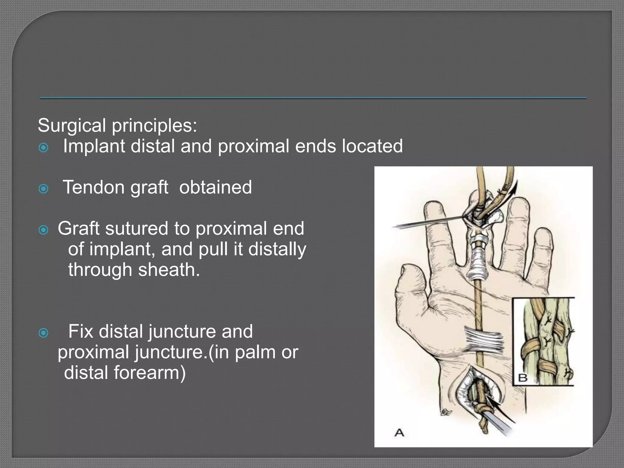
![ Palmaris longus[1] tendon present in approximately 85%
of all individuals of sufficient length and size .
Plantaris [2] when graft length is important.
present in about 93% of population
EDL[3]
EI [3]
EDM[3]
FDS of unaffected finger[4]
Ref: 1. MARTIN I. BOYER.JBJS 2002
2.Morrison WA J Hand Surg [Br] 1992
3. Harvey FJ, J Hand Surg [Am] 1983
4.Snow JW: Plast Reconstr Surg 1968](https://image.slidesharecdn.com/flexortendoninjury-161218170549/75/Flexor-tendon-injury-30-2048.jpg)


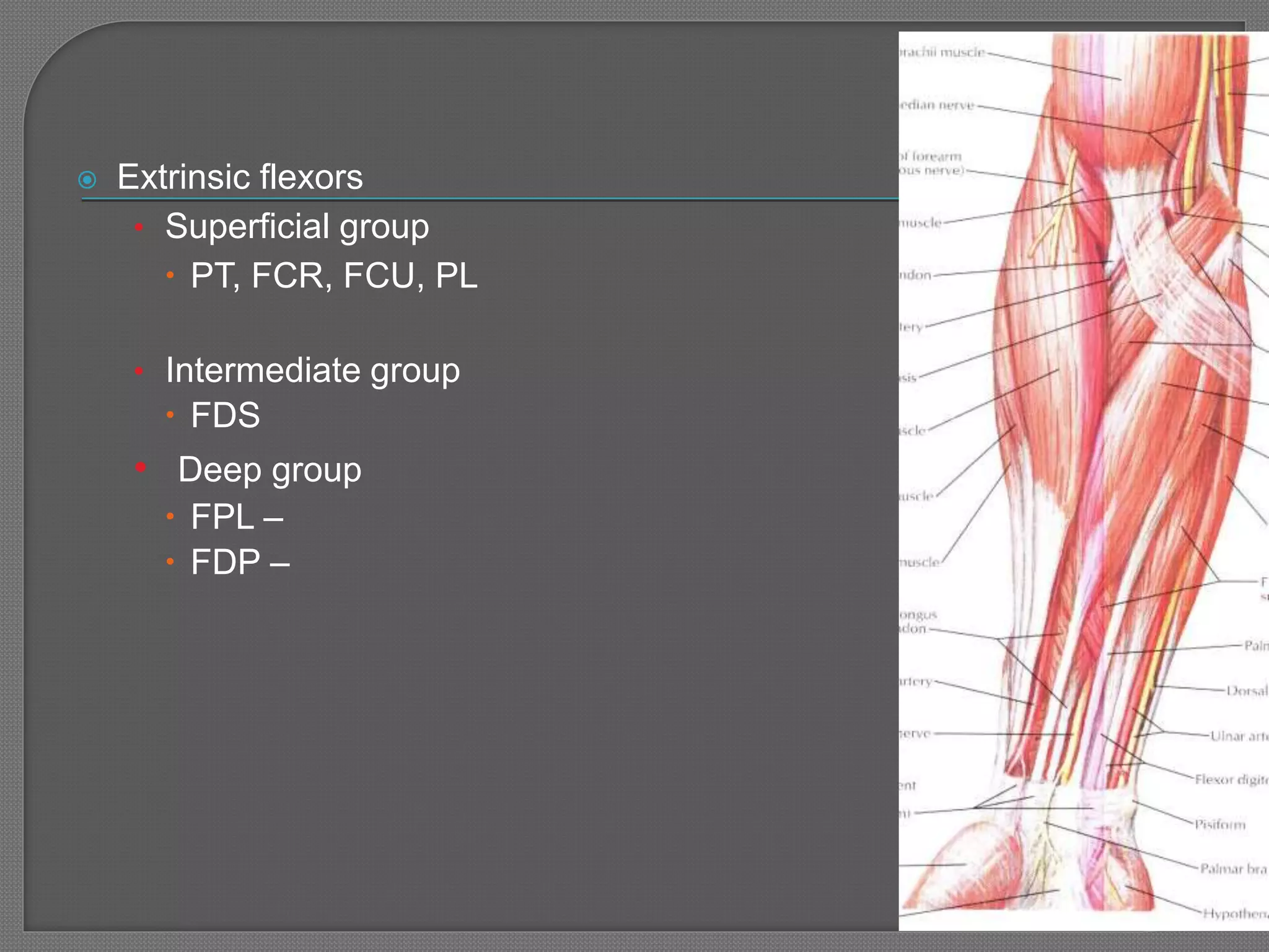
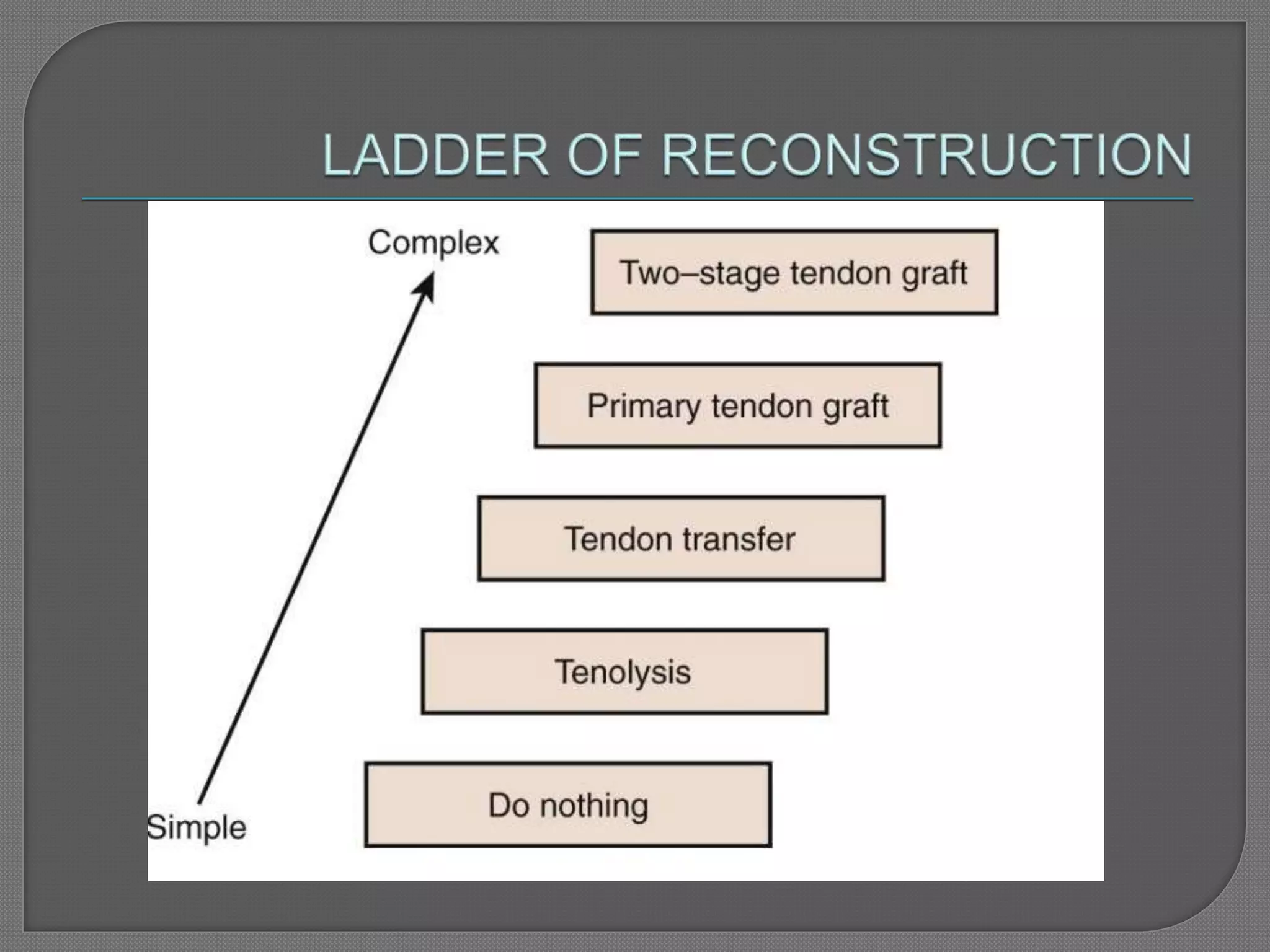
1. Flexor tendon injuries can occur in any of the 5 zones defined by Kleinert and Verdan and require different surgical approaches depending on the location and severity of the injury. 2. Primary repair within 12-24 hours of injury provides the best functional outcomes while delayed or secondary repairs have higher risks of adhesion formation. 3. Flexor tendon repair techniques aim to accurately approximate the tendon ends with core sutures while minimizing handling and restoring the normal gliding relationship between tendons. Postoperative rehabilitation is crucial. 4. Flexor tendon grafting is indicated for injuries with segment


