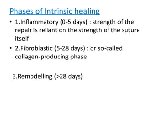
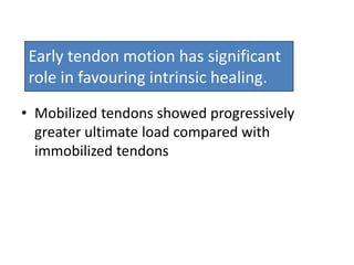
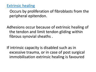
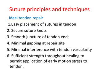
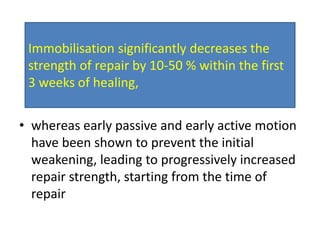
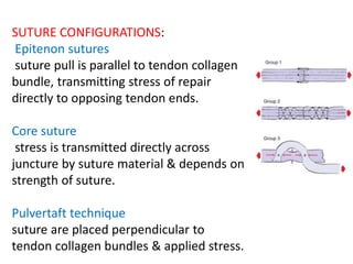
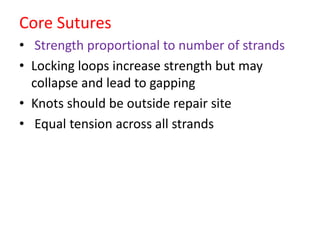
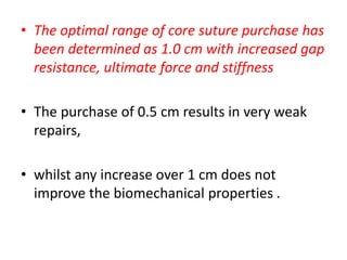
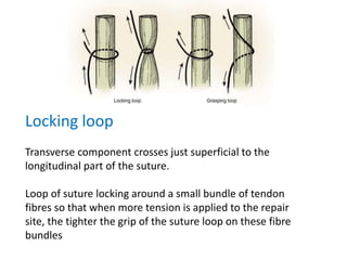
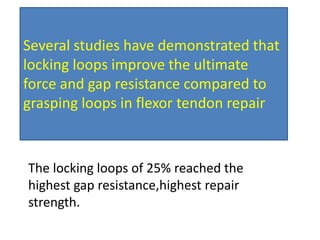
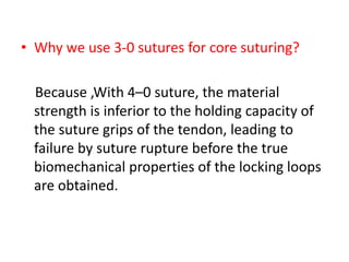
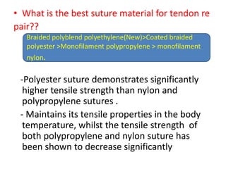
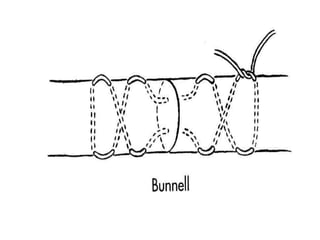
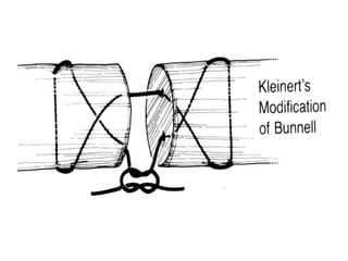
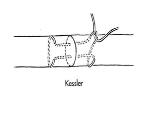
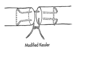
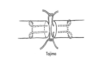
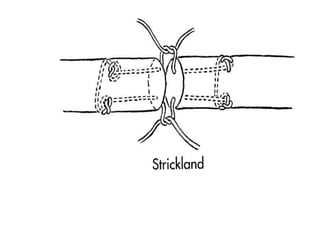
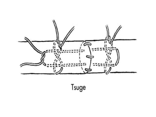
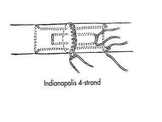
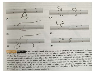
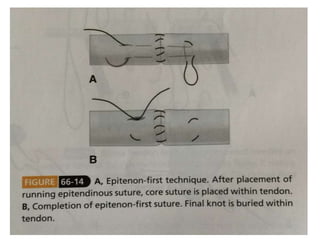
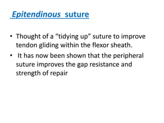
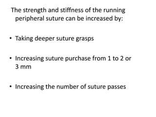
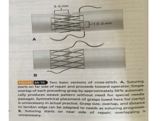
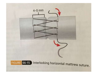
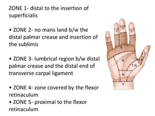
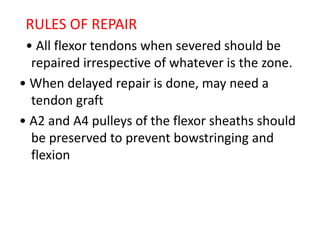
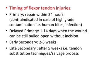
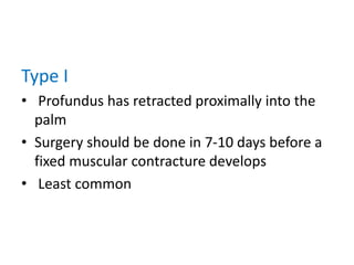
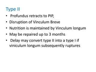
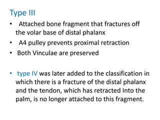
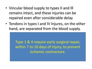
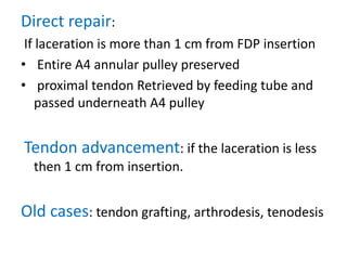
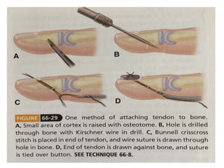
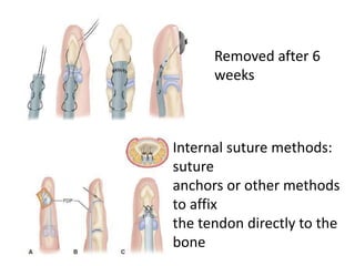
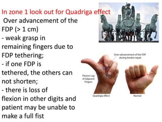
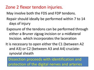
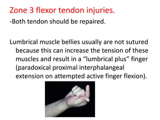
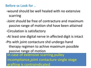
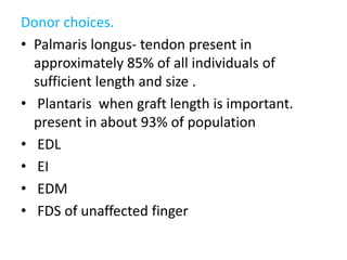
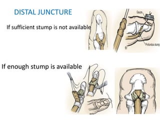
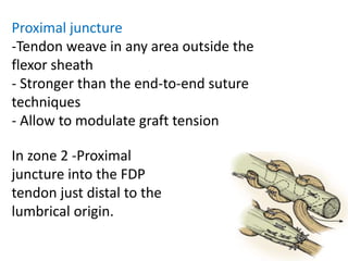
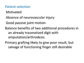
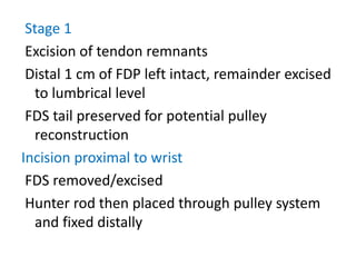
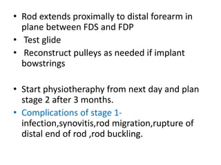
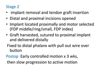
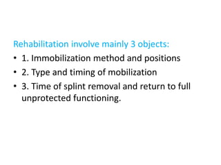
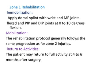
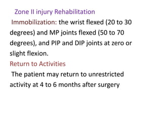
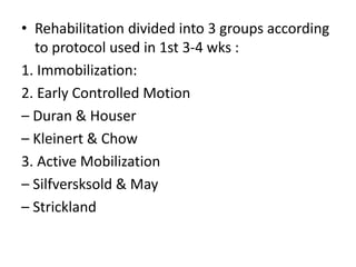
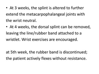
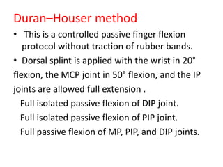
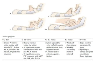
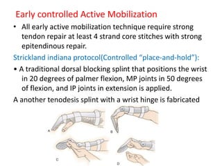
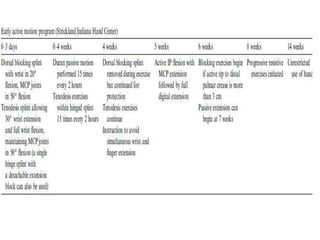
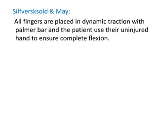
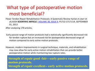
SlideShare a Scribd company logo
1 of 99
FLEXOR TENDON REPAIR.
DR.B.PUNITHAVASANTHAN
HAND AND MICRO SURGERY FELLOW
DEPARTMENT OF PLASTIC SURGERY
SKIMS-SOURA
DILEMMAS SURROUNDING
FLEXOR TENDON REPAIR
• Should both tendons be repaired or just the
FDP?
• Should the sheath be excised or repaired?
• What type of sutures should be utilized?
• What type of postoperative motion most
beneficial?
SYNOPSIS.
• FLEXOR TENDON HEALING.
• SUTURE TECHNIQUES.
• TENDON REPAIR ACCORDING TO ZONES.
• FLEXOR TENDON RECONSTRUCTION.
• REHABILITATION PROTOCOLS.
Flexor tendon healing
Intrinsic healing
- From prolifaration of tenocytes and
extracellular matrix from intrinsic cells.
- occurs without direct blood flow to the
tendon
Phases of Intrinsic healing
• 1.Inflammatory (0-5 days) : strength of the
repair is reliant on the strength of the suture
itself
• 2.Fibroblastic (5-28 days) : or so-called
collagen-producing phase
3.Remodelling (>28 days)
• Mobilized tendons showed progressively
greater ultimate load compared with
immobilized tendons
Early tendon motion has significant
role in favouring intrinsic healing.
Extrinsic healing
Occurs by proliferation of fibroblasts from the
peripheral epitendon.
Adhesions occur because of extrinsic healing of
the tendon and limit tendon gliding within
fibrous synovial sheaths .
If intrinsic capacity is disabled such as in
excessive trauma, or in case of post surgical
immobilisation extrinsic healing is favoured
Suture principles and techniques
Ideal tendon repair
1.Easy placement of sutures in tendon
2. Secure suture knots
3. Smooth juncture of tendon ends
4. Minimal gapping at repair site
5. Minimal interference with tendon vascularity
6. Sufficient strength throughout healing to
permit application of early motion stress to
tendon.
• whereas early passive and early active motion
have been shown to prevent the initial
weakening, leading to progressively increased
repair strength, starting from the time of
repair
Immobilisation significantly decreases the
strength of repair by 10-50 % within the first
3 weeks of healing,
SUTURE CONFIGURATIONS:
Epitenon sutures
suture pull is parallel to tendon collagen
bundle, transmitting stress of repair
directly to opposing tendon ends.
Core suture
stress is transmitted directly across
juncture by suture material & depends on
strength of suture.
Pulvertaft technique
suture are placed perpendicular to
tendon collagen bundles & applied stress.
Core Sutures
• Strength proportional to number of strands
• Locking loops increase strength but may
collapse and lead to gapping
• Knots should be outside repair site
• Equal tension across all strands
• The optimal range of core suture purchase has
been determined as 1.0 cm with increased gap
resistance, ultimate force and stiffness
• The purchase of 0.5 cm results in very weak
repairs,
• whilst any increase over 1 cm does not
improve the biomechanical properties .
Locking loop
Transverse component crosses just superficial to the
longitudinal part of the suture.
Loop of suture locking around a small bundle of tendon
fibres so that when more tension is applied to the repair
site, the tighter the grip of the suture loop on these fibre
bundles
Several studies have demonstrated that
locking loops improve the ultimate
force and gap resistance compared to
grasping loops in flexor tendon repair
The locking loops of 25% reached the
highest gap resistance,highest repair
strength.
• Why we use 3-0 sutures for core suturing?
Because ,With 4–0 suture, the material
strength is inferior to the holding capacity of
the suture grips of the tendon, leading to
failure by suture rupture before the true
biomechanical properties of the locking loops
are obtained.
• What is the best suture material for tendon re
pair??
-Polyester suture demonstrates significantly
higher tensile strength than nylon and
polypropylene sutures .
- Maintains its tensile properties in the body
temperature, whilst the tensile strength of
both polypropylene and nylon suture has
been shown to decrease significantly
Braided polyblend polyethylene(New)>Coated braided
polyester >Monofilament polypropylene > monofilament
nylon.
Epitendinous suture
• Thought of a “tidying up” suture to improve
tendon gliding within the flexor sheath.
• It has now been shown that the peripheral
suture improves the gap resistance and
strength of repair
The strength and stiffness of the running
peripheral suture can be increased by:
• Taking deeper suture grasps
• Increasing suture purchase from 1 to 2 or
3 mm
• Increasing the number of suture passes
• It helps to decrease bulk by smoothing out the
repair site
• addition of peripheral suture itself contributes
up to 50% of ultimate load to failure and
potentially up to 50 N of strength.
• Several epitendinous configurations have
been suggested, Including simple running,
cross-stitch, interlocking cross-stitch, and
Interlocking horizontal mattress.
• -
It should be noted that the
repair strength of the core
stitch and epitendinous
suture are not summative
and can fail independently.
Tendon repair according to zones
ZONE 1- distal to the insertion of
superficialis
• ZONE 2- no mans land b/w the
distal palmar crease and insertion of
the sublimis
• ZONE 3- lumbrical region b/w distal
palmar crease and the distal end of
transverse carpal ligament
• ZONE 4- zone covered by the flexor
retinaculum
• ZONE 5- proximal to the flexor
retinaculum
RULES OF REPAIR
• All flexor tendons when severed should be
repaired irrespective of whatever is the zone.
• When delayed repair is done, may need a
tendon graft
• A2 and A4 pulleys of the flexor sheaths should
be preserved to prevent bowstringing and
flexion
• Timing of flexor tendon injuries:
• Primary: repair within 24 hours
(contraindicated in case of high grade
contamination i.e. human bites, infection)
• Delayed Primary: 1-14 days when the wound
can be still pulled open without incision
• Early Secondary: 2-5 weeks.
• Late Secondary : after 5 weeks i.e. tendon
substitution techniques/salvage process
Incisions
Avoid crossing joints and
creases at 90 deg.
Existing lacerations
-zigzag
-mid lateral
Should we do tendon sheath repair??
Advantages
• Barrier to extrinsic adhesion formation
• More rapid return of synovial nutrition
• Tendon within the sheath have an intrinsic
capacity for healing
Disadvantages
Technically difficult
• Presently, no clearcut advantage to sheath repair
has been established
ZONE 1 flexor
Tendon injuries.
Profundus Tendon Avulsion
• Avulsion of FDP from its insertion by forced
hyperextension
• Most common in the ring finger
Leddy and Parker classification
• Based on the level to which the tendon
retracts
• Status of the tendon vascular anatomy
Type I
• Profundus has retracted proximally into the
palm
• Surgery should be done in 7-10 days before a
fixed muscular contracture develops
• Least common
Type II
• Profundus retracts to PIP,
• Disruption of Vinculum Breve
• Nutrition is maintained by Vinculum longum
• May be repaired up to 3 months
• Delay may convert type II into a type I if
viniculum longum subsequently ruptures
Type III
• Attached bone fragment that fractures off
the volar base of distal phalanx
• A4 pulley prevents proximal retraction
• Both Vinculae are preserved
• type IV was later added to the classification in
which there is a fracture of the distal phalanx
and the tendon, which has retracted Into the
palm, is no longer attached to this fragment.
• Vincular blood supply to types II and Ill
remains intact, and these injuries can be
repaired even after considerable delay
• Tendons in types I and IV Injures, on the other
hand, are separated from the blood supply.
Type 1 & 4 require early surgical repair,
within 7 to 10 days of injury, to prevent
ischemic contracture.
Direct repair:
If laceration is more than 1 cm from FDP insertion
• Entire A4 annular pulley preserved
• proximal tendon Retrieved by feeding tube and
passed underneath A4 pulley
Tendon advancement: if the laceration is less
then 1 cm from insertion.
Old cases: tendon grafting, arthrodesis, tenodesis
Removed after 6
weeks
Internal suture methods:
suture
anchors or other methods
to affix
the tendon directly to the
bone
In zone 1 look out for Quadriga effect
Over advancement of the
FDP (> 1 cm)
- weak grasp in
remaining fingers due to
FDP tethering;
- if one FDP is
tethered, the others can
not shorten;
- there is loss of
flexion in other digits and
patient may be unable to
make a full fist
Zone 2 flexor tendon injuries.
May involve both the FDS and FDP tendons.
Repair should ideally be performed within 7 to 14
days of injury
Exposure of the tendons can be performed through
either a Bruner zigzag incision or a midlateral
Incision. which incorporates the laceration
It is necessary to open either the C1 (between A2
and A3) or C2 (between A3 and A4) cruciate-
synovial sheath
Dissection proceeds with identification and
protection of the digital nerves and arteries
Should both tendons be repaired or only FDP??
Henry M et al, Zone II: Repair or reset the flexor
digitorum superficialis? J Hand Surg Am. 2011
• repair only the FDP tendon if both the slips of FDS
tendon are to be repaired underneath A2 pulley
• Repairs FDS if the injury is proximal or distal to A2
pulley in zone 2.
• Most hand surgeons prefer to repair the FDP and one
slip of FDS as the repair of both slips of FDS may
produce overcrowding within the sheath and pulleys
and compromise the results.
• If only one slip of FDS is cut then the chiasma should
be opened from that side and the FDP should be
delivered from the same side.
Zone 3 flexor tendon injuries.
-Both tendon should be repaired.
Lumbrical muscle bellies usually are not sutured
because this can increase the tension of these
muscles and result in a “lumbrical plus” finger
(paradoxical proximal interphalangeal
extension on attempted active finger flexion).
Zone 4 and 5 flexor tendon injuries.
• Be aware of median ,ulnar nerve or ulnar or
radial vessel injury.
• Here vessels and nerves should be repaired
first and the tendons last
Complications
Adhesion
• Most common complication despite early
motion protocols
Tendon Rupture
• Noted by the patient at “popping” in the hand
• 7-10 days postop when tensile strength is
weakest
• MRI may help in diagnosis
Triggering and entrapment
• Especially when sheath is not repaired
Post traumatic regional pain syndromes
• Cold intolerance
• RSD
FDP advancement more than 1 cm may
lead to flexion contracture and weakened
hand grip because of quadrigia effect
FLEXOR TENDON RECONSTRUCTION
Tenolysis
Indication -tendon adhesion
Preop evaluation
Discripency exists between passive and active
range of motion after hand therapy has
platued
Timing – 3-6 mos. Post repair (minimum)
Anesthesia – Local with sedation , Allows
patient participation
Technique
• Zig zag incisions
• Adhesions divided maintaining non-limiting
adhesions
• Pulleys reconstructed as needed
• On post op- active range of motion should
begin immediately
If extensive or not possible convert to
staged reconstruction
Boyes’ grading scale of
flexor tendon injury
provides a guideline in
determining the
achievable outcome
after flexion tendon
reconstruction
Single Stage Tendon Grafting
Principles of tendon grafting include
• Place only one graft in each finger
• Never sacrifice intact FDS
• Use a graft of smaller caliber
• Perform the junctions outside the tndon
sheath
• Ensure adequate graft tension.
Indications
-Injuries resulting in segmental tendon loss.
- Delay in repair that obviates primary repair, such
as laceration that have been neglected for 3 to
6 weeks and show tendon degenerations
-Zone 2 injury where large section of tendon
have been damaged and surgeon belives
delayed grafting is better option
- Delay presentation of FDP avulsion injury
associated with significant tendon retraction
-Supple joints with adequate passive ROM
Before sx Look for ..
-wound should be well healed with no extensive
scarring
-Joint should be free of contracture and maximum
passive range of motion shd have been attained
-Circulation is satisfactory
-At least one digital nerve in affected digit is intact
-Pts with joint contacture shd undergo hand
therapy regimen to achive maximum possible
passive range of motion
In case of extensive scarring,pulley
incompitance,joint contacture single stage
grafting is contraindicated.
Donor choices.
• Palmaris longus- tendon present in
approximately 85% of all individuals of
sufficient length and size .
• Plantaris when graft length is important.
present in about 93% of population
• EDL
• EI
• EDM
• FDS of unaffected finger
DISTAL JUNCTURE
If enough stump is available
If sufficient stump is not available
Proximal juncture
-Tendon weave in any area outside the
flexor sheath
- Stronger than the end-to-end suture
techniques
- Allow to modulate graft tension
In zone 2 -Proximal
juncture into the FDP
tendon just distal to the
lumbrical origin.
• If FDP is of poor quality,FDS may be used for
proximal juncture.
• Occasionaly FDP of uninjured neigbour digit may
be used as end to side proximal juncture.
• For zone 3,4,5 distal end junction is taken just
proximal to lumbrical origin.
• If intact FDS,and FDP injured ,arthrodesis of DIP is
done in OLD age group.
In young FDP reconstruction is offered
Keep 1-2 cm of distal fds stump as it provides
a favourable bed for graft,stability to PIP,and
prevents swan neck
Tension adjustment
-Proximal weave is
adjusted.
-Wrist is extended and
flex
the fingers into the
cascade of the hand
-Overcorrect slightly
because some stretching
occurs after surgery
• Post op care
• Early motion exercise have been used
• Protected passive range of motion can be
instituted after 2 to 3 days of sx if the juncture
are strong enough to withstand the tesile
strength
• Post op splint- static dorsal bloking splint with
wrist positioned in neutral,MP in 45 degree
flexion, Ipin neutral. And worn for 6 weeks
after sx
Two Stage Reconstruction
Hunter and salisbury introduced in 1971.
They noted silicon rod placement helped in
forming soft,flexible,translucent sheath.
Indications
Crushing injuries a/w # or skin damage
Damaged pulley system
Excessive scarring of the tendon bed
Failure of previous operations
Contracted joint.
Patient selection
Motivated
Absence of neurovascular injury
Good passive joint motion
Balance benefits of two additional procedures in
an already traumatized digit with
amputation/arthrodesis.
Primary grafting likely to give poor result, but
salvage of functioning finger still desirable
Stage 1
Excision of tendon remnants
Distal 1 cm of FDP left intact, remainder excised
to lumbrical level
FDS tail preserved for potential pulley
reconstruction
Incision proximal to wrist
FDS removed/excised
Hunter rod then placed through pulley system
and fixed distally
• Rod extends proximally to distal forearm in
plane between FDS and FDP
• Test glide
• Reconstruct pulleys as needed if implant
bowstrings
• Start physiotheraphy from next day and plan
stage 2 after 3 months.
• Complications of stage 1-
infection,synovitis,rod migration,rupture of
distal end of rod ,rod buckling.
Stage 2
• implant removal and tendon graft insertion
• Distal and proximal incisions opened
• Implant located proximally and motor selected
(FDP middle/ring/small, FDP index)
• Graft harvested, sutured to proximal implant
and delivered distally
Fixed to distal phalanx with pull out wire over
button
Postop Early controlled motion x 3 wks,
then slow progression to active motion
Thumb flexor tendon reconstruction.
Direct repair is possible upto 3-6 weeks if the
tendon ends have not widely seperated.
Recostruction is indicated when satisfactory IP
joint motion is present.
Options available ;
1)Single stage tendon grafting.
Tension setting:wrist neutral.IP joint 30 degree
flexion.
2)Staged tendon reconstruction;
when pulleys are damaged,
3 ) single stage FDS transfer
from RF,
In stage 2 a FDS transfer can also be done
after removing silicon rod.
For Precise tensioning at distal juncture ,the
FDS is woven through the stump of FDP
Secondary Reconstruction
• Zone 1 (functioning FDS) – Eg. Late
presentation of FDP avulsion
DIP fusion
• Tendon graft Risks damaging FDS function
through injury/adhesions in a very functional
finger
• Young patients, supple joints, need for active
DIP flexion
• Zones 2,3, 4 and 5
Usually associated with 3 – 5 cm gap
Interposition graft
FDS to FDP transfer
End to side profundus juncture
• FDS finger or PIP joint finger,
Salvage procedure in cases of DIP joint
damage,pulley failure even after 2 attempts at
pulley reconsrution,
Tendon graft is attached to middle phalynx
through pullout sutures.
• Complication after flexor tendon reconstruction.
• Adhesions
• Mechanical failure of implant(Dacron reinforced
silicon helps in suture pull out)
• Graft rupture
• Pulley disruption,
• Quadriga syndrome(too much tension)
• Hyper extension of PIP (due to Absent FDS)
• Lumbrical plus finger(insufficient tension)
• Synovitis,infection,
• Late flexion deformity,(inadequate pulleys,wound
contractures,splinting )
Flexor tendon rehabilitation
• All protocols employ a dorsal shell with the wrist,
MCPs, and IP joints extended or flexed.
• Active extension and passive flexion are encouraged
after the first postoperative day,
• After 2 to 3 weeks, “place and hold no power” can be
initiated along with protected passive motion to
maintain joint mobility and avoid contractures
especially the PIP joint.
Differential gliding of the uninjured tendons can also
start at this time.
• Isolated active tendon differential gliding of
the injured tendon begins by atleast 4 to 6
weeks and active, assisted, complete fist and
passive range of motion.
• Protective splinting may continue with
progressive resistance exercises for upto 8
weeks total.
• Gradually increased active resistance-
strengthening exercises up to 12 weeks
Rehabilitation involve mainly 3 objects:
• 1. Immobilization method and positions
• 2. Type and timing of mobilization
• 3. Time of splint removal and return to full
unprotected functioning.
Zone 1 Rehabilitation
Immobilization:
Apply dorsal splint with wrist and MP joints
flexed and PIP and DIP joints at 0 to 10 degrees
flexion.
Mobilization:
The rehabilitation protocol generally follows the
same progression as for zone 2 injuries.
Return to Activities:
The patient may return to full activity at 4 to 6
months after surgery.
Zone II injury Rehabilitation
Immobilization: the wrist flexed (20 to 30
degrees) and MP joints flexed (50 to 70
degrees), and PIP and DIP joints at zero or
slight flexion.
Return to Activities
The patient may return to unrestricted
activity at 4 to 6 months after surgery
• Rehabilitation divided into 3 groups according
to protocol used in 1st 3-4 wks :
1. Immobilization:
2. Early Controlled Motion
– Duran & Houser
– Kleinert & Chow
3. Active Mobilization
– Silfversksold & May
– Strickland
Kleinert & Chow protocol:
• A controlled active extension–passive flexion
motion protocol.
• The wrist is palmary flexed with a dorsal
protective splint with 30–40° wrist flexion,
50–70° MCP joint flexion, and the IP joints are
allowed full extension.
• Original Kleinert regime had been changed to Modified
Kleinert regime (Chow) due to rubber band traction
was found to lead to flexion contractures of the finger.
Modified Kleinert regime (Chow) differ by:
1. a palmer bar at the level of the MCP joint as a pulley
for the rubber bands to create greater flexion of both
the PIP and DIP joints.
2. the elastic band is detached at night and the fingers
are strapped into extension within the splint to
minimize the risk of flexion contractures of the
fingers.
• All fingers should be included in the rubber band
traction to ensure added FDP protection and to
promote better tendon excursion also decrease
risk of PIP contracture through more efficient
action of EDC.
• mobilization program is begun 1 to 3 days
postoperatively.
• The finger to be actively extended within the
limitations of the splint.
The patient is instructed to perform this exercise 10
times every waking hour.
• At 3 weeks, the splint is altered to further
extend the metacarpophalangeal joints with
the wrist neutral.
• At 4 weeks, the dorsal splint can be removed,
leaving the line/rubber band attached to a
wristlet. Wrist exercises are encouraged.
at 5th week, the rubber band is discontinued;
the patient actively flexes without resistance.
Duran–Houser method
• This is a controlled passive finger flexion
protocol without traction of rubber bands.
• Dorsal splint is applied with the wrist in 20°
flexion, the MCP joint in 50° flexion, and the IP
joints are allowed full extension .
Full isolated passive flexion of DIP joint.
Full isolated passive flexion of PIP joint.
Full passive flexion of MP, PIP, and DIP joints.
Advantages:
• decreased the frequency of PIP joint
contracture seen with Kleinert’s rubber band
traction.
• Better differential gliding.
• Improved protection between exercise time.
Disadvantages:
• Greater tension on repair site.
• Patient may not be able to resist temptation
to flex against volar strap.
Early controlled Active Mobilization
• All early active mobilization technique require strong
tendon repair at least 4 strand core stitches with strong
epitendinous repair.
Strickland indiana protocol(Controlled “place-and-hold”):
• A traditional dorsal blocking splint that positions the wrist
in 20 degrees of palmer flexion, MP joints in 50 degrees
of flexion, and IP joints in extension is applied.
A another tenodesis splint with a wrist hinge is fabricated
Silfversksold & May:
All fingers are placed in dynamic traction with
palmer bar and the patient use their uninjured
hand to ensure complete flexion.
Differential gliding exercises
What type of postoperative motion
most beneficial?
Flexor Tendon Repair Rehabilitation Protocols: A Systematic Review harlan.m.starr et
all,ASSH SCIENTIFIC ARTICLE| VOLUME 38, ISSUE 9, P1712-1717.E14, SEPTEMBER
01, 2013
After analysing 170 articles,
Early passive range of motion protocols had a statistically significantly decreased risk
for tendon rupture but an increased risk for postoperative decreased range of
motion compared to early active motion protocols.
However, modern improvements in surgical technique, materials, and rehabilitation
may now allow for early active motion rehabilitation that can provide better
postoperative motion while maintaining low rupture rates.
Strength of repair good -fair—early passive range of
motion protocols.
Strength of repair excellent-- early active motion protocols.
1)Early tendon motion has significant role in favouring intrinsic healing.
2)Immobilisation significantly decreases the strength of repair by 10-50 %
within the first 3 weeks of healing,
3)Dissection proceeds with identification and protection of the digital nerves
and arteries.
4)In cases of extensive injuries ,do staged tendon reconstruction.
5)Quadriga syndrome(too much tension)
6)Hyper extension of PIP (due to Absent FDS)
7)Lumbrical plus finger(insufficient tension)
8)Early active vs early passive is (excellent vs good repair)
Take home messages
Flexor Tendon Repair Techniques and Rehabilitation

More Related Content

What's hot

Knee Portal Placement & Diagnostic arthroscopy
Knee Portal Placement & Diagnostic arthroscopyKnee Portal Placement & Diagnostic arthroscopy
Knee Portal Placement & Diagnostic arthroscopyAsish Rajak
 
Dhs principles
Dhs principlesDhs principles
Dhs principlesAhmad Sulong
 
Masquelet technique ppt
Masquelet technique pptMasquelet technique ppt
Masquelet technique pptApoorv Garg
 
Common hand injuries
Common hand injuriesCommon hand injuries
Common hand injuriestbf413
 
Management of TendoAchillis rupture
Management of TendoAchillis ruptureManagement of TendoAchillis rupture
Management of TendoAchillis ruptureAnkur Mittal
 
Septic arthritis sequelae
Septic arthritis sequelaeSeptic arthritis sequelae
Septic arthritis sequelaeorthoprince
 
Flexor tendon injury, management and rehabilitation
Flexor tendon injury, management and rehabilitationFlexor tendon injury, management and rehabilitation
Flexor tendon injury, management and rehabilitationKhadijah Nordin
 
Triple arthrodesis seminar by Dr Chirag Patel
Triple arthrodesis seminar by Dr Chirag PatelTriple arthrodesis seminar by Dr Chirag Patel
Triple arthrodesis seminar by Dr Chirag PatelChirag Patel
 
unilateral knee replacement vs high tibial osteotomy.
unilateral knee replacement vs high tibial osteotomy.unilateral knee replacement vs high tibial osteotomy.
unilateral knee replacement vs high tibial osteotomy.drabhichaudhary88
 
Evolution of Intramedullary Nails
Evolution of Intramedullary NailsEvolution of Intramedullary Nails
Evolution of Intramedullary NailsPrateek Goel
 
Arthroscopic Meniscus Surgery: Resect or Repair 2014
Arthroscopic Meniscus Surgery: Resect or Repair 2014Arthroscopic Meniscus Surgery: Resect or Repair 2014
Arthroscopic Meniscus Surgery: Resect or Repair 2014Dhananjaya Sabat
 
Complications after THR
Complications after THRComplications after THR
Complications after THRHiren Divecha
 
MEDIAN NERVE PALSY AND TENDON TRANSFERS
MEDIAN NERVE PALSY AND TENDON TRANSFERSMEDIAN NERVE PALSY AND TENDON TRANSFERS
MEDIAN NERVE PALSY AND TENDON TRANSFERSBenthungo Tungoe
 
Telescopic nails in Osteogenesis Imperfecta
Telescopic nails in Osteogenesis ImperfectaTelescopic nails in Osteogenesis Imperfecta
Telescopic nails in Osteogenesis ImperfectaShady Mahmoud
 
CORA (center of rotation of angulation)
CORA (center of rotation of angulation)CORA (center of rotation of angulation)
CORA (center of rotation of angulation)Morshed Abir
 

What's hot (20)

Knee Portal Placement & Diagnostic arthroscopy
Knee Portal Placement & Diagnostic arthroscopyKnee Portal Placement & Diagnostic arthroscopy
Knee Portal Placement & Diagnostic arthroscopy
 
Dhs principles
Dhs principlesDhs principles
Dhs principles
 
Masquelet technique ppt
Masquelet technique pptMasquelet technique ppt
Masquelet technique ppt
 
Suzukiframepaper
SuzukiframepaperSuzukiframepaper
Suzukiframepaper
 
Common hand injuries
Common hand injuriesCommon hand injuries
Common hand injuries
 
Management of TendoAchillis rupture
Management of TendoAchillis ruptureManagement of TendoAchillis rupture
Management of TendoAchillis rupture
 
Septic arthritis sequelae
Septic arthritis sequelaeSeptic arthritis sequelae
Septic arthritis sequelae
 
Flexor tendon injury, management and rehabilitation
Flexor tendon injury, management and rehabilitationFlexor tendon injury, management and rehabilitation
Flexor tendon injury, management and rehabilitation
 
Triple arthrodesis seminar by Dr Chirag Patel
Triple arthrodesis seminar by Dr Chirag PatelTriple arthrodesis seminar by Dr Chirag Patel
Triple arthrodesis seminar by Dr Chirag Patel
 
unilateral knee replacement vs high tibial osteotomy.
unilateral knee replacement vs high tibial osteotomy.unilateral knee replacement vs high tibial osteotomy.
unilateral knee replacement vs high tibial osteotomy.
 
Evolution of Intramedullary Nails
Evolution of Intramedullary NailsEvolution of Intramedullary Nails
Evolution of Intramedullary Nails
 
Arthroscopic Meniscus Surgery: Resect or Repair 2014
Arthroscopic Meniscus Surgery: Resect or Repair 2014Arthroscopic Meniscus Surgery: Resect or Repair 2014
Arthroscopic Meniscus Surgery: Resect or Repair 2014
 
Complications after THR
Complications after THRComplications after THR
Complications after THR
 
MEDIAN NERVE PALSY AND TENDON TRANSFERS
MEDIAN NERVE PALSY AND TENDON TRANSFERSMEDIAN NERVE PALSY AND TENDON TRANSFERS
MEDIAN NERVE PALSY AND TENDON TRANSFERS
 
Extensor tendon injury
Extensor tendon injuryExtensor tendon injury
Extensor tendon injury
 
Oberlin Transfer
Oberlin TransferOberlin Transfer
Oberlin Transfer
 
Poller screw
Poller screwPoller screw
Poller screw
 
Telescopic nails in Osteogenesis Imperfecta
Telescopic nails in Osteogenesis ImperfectaTelescopic nails in Osteogenesis Imperfecta
Telescopic nails in Osteogenesis Imperfecta
 
Gap nonunion
Gap nonunionGap nonunion
Gap nonunion
 
CORA (center of rotation of angulation)
CORA (center of rotation of angulation)CORA (center of rotation of angulation)
CORA (center of rotation of angulation)
 

Similar to Flexor Tendon Repair Techniques and Rehabilitation

Flexor tendon injuries
Flexor tendon injuriesFlexor tendon injuries
Flexor tendon injuriesMohamed Eldeeb
 
Flexor tendon injuries
Flexor tendon injuriesFlexor tendon injuries
Flexor tendon injuriesIbrahim ibrahim
 
Tendon repair
Tendon repairTendon repair
Tendon repairMonitoshPaul
 
Flexortendoninjuries1 140504003542-phpapp01
Flexortendoninjuries1 140504003542-phpapp01Flexortendoninjuries1 140504003542-phpapp01
Flexortendoninjuries1 140504003542-phpapp01ssanjibanisudha
 
Tendon injuries of hand
Tendon injuries of handTendon injuries of hand
Tendon injuries of handrohit raj
 
Flexor Tendon injury.pptx
Flexor Tendon injury.pptxFlexor Tendon injury.pptx
Flexor Tendon injury.pptxAthishjaims1
 
Acute Extensor tendon injuries diagnosis and management.pptx
Acute Extensor tendon injuries diagnosis and management.pptxAcute Extensor tendon injuries diagnosis and management.pptx
Acute Extensor tendon injuries diagnosis and management.pptxRohie3
 
Acute Tendon injury of the Hand and its Repair.pptx
Acute Tendon injury of the Hand and its Repair.pptxAcute Tendon injury of the Hand and its Repair.pptx
Acute Tendon injury of the Hand and its Repair.pptxOkpako Isaac
 
Meniscus repair
Meniscus repairMeniscus repair
Meniscus repairAvik Sarkar
 
RME by Dr.Himali Gupta for PG Students
RME by Dr.Himali Gupta for PG StudentsRME by Dr.Himali Gupta for PG Students
RME by Dr.Himali Gupta for PG StudentsHimali Gupta
 
Fracture Fixation Techniques.pptx
Fracture Fixation Techniques.pptxFracture Fixation Techniques.pptx
Fracture Fixation Techniques.pptxRekha Pathak
 
Extensor tendon injury
Extensor tendon injuryExtensor tendon injury
Extensor tendon injuryJaskaran Sandhu
 
extensortendoninjury-210227171109.pdf
extensortendoninjury-210227171109.pdfextensortendoninjury-210227171109.pdf
extensortendoninjury-210227171109.pdfKareemElsharkawy6
 
Bennett , rolando , tendon injuries
Bennett , rolando , tendon injuriesBennett , rolando , tendon injuries
Bennett , rolando , tendon injuriesSagar Savsani
 
Fractures of the olecranon
Fractures of the olecranonFractures of the olecranon
Fractures of the olecranonMuhammad Abdelghani
 
Principles of external fixation
Principles of external fixationPrinciples of external fixation
Principles of external fixationMohamoud Abdulahi
 

Similar to Flexor Tendon Repair Techniques and Rehabilitation (20)

Flexor tendon injuries
Flexor tendon injuriesFlexor tendon injuries
Flexor tendon injuries
 
Flexor tendon injuries
Flexor tendon injuriesFlexor tendon injuries
Flexor tendon injuries
 
Tendon repair
Tendon repairTendon repair
Tendon repair
 
Flexortendoninjuries1 140504003542-phpapp01
Flexortendoninjuries1 140504003542-phpapp01Flexortendoninjuries1 140504003542-phpapp01
Flexortendoninjuries1 140504003542-phpapp01
 
Tendon injuries of hand
Tendon injuries of handTendon injuries of hand
Tendon injuries of hand
 
Flexor Tendon injury.pptx
Flexor Tendon injury.pptxFlexor Tendon injury.pptx
Flexor Tendon injury.pptx
 
Tendon injuries of hand by Dr Saumya Agarwal
Tendon injuries of hand by Dr Saumya AgarwalTendon injuries of hand by Dr Saumya Agarwal
Tendon injuries of hand by Dr Saumya Agarwal
 
Flexor tendon injuries slideshare
Flexor  tendon injuries slideshareFlexor  tendon injuries slideshare
Flexor tendon injuries slideshare
 
Flexor tendon injury
Flexor tendon injuryFlexor tendon injury
Flexor tendon injury
 
Acute Extensor tendon injuries diagnosis and management.pptx
Acute Extensor tendon injuries diagnosis and management.pptxAcute Extensor tendon injuries diagnosis and management.pptx
Acute Extensor tendon injuries diagnosis and management.pptx
 
Acute Tendon injury of the Hand and its Repair.pptx
Acute Tendon injury of the Hand and its Repair.pptxAcute Tendon injury of the Hand and its Repair.pptx
Acute Tendon injury of the Hand and its Repair.pptx
 
Meniscus repair
Meniscus repairMeniscus repair
Meniscus repair
 
RME by Dr.Himali Gupta for PG Students
RME by Dr.Himali Gupta for PG StudentsRME by Dr.Himali Gupta for PG Students
RME by Dr.Himali Gupta for PG Students
 
Fracture Fixation Techniques.pptx
Fracture Fixation Techniques.pptxFracture Fixation Techniques.pptx
Fracture Fixation Techniques.pptx
 
Extensor tendon injury
Extensor tendon injuryExtensor tendon injury
Extensor tendon injury
 
extensortendoninjury-210227171109.pdf
extensortendoninjury-210227171109.pdfextensortendoninjury-210227171109.pdf
extensortendoninjury-210227171109.pdf
 
Bennett , rolando , tendon injuries
Bennett , rolando , tendon injuriesBennett , rolando , tendon injuries
Bennett , rolando , tendon injuries
 
Fractures of the olecranon
Fractures of the olecranonFractures of the olecranon
Fractures of the olecranon
 
Principles of external fixation
Principles of external fixationPrinciples of external fixation
Principles of external fixation
 
Ortho Journal Club 1 by Dr Saumya Agarwal
Ortho Journal Club 1 by Dr Saumya AgarwalOrtho Journal Club 1 by Dr Saumya Agarwal
Ortho Journal Club 1 by Dr Saumya Agarwal
 

Recently uploaded

Call Girls Service In Shyam Nagar Whatsapp 8445551418 Independent Escort Service
Call Girls Service In Shyam Nagar Whatsapp 8445551418 Independent Escort ServiceCall Girls Service In Shyam Nagar Whatsapp 8445551418 Independent Escort Service
Call Girls Service In Shyam Nagar Whatsapp 8445551418 Independent Escort Serviceparulsinha
 
Book Paid Powai Call Girls Mumbai 𖠋 9930245274 𖠋Low Budget Full Independent H...
Book Paid Powai Call Girls Mumbai 𖠋 9930245274 𖠋Low Budget Full Independent H...Book Paid Powai Call Girls Mumbai 𖠋 9930245274 𖠋Low Budget Full Independent H...
Book Paid Powai Call Girls Mumbai 𖠋 9930245274 𖠋Low Budget Full Independent H...Call Girls in Nagpur High Profile
 
Russian Call Girls in Pune Tanvi 9907093804 Short 1500 Night 6000 Best call g...
Russian Call Girls in Pune Tanvi 9907093804 Short 1500 Night 6000 Best call g...Russian Call Girls in Pune Tanvi 9907093804 Short 1500 Night 6000 Best call g...
Russian Call Girls in Pune Tanvi 9907093804 Short 1500 Night 6000 Best call g...Miss joya
 
Bangalore Call Girls Majestic 📞 9907093804 High Profile Service 100% Safe
Bangalore Call Girls Majestic 📞 9907093804 High Profile Service 100% SafeBangalore Call Girls Majestic 📞 9907093804 High Profile Service 100% Safe
Bangalore Call Girls Majestic 📞 9907093804 High Profile Service 100% Safenarwatsonia7
 
Artifacts in Nuclear Medicine with Identifying and resolving artifacts.
Artifacts in Nuclear Medicine with Identifying and resolving artifacts.Artifacts in Nuclear Medicine with Identifying and resolving artifacts.
Artifacts in Nuclear Medicine with Identifying and resolving artifacts.MiadAlsulami
 
Lucknow Call girls - 8800925952 - 24x7 service with hotel room
Lucknow Call girls - 8800925952 - 24x7 service with hotel roomLucknow Call girls - 8800925952 - 24x7 service with hotel room
Lucknow Call girls - 8800925952 - 24x7 service with hotel roomdiscovermytutordmt
 
CALL ON ➥9907093804 🔝 Call Girls Hadapsar ( Pune) Girls Service
CALL ON ➥9907093804 🔝 Call Girls Hadapsar ( Pune)  Girls ServiceCALL ON ➥9907093804 🔝 Call Girls Hadapsar ( Pune)  Girls Service
CALL ON ➥9907093804 🔝 Call Girls Hadapsar ( Pune) Girls ServiceMiss joya
 
Call Girls Service Pune Vaishnavi 9907093804 Short 1500 Night 6000 Best call ...
Call Girls Service Pune Vaishnavi 9907093804 Short 1500 Night 6000 Best call ...Call Girls Service Pune Vaishnavi 9907093804 Short 1500 Night 6000 Best call ...
Call Girls Service Pune Vaishnavi 9907093804 Short 1500 Night 6000 Best call ...Miss joya
 
High Profile Call Girls Coimbatore Saanvi☎️ 8250192130 Independent Escort Se...
High Profile Call Girls Coimbatore Saanvi☎️  8250192130 Independent Escort Se...High Profile Call Girls Coimbatore Saanvi☎️  8250192130 Independent Escort Se...
High Profile Call Girls Coimbatore Saanvi☎️ 8250192130 Independent Escort Se...narwatsonia7
 
Aspirin presentation slides by Dr. Rewas Ali
Aspirin presentation slides by Dr. Rewas AliAspirin presentation slides by Dr. Rewas Ali
Aspirin presentation slides by Dr. Rewas AliRewAs ALI
 
VIP Call Girls Tirunelveli Aaradhya 8250192130 Independent Escort Service Tir...
VIP Call Girls Tirunelveli Aaradhya 8250192130 Independent Escort Service Tir...VIP Call Girls Tirunelveli Aaradhya 8250192130 Independent Escort Service Tir...
VIP Call Girls Tirunelveli Aaradhya 8250192130 Independent Escort Service Tir...narwatsonia7
 
Call Girls Darjeeling Just Call 9907093804 Top Class Call Girl Service Available
Call Girls Darjeeling Just Call 9907093804 Top Class Call Girl Service AvailableCall Girls Darjeeling Just Call 9907093804 Top Class Call Girl Service Available
Call Girls Darjeeling Just Call 9907093804 Top Class Call Girl Service AvailableDipal Arora
 
(👑VVIP ISHAAN ) Russian Call Girls Service Navi Mumbai🖕9920874524🖕Independent...
(👑VVIP ISHAAN ) Russian Call Girls Service Navi Mumbai🖕9920874524🖕Independent...(👑VVIP ISHAAN ) Russian Call Girls Service Navi Mumbai🖕9920874524🖕Independent...
(👑VVIP ISHAAN ) Russian Call Girls Service Navi Mumbai🖕9920874524🖕Independent...Taniya Sharma
 
Russian Escorts Girls Nehru Place ZINATHI 🔝9711199012 ☪ 24/7 Call Girls Delhi
Russian Escorts Girls  Nehru Place ZINATHI 🔝9711199012 ☪ 24/7 Call Girls DelhiRussian Escorts Girls  Nehru Place ZINATHI 🔝9711199012 ☪ 24/7 Call Girls Delhi
Russian Escorts Girls Nehru Place ZINATHI 🔝9711199012 ☪ 24/7 Call Girls DelhiAlinaDevecerski
 
VIP Mumbai Call Girls Hiranandani Gardens Just Call 9920874524 with A/C Room ...
VIP Mumbai Call Girls Hiranandani Gardens Just Call 9920874524 with A/C Room ...VIP Mumbai Call Girls Hiranandani Gardens Just Call 9920874524 with A/C Room ...
VIP Mumbai Call Girls Hiranandani Gardens Just Call 9920874524 with A/C Room ...Garima Khatri
 
College Call Girls Pune Mira 9907093804 Short 1500 Night 6000 Best call girls...
College Call Girls Pune Mira 9907093804 Short 1500 Night 6000 Best call girls...College Call Girls Pune Mira 9907093804 Short 1500 Night 6000 Best call girls...
College Call Girls Pune Mira 9907093804 Short 1500 Night 6000 Best call girls...Miss joya
 
Best Rate (Hyderabad) Call Girls Jahanuma ⟟ 8250192130 ⟟ High Class Call Girl...
Best Rate (Hyderabad) Call Girls Jahanuma ⟟ 8250192130 ⟟ High Class Call Girl...Best Rate (Hyderabad) Call Girls Jahanuma ⟟ 8250192130 ⟟ High Class Call Girl...
Best Rate (Hyderabad) Call Girls Jahanuma ⟟ 8250192130 ⟟ High Class Call Girl...astropune
 
Bangalore Call Girls Hebbal Kempapura Number 7001035870 Meetin With Bangalor...
Bangalore Call Girls Hebbal Kempapura Number 7001035870  Meetin With Bangalor...Bangalore Call Girls Hebbal Kempapura Number 7001035870  Meetin With Bangalor...
Bangalore Call Girls Hebbal Kempapura Number 7001035870 Meetin With Bangalor...narwatsonia7
 
Call Girl Number in Vashi Mumbai📲 9833363713 💞 Full Night Enjoy
Call Girl Number in Vashi Mumbai📲 9833363713 💞 Full Night EnjoyCall Girl Number in Vashi Mumbai📲 9833363713 💞 Full Night Enjoy
Call Girl Number in Vashi Mumbai📲 9833363713 💞 Full Night Enjoybabeytanya
 
Bangalore Call Girls Marathahalli 📞 9907093804 High Profile Service 100% Safe
Bangalore Call Girls Marathahalli 📞 9907093804 High Profile Service 100% SafeBangalore Call Girls Marathahalli 📞 9907093804 High Profile Service 100% Safe
Bangalore Call Girls Marathahalli 📞 9907093804 High Profile Service 100% Safenarwatsonia7
 

Recently uploaded (20)

Call Girls Service In Shyam Nagar Whatsapp 8445551418 Independent Escort Service
Call Girls Service In Shyam Nagar Whatsapp 8445551418 Independent Escort ServiceCall Girls Service In Shyam Nagar Whatsapp 8445551418 Independent Escort Service
Call Girls Service In Shyam Nagar Whatsapp 8445551418 Independent Escort Service
 
Book Paid Powai Call Girls Mumbai 𖠋 9930245274 𖠋Low Budget Full Independent H...
Book Paid Powai Call Girls Mumbai 𖠋 9930245274 𖠋Low Budget Full Independent H...Book Paid Powai Call Girls Mumbai 𖠋 9930245274 𖠋Low Budget Full Independent H...
Book Paid Powai Call Girls Mumbai 𖠋 9930245274 𖠋Low Budget Full Independent H...
 
Russian Call Girls in Pune Tanvi 9907093804 Short 1500 Night 6000 Best call g...
Russian Call Girls in Pune Tanvi 9907093804 Short 1500 Night 6000 Best call g...Russian Call Girls in Pune Tanvi 9907093804 Short 1500 Night 6000 Best call g...
Russian Call Girls in Pune Tanvi 9907093804 Short 1500 Night 6000 Best call g...
 
Bangalore Call Girls Majestic 📞 9907093804 High Profile Service 100% Safe
Bangalore Call Girls Majestic 📞 9907093804 High Profile Service 100% SafeBangalore Call Girls Majestic 📞 9907093804 High Profile Service 100% Safe
Bangalore Call Girls Majestic 📞 9907093804 High Profile Service 100% Safe
 
Artifacts in Nuclear Medicine with Identifying and resolving artifacts.
Artifacts in Nuclear Medicine with Identifying and resolving artifacts.Artifacts in Nuclear Medicine with Identifying and resolving artifacts.
Artifacts in Nuclear Medicine with Identifying and resolving artifacts.
 
Lucknow Call girls - 8800925952 - 24x7 service with hotel room
Lucknow Call girls - 8800925952 - 24x7 service with hotel roomLucknow Call girls - 8800925952 - 24x7 service with hotel room
Lucknow Call girls - 8800925952 - 24x7 service with hotel room
 
CALL ON ➥9907093804 🔝 Call Girls Hadapsar ( Pune) Girls Service
CALL ON ➥9907093804 🔝 Call Girls Hadapsar ( Pune)  Girls ServiceCALL ON ➥9907093804 🔝 Call Girls Hadapsar ( Pune)  Girls Service
CALL ON ➥9907093804 🔝 Call Girls Hadapsar ( Pune) Girls Service
 
Call Girls Service Pune Vaishnavi 9907093804 Short 1500 Night 6000 Best call ...
Call Girls Service Pune Vaishnavi 9907093804 Short 1500 Night 6000 Best call ...Call Girls Service Pune Vaishnavi 9907093804 Short 1500 Night 6000 Best call ...
Call Girls Service Pune Vaishnavi 9907093804 Short 1500 Night 6000 Best call ...
 
High Profile Call Girls Coimbatore Saanvi☎️ 8250192130 Independent Escort Se...
High Profile Call Girls Coimbatore Saanvi☎️  8250192130 Independent Escort Se...High Profile Call Girls Coimbatore Saanvi☎️  8250192130 Independent Escort Se...
High Profile Call Girls Coimbatore Saanvi☎️ 8250192130 Independent Escort Se...
 
Aspirin presentation slides by Dr. Rewas Ali
Aspirin presentation slides by Dr. Rewas AliAspirin presentation slides by Dr. Rewas Ali
Aspirin presentation slides by Dr. Rewas Ali
 
VIP Call Girls Tirunelveli Aaradhya 8250192130 Independent Escort Service Tir...
VIP Call Girls Tirunelveli Aaradhya 8250192130 Independent Escort Service Tir...VIP Call Girls Tirunelveli Aaradhya 8250192130 Independent Escort Service Tir...
VIP Call Girls Tirunelveli Aaradhya 8250192130 Independent Escort Service Tir...
 
Call Girls Darjeeling Just Call 9907093804 Top Class Call Girl Service Available
Call Girls Darjeeling Just Call 9907093804 Top Class Call Girl Service AvailableCall Girls Darjeeling Just Call 9907093804 Top Class Call Girl Service Available
Call Girls Darjeeling Just Call 9907093804 Top Class Call Girl Service Available
 
(👑VVIP ISHAAN ) Russian Call Girls Service Navi Mumbai🖕9920874524🖕Independent...
(👑VVIP ISHAAN ) Russian Call Girls Service Navi Mumbai🖕9920874524🖕Independent...(👑VVIP ISHAAN ) Russian Call Girls Service Navi Mumbai🖕9920874524🖕Independent...
(👑VVIP ISHAAN ) Russian Call Girls Service Navi Mumbai🖕9920874524🖕Independent...
 
Russian Escorts Girls Nehru Place ZINATHI 🔝9711199012 ☪ 24/7 Call Girls Delhi
Russian Escorts Girls  Nehru Place ZINATHI 🔝9711199012 ☪ 24/7 Call Girls DelhiRussian Escorts Girls  Nehru Place ZINATHI 🔝9711199012 ☪ 24/7 Call Girls Delhi
Russian Escorts Girls Nehru Place ZINATHI 🔝9711199012 ☪ 24/7 Call Girls Delhi
 
VIP Mumbai Call Girls Hiranandani Gardens Just Call 9920874524 with A/C Room ...
VIP Mumbai Call Girls Hiranandani Gardens Just Call 9920874524 with A/C Room ...VIP Mumbai Call Girls Hiranandani Gardens Just Call 9920874524 with A/C Room ...
VIP Mumbai Call Girls Hiranandani Gardens Just Call 9920874524 with A/C Room ...
 
College Call Girls Pune Mira 9907093804 Short 1500 Night 6000 Best call girls...
College Call Girls Pune Mira 9907093804 Short 1500 Night 6000 Best call girls...College Call Girls Pune Mira 9907093804 Short 1500 Night 6000 Best call girls...
College Call Girls Pune Mira 9907093804 Short 1500 Night 6000 Best call girls...
 
Best Rate (Hyderabad) Call Girls Jahanuma ⟟ 8250192130 ⟟ High Class Call Girl...
Best Rate (Hyderabad) Call Girls Jahanuma ⟟ 8250192130 ⟟ High Class Call Girl...Best Rate (Hyderabad) Call Girls Jahanuma ⟟ 8250192130 ⟟ High Class Call Girl...
Best Rate (Hyderabad) Call Girls Jahanuma ⟟ 8250192130 ⟟ High Class Call Girl...
 
Bangalore Call Girls Hebbal Kempapura Number 7001035870 Meetin With Bangalor...
Bangalore Call Girls Hebbal Kempapura Number 7001035870  Meetin With Bangalor...Bangalore Call Girls Hebbal Kempapura Number 7001035870  Meetin With Bangalor...
Bangalore Call Girls Hebbal Kempapura Number 7001035870 Meetin With Bangalor...
 
Call Girl Number in Vashi Mumbai📲 9833363713 💞 Full Night Enjoy
Call Girl Number in Vashi Mumbai📲 9833363713 💞 Full Night EnjoyCall Girl Number in Vashi Mumbai📲 9833363713 💞 Full Night Enjoy
Call Girl Number in Vashi Mumbai📲 9833363713 💞 Full Night Enjoy
 
Bangalore Call Girls Marathahalli 📞 9907093804 High Profile Service 100% Safe
Bangalore Call Girls Marathahalli 📞 9907093804 High Profile Service 100% SafeBangalore Call Girls Marathahalli 📞 9907093804 High Profile Service 100% Safe
Bangalore Call Girls Marathahalli 📞 9907093804 High Profile Service 100% Safe
 

Flexor Tendon Repair Techniques and Rehabilitation

  • 1. FLEXOR TENDON REPAIR. DR.B.PUNITHAVASANTHAN HAND AND MICRO SURGERY FELLOW DEPARTMENT OF PLASTIC SURGERY SKIMS-SOURA
  • 2. DILEMMAS SURROUNDING FLEXOR TENDON REPAIR • Should both tendons be repaired or just the FDP? • Should the sheath be excised or repaired? • What type of sutures should be utilized? • What type of postoperative motion most beneficial?
  • 3. SYNOPSIS. • FLEXOR TENDON HEALING. • SUTURE TECHNIQUES. • TENDON REPAIR ACCORDING TO ZONES. • FLEXOR TENDON RECONSTRUCTION. • REHABILITATION PROTOCOLS.
  • 4. Flexor tendon healing Intrinsic healing - From prolifaration of tenocytes and extracellular matrix from intrinsic cells. - occurs without direct blood flow to the tendon
  • 5. Phases of Intrinsic healing • 1.Inflammatory (0-5 days) : strength of the repair is reliant on the strength of the suture itself • 2.Fibroblastic (5-28 days) : or so-called collagen-producing phase 3.Remodelling (>28 days)
  • 6. • Mobilized tendons showed progressively greater ultimate load compared with immobilized tendons Early tendon motion has significant role in favouring intrinsic healing.
  • 7. Extrinsic healing Occurs by proliferation of fibroblasts from the peripheral epitendon. Adhesions occur because of extrinsic healing of the tendon and limit tendon gliding within fibrous synovial sheaths . If intrinsic capacity is disabled such as in excessive trauma, or in case of post surgical immobilisation extrinsic healing is favoured
  • 8. Suture principles and techniques Ideal tendon repair 1.Easy placement of sutures in tendon 2. Secure suture knots 3. Smooth juncture of tendon ends 4. Minimal gapping at repair site 5. Minimal interference with tendon vascularity 6. Sufficient strength throughout healing to permit application of early motion stress to tendon.
  • 9. • whereas early passive and early active motion have been shown to prevent the initial weakening, leading to progressively increased repair strength, starting from the time of repair Immobilisation significantly decreases the strength of repair by 10-50 % within the first 3 weeks of healing,
  • 10. SUTURE CONFIGURATIONS: Epitenon sutures suture pull is parallel to tendon collagen bundle, transmitting stress of repair directly to opposing tendon ends. Core suture stress is transmitted directly across juncture by suture material & depends on strength of suture. Pulvertaft technique suture are placed perpendicular to tendon collagen bundles & applied stress.
  • 11. Core Sutures • Strength proportional to number of strands • Locking loops increase strength but may collapse and lead to gapping • Knots should be outside repair site • Equal tension across all strands
  • 12. • The optimal range of core suture purchase has been determined as 1.0 cm with increased gap resistance, ultimate force and stiffness • The purchase of 0.5 cm results in very weak repairs, • whilst any increase over 1 cm does not improve the biomechanical properties .
  • 13. Locking loop Transverse component crosses just superficial to the longitudinal part of the suture. Loop of suture locking around a small bundle of tendon fibres so that when more tension is applied to the repair site, the tighter the grip of the suture loop on these fibre bundles
  • 14. Several studies have demonstrated that locking loops improve the ultimate force and gap resistance compared to grasping loops in flexor tendon repair The locking loops of 25% reached the highest gap resistance,highest repair strength.
  • 15. • Why we use 3-0 sutures for core suturing? Because ,With 4–0 suture, the material strength is inferior to the holding capacity of the suture grips of the tendon, leading to failure by suture rupture before the true biomechanical properties of the locking loops are obtained.
  • 16. • What is the best suture material for tendon re pair?? -Polyester suture demonstrates significantly higher tensile strength than nylon and polypropylene sutures . - Maintains its tensile properties in the body temperature, whilst the tensile strength of both polypropylene and nylon suture has been shown to decrease significantly Braided polyblend polyethylene(New)>Coated braided polyester >Monofilament polypropylene > monofilament nylon.
  • 17.
  • 18.
  • 19.
  • 20.
  • 21.
  • 22.
  • 23.
  • 24.
  • 25.
  • 26.
  • 27. Epitendinous suture • Thought of a “tidying up” suture to improve tendon gliding within the flexor sheath. • It has now been shown that the peripheral suture improves the gap resistance and strength of repair
  • 28. The strength and stiffness of the running peripheral suture can be increased by: • Taking deeper suture grasps • Increasing suture purchase from 1 to 2 or 3 mm • Increasing the number of suture passes
  • 29. • It helps to decrease bulk by smoothing out the repair site • addition of peripheral suture itself contributes up to 50% of ultimate load to failure and potentially up to 50 N of strength. • Several epitendinous configurations have been suggested, Including simple running, cross-stitch, interlocking cross-stitch, and Interlocking horizontal mattress.
  • 30.
  • 31.
  • 32. • - It should be noted that the repair strength of the core stitch and epitendinous suture are not summative and can fail independently.
  • 34. ZONE 1- distal to the insertion of superficialis • ZONE 2- no mans land b/w the distal palmar crease and insertion of the sublimis • ZONE 3- lumbrical region b/w distal palmar crease and the distal end of transverse carpal ligament • ZONE 4- zone covered by the flexor retinaculum • ZONE 5- proximal to the flexor retinaculum
  • 35. RULES OF REPAIR • All flexor tendons when severed should be repaired irrespective of whatever is the zone. • When delayed repair is done, may need a tendon graft • A2 and A4 pulleys of the flexor sheaths should be preserved to prevent bowstringing and flexion
  • 36. • Timing of flexor tendon injuries: • Primary: repair within 24 hours (contraindicated in case of high grade contamination i.e. human bites, infection) • Delayed Primary: 1-14 days when the wound can be still pulled open without incision • Early Secondary: 2-5 weeks. • Late Secondary : after 5 weeks i.e. tendon substitution techniques/salvage process
  • 37. Incisions Avoid crossing joints and creases at 90 deg. Existing lacerations -zigzag -mid lateral
  • 38. Should we do tendon sheath repair?? Advantages • Barrier to extrinsic adhesion formation • More rapid return of synovial nutrition • Tendon within the sheath have an intrinsic capacity for healing Disadvantages Technically difficult • Presently, no clearcut advantage to sheath repair has been established
  • 39. ZONE 1 flexor Tendon injuries.
  • 40. Profundus Tendon Avulsion • Avulsion of FDP from its insertion by forced hyperextension • Most common in the ring finger Leddy and Parker classification • Based on the level to which the tendon retracts • Status of the tendon vascular anatomy
  • 41. Type I • Profundus has retracted proximally into the palm • Surgery should be done in 7-10 days before a fixed muscular contracture develops • Least common
  • 42. Type II • Profundus retracts to PIP, • Disruption of Vinculum Breve • Nutrition is maintained by Vinculum longum • May be repaired up to 3 months • Delay may convert type II into a type I if viniculum longum subsequently ruptures
  • 43. Type III • Attached bone fragment that fractures off the volar base of distal phalanx • A4 pulley prevents proximal retraction • Both Vinculae are preserved • type IV was later added to the classification in which there is a fracture of the distal phalanx and the tendon, which has retracted Into the palm, is no longer attached to this fragment.
  • 44. • Vincular blood supply to types II and Ill remains intact, and these injuries can be repaired even after considerable delay • Tendons in types I and IV Injures, on the other hand, are separated from the blood supply. Type 1 & 4 require early surgical repair, within 7 to 10 days of injury, to prevent ischemic contracture.
  • 45. Direct repair: If laceration is more than 1 cm from FDP insertion • Entire A4 annular pulley preserved • proximal tendon Retrieved by feeding tube and passed underneath A4 pulley Tendon advancement: if the laceration is less then 1 cm from insertion. Old cases: tendon grafting, arthrodesis, tenodesis
  • 46.
  • 47. Removed after 6 weeks Internal suture methods: suture anchors or other methods to affix the tendon directly to the bone
  • 48. In zone 1 look out for Quadriga effect Over advancement of the FDP (> 1 cm) - weak grasp in remaining fingers due to FDP tethering; - if one FDP is tethered, the others can not shorten; - there is loss of flexion in other digits and patient may be unable to make a full fist
  • 49. Zone 2 flexor tendon injuries. May involve both the FDS and FDP tendons. Repair should ideally be performed within 7 to 14 days of injury Exposure of the tendons can be performed through either a Bruner zigzag incision or a midlateral Incision. which incorporates the laceration It is necessary to open either the C1 (between A2 and A3) or C2 (between A3 and A4) cruciate- synovial sheath Dissection proceeds with identification and protection of the digital nerves and arteries
  • 50. Should both tendons be repaired or only FDP?? Henry M et al, Zone II: Repair or reset the flexor digitorum superficialis? J Hand Surg Am. 2011 • repair only the FDP tendon if both the slips of FDS tendon are to be repaired underneath A2 pulley • Repairs FDS if the injury is proximal or distal to A2 pulley in zone 2. • Most hand surgeons prefer to repair the FDP and one slip of FDS as the repair of both slips of FDS may produce overcrowding within the sheath and pulleys and compromise the results. • If only one slip of FDS is cut then the chiasma should be opened from that side and the FDP should be delivered from the same side.
  • 51. Zone 3 flexor tendon injuries. -Both tendon should be repaired. Lumbrical muscle bellies usually are not sutured because this can increase the tension of these muscles and result in a “lumbrical plus” finger (paradoxical proximal interphalangeal extension on attempted active finger flexion).
  • 52. Zone 4 and 5 flexor tendon injuries. • Be aware of median ,ulnar nerve or ulnar or radial vessel injury. • Here vessels and nerves should be repaired first and the tendons last
  • 53. Complications Adhesion • Most common complication despite early motion protocols Tendon Rupture • Noted by the patient at “popping” in the hand • 7-10 days postop when tensile strength is weakest • MRI may help in diagnosis
  • 54. Triggering and entrapment • Especially when sheath is not repaired Post traumatic regional pain syndromes • Cold intolerance • RSD FDP advancement more than 1 cm may lead to flexion contracture and weakened hand grip because of quadrigia effect
  • 56. Tenolysis Indication -tendon adhesion Preop evaluation Discripency exists between passive and active range of motion after hand therapy has platued Timing – 3-6 mos. Post repair (minimum) Anesthesia – Local with sedation , Allows patient participation
  • 57. Technique • Zig zag incisions • Adhesions divided maintaining non-limiting adhesions • Pulleys reconstructed as needed • On post op- active range of motion should begin immediately If extensive or not possible convert to staged reconstruction
  • 58. Boyes’ grading scale of flexor tendon injury provides a guideline in determining the achievable outcome after flexion tendon reconstruction Single Stage Tendon Grafting
  • 59. Principles of tendon grafting include • Place only one graft in each finger • Never sacrifice intact FDS • Use a graft of smaller caliber • Perform the junctions outside the tndon sheath • Ensure adequate graft tension.
  • 60. Indications -Injuries resulting in segmental tendon loss. - Delay in repair that obviates primary repair, such as laceration that have been neglected for 3 to 6 weeks and show tendon degenerations -Zone 2 injury where large section of tendon have been damaged and surgeon belives delayed grafting is better option - Delay presentation of FDP avulsion injury associated with significant tendon retraction -Supple joints with adequate passive ROM
  • 61. Before sx Look for .. -wound should be well healed with no extensive scarring -Joint should be free of contracture and maximum passive range of motion shd have been attained -Circulation is satisfactory -At least one digital nerve in affected digit is intact -Pts with joint contacture shd undergo hand therapy regimen to achive maximum possible passive range of motion In case of extensive scarring,pulley incompitance,joint contacture single stage grafting is contraindicated.
  • 62. Donor choices. • Palmaris longus- tendon present in approximately 85% of all individuals of sufficient length and size . • Plantaris when graft length is important. present in about 93% of population • EDL • EI • EDM • FDS of unaffected finger
  • 63. DISTAL JUNCTURE If enough stump is available If sufficient stump is not available
  • 64. Proximal juncture -Tendon weave in any area outside the flexor sheath - Stronger than the end-to-end suture techniques - Allow to modulate graft tension In zone 2 -Proximal juncture into the FDP tendon just distal to the lumbrical origin.
  • 65. • If FDP is of poor quality,FDS may be used for proximal juncture. • Occasionaly FDP of uninjured neigbour digit may be used as end to side proximal juncture. • For zone 3,4,5 distal end junction is taken just proximal to lumbrical origin. • If intact FDS,and FDP injured ,arthrodesis of DIP is done in OLD age group. In young FDP reconstruction is offered Keep 1-2 cm of distal fds stump as it provides a favourable bed for graft,stability to PIP,and prevents swan neck
  • 66. Tension adjustment -Proximal weave is adjusted. -Wrist is extended and flex the fingers into the cascade of the hand -Overcorrect slightly because some stretching occurs after surgery
  • 67. • Post op care • Early motion exercise have been used • Protected passive range of motion can be instituted after 2 to 3 days of sx if the juncture are strong enough to withstand the tesile strength • Post op splint- static dorsal bloking splint with wrist positioned in neutral,MP in 45 degree flexion, Ipin neutral. And worn for 6 weeks after sx
  • 68. Two Stage Reconstruction Hunter and salisbury introduced in 1971. They noted silicon rod placement helped in forming soft,flexible,translucent sheath. Indications Crushing injuries a/w # or skin damage Damaged pulley system Excessive scarring of the tendon bed Failure of previous operations Contracted joint.
  • 69. Patient selection Motivated Absence of neurovascular injury Good passive joint motion Balance benefits of two additional procedures in an already traumatized digit with amputation/arthrodesis. Primary grafting likely to give poor result, but salvage of functioning finger still desirable
  • 70. Stage 1 Excision of tendon remnants Distal 1 cm of FDP left intact, remainder excised to lumbrical level FDS tail preserved for potential pulley reconstruction Incision proximal to wrist FDS removed/excised Hunter rod then placed through pulley system and fixed distally
  • 71. • Rod extends proximally to distal forearm in plane between FDS and FDP • Test glide • Reconstruct pulleys as needed if implant bowstrings • Start physiotheraphy from next day and plan stage 2 after 3 months. • Complications of stage 1- infection,synovitis,rod migration,rupture of distal end of rod ,rod buckling.
  • 72. Stage 2 • implant removal and tendon graft insertion • Distal and proximal incisions opened • Implant located proximally and motor selected (FDP middle/ring/small, FDP index) • Graft harvested, sutured to proximal implant and delivered distally Fixed to distal phalanx with pull out wire over button Postop Early controlled motion x 3 wks, then slow progression to active motion
  • 73. Thumb flexor tendon reconstruction. Direct repair is possible upto 3-6 weeks if the tendon ends have not widely seperated. Recostruction is indicated when satisfactory IP joint motion is present. Options available ; 1)Single stage tendon grafting. Tension setting:wrist neutral.IP joint 30 degree flexion.
  • 74. 2)Staged tendon reconstruction; when pulleys are damaged, 3 ) single stage FDS transfer from RF, In stage 2 a FDS transfer can also be done after removing silicon rod. For Precise tensioning at distal juncture ,the FDS is woven through the stump of FDP
  • 75. Secondary Reconstruction • Zone 1 (functioning FDS) – Eg. Late presentation of FDP avulsion DIP fusion • Tendon graft Risks damaging FDS function through injury/adhesions in a very functional finger • Young patients, supple joints, need for active DIP flexion
  • 76. • Zones 2,3, 4 and 5 Usually associated with 3 – 5 cm gap Interposition graft FDS to FDP transfer End to side profundus juncture
  • 77. • FDS finger or PIP joint finger, Salvage procedure in cases of DIP joint damage,pulley failure even after 2 attempts at pulley reconsrution, Tendon graft is attached to middle phalynx through pullout sutures.
  • 78. • Complication after flexor tendon reconstruction. • Adhesions • Mechanical failure of implant(Dacron reinforced silicon helps in suture pull out) • Graft rupture • Pulley disruption, • Quadriga syndrome(too much tension) • Hyper extension of PIP (due to Absent FDS) • Lumbrical plus finger(insufficient tension) • Synovitis,infection, • Late flexion deformity,(inadequate pulleys,wound contractures,splinting )
  • 79. Flexor tendon rehabilitation • All protocols employ a dorsal shell with the wrist, MCPs, and IP joints extended or flexed. • Active extension and passive flexion are encouraged after the first postoperative day, • After 2 to 3 weeks, “place and hold no power” can be initiated along with protected passive motion to maintain joint mobility and avoid contractures especially the PIP joint. Differential gliding of the uninjured tendons can also start at this time.
  • 80. • Isolated active tendon differential gliding of the injured tendon begins by atleast 4 to 6 weeks and active, assisted, complete fist and passive range of motion. • Protective splinting may continue with progressive resistance exercises for upto 8 weeks total. • Gradually increased active resistance- strengthening exercises up to 12 weeks
  • 81. Rehabilitation involve mainly 3 objects: • 1. Immobilization method and positions • 2. Type and timing of mobilization • 3. Time of splint removal and return to full unprotected functioning.
  • 82. Zone 1 Rehabilitation Immobilization: Apply dorsal splint with wrist and MP joints flexed and PIP and DIP joints at 0 to 10 degrees flexion. Mobilization: The rehabilitation protocol generally follows the same progression as for zone 2 injuries. Return to Activities: The patient may return to full activity at 4 to 6 months after surgery.
  • 83. Zone II injury Rehabilitation Immobilization: the wrist flexed (20 to 30 degrees) and MP joints flexed (50 to 70 degrees), and PIP and DIP joints at zero or slight flexion. Return to Activities The patient may return to unrestricted activity at 4 to 6 months after surgery
  • 84. • Rehabilitation divided into 3 groups according to protocol used in 1st 3-4 wks : 1. Immobilization: 2. Early Controlled Motion – Duran & Houser – Kleinert & Chow 3. Active Mobilization – Silfversksold & May – Strickland
  • 85. Kleinert & Chow protocol: • A controlled active extension–passive flexion motion protocol. • The wrist is palmary flexed with a dorsal protective splint with 30–40° wrist flexion, 50–70° MCP joint flexion, and the IP joints are allowed full extension.
  • 86. • Original Kleinert regime had been changed to Modified Kleinert regime (Chow) due to rubber band traction was found to lead to flexion contractures of the finger. Modified Kleinert regime (Chow) differ by: 1. a palmer bar at the level of the MCP joint as a pulley for the rubber bands to create greater flexion of both the PIP and DIP joints. 2. the elastic band is detached at night and the fingers are strapped into extension within the splint to minimize the risk of flexion contractures of the fingers.
  • 87.
  • 88. • All fingers should be included in the rubber band traction to ensure added FDP protection and to promote better tendon excursion also decrease risk of PIP contracture through more efficient action of EDC. • mobilization program is begun 1 to 3 days postoperatively. • The finger to be actively extended within the limitations of the splint. The patient is instructed to perform this exercise 10 times every waking hour.
  • 89. • At 3 weeks, the splint is altered to further extend the metacarpophalangeal joints with the wrist neutral. • At 4 weeks, the dorsal splint can be removed, leaving the line/rubber band attached to a wristlet. Wrist exercises are encouraged. at 5th week, the rubber band is discontinued; the patient actively flexes without resistance.
  • 90. Duran–Houser method • This is a controlled passive finger flexion protocol without traction of rubber bands. • Dorsal splint is applied with the wrist in 20° flexion, the MCP joint in 50° flexion, and the IP joints are allowed full extension . Full isolated passive flexion of DIP joint. Full isolated passive flexion of PIP joint. Full passive flexion of MP, PIP, and DIP joints.
  • 91.
  • 92. Advantages: • decreased the frequency of PIP joint contracture seen with Kleinert’s rubber band traction. • Better differential gliding. • Improved protection between exercise time. Disadvantages: • Greater tension on repair site. • Patient may not be able to resist temptation to flex against volar strap.
  • 93. Early controlled Active Mobilization • All early active mobilization technique require strong tendon repair at least 4 strand core stitches with strong epitendinous repair. Strickland indiana protocol(Controlled “place-and-hold”): • A traditional dorsal blocking splint that positions the wrist in 20 degrees of palmer flexion, MP joints in 50 degrees of flexion, and IP joints in extension is applied. A another tenodesis splint with a wrist hinge is fabricated
  • 94.
  • 95. Silfversksold & May: All fingers are placed in dynamic traction with palmer bar and the patient use their uninjured hand to ensure complete flexion.
  • 97. What type of postoperative motion most beneficial? Flexor Tendon Repair Rehabilitation Protocols: A Systematic Review harlan.m.starr et all,ASSH SCIENTIFIC ARTICLE| VOLUME 38, ISSUE 9, P1712-1717.E14, SEPTEMBER 01, 2013 After analysing 170 articles, Early passive range of motion protocols had a statistically significantly decreased risk for tendon rupture but an increased risk for postoperative decreased range of motion compared to early active motion protocols. However, modern improvements in surgical technique, materials, and rehabilitation may now allow for early active motion rehabilitation that can provide better postoperative motion while maintaining low rupture rates. Strength of repair good -fair—early passive range of motion protocols. Strength of repair excellent-- early active motion protocols.
  • 98. 1)Early tendon motion has significant role in favouring intrinsic healing. 2)Immobilisation significantly decreases the strength of repair by 10-50 % within the first 3 weeks of healing, 3)Dissection proceeds with identification and protection of the digital nerves and arteries. 4)In cases of extensive injuries ,do staged tendon reconstruction. 5)Quadriga syndrome(too much tension) 6)Hyper extension of PIP (due to Absent FDS) 7)Lumbrical plus finger(insufficient tension) 8)Early active vs early passive is (excellent vs good repair) Take home messages