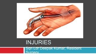
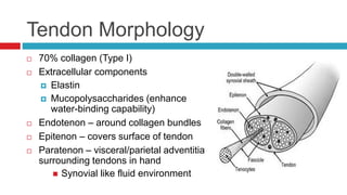
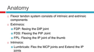
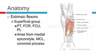
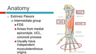
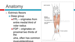
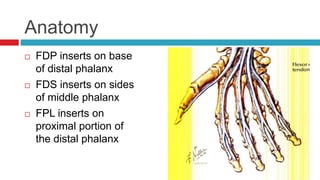
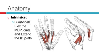
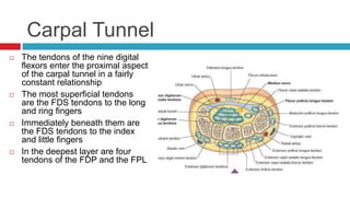
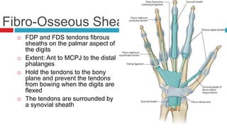
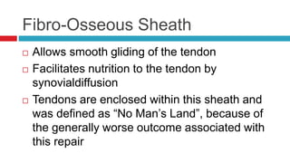
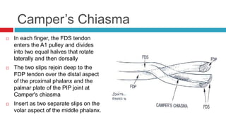
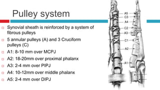
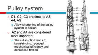
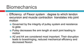
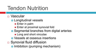
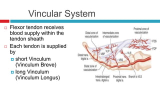
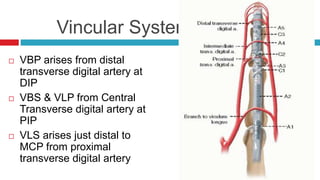
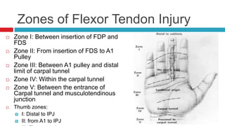
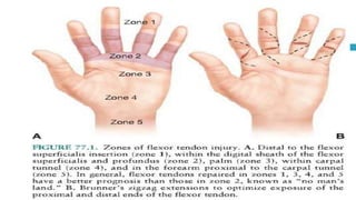
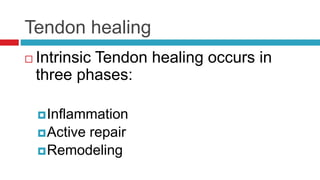
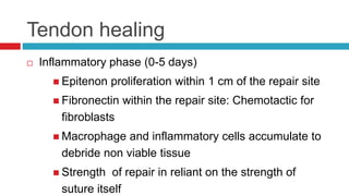
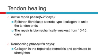
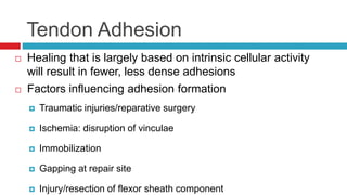
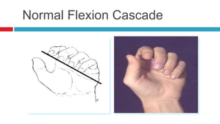
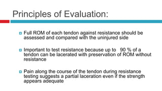
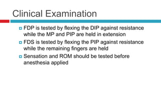
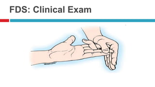
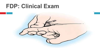
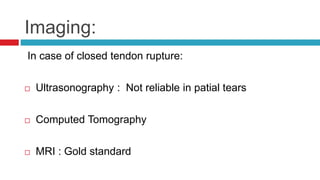
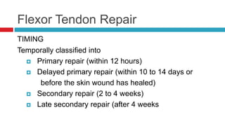
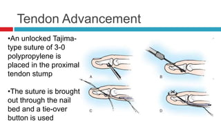
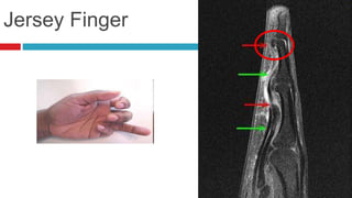
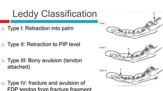
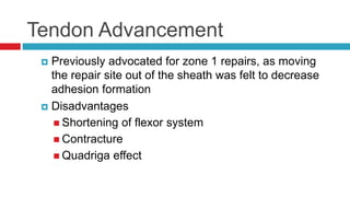
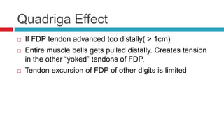
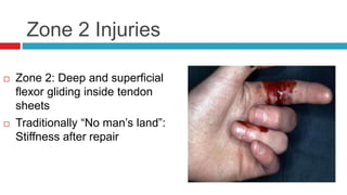
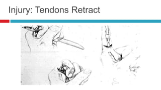
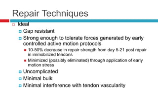
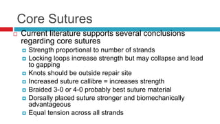
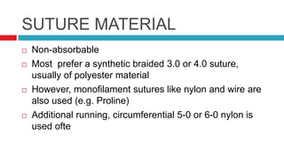
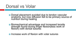
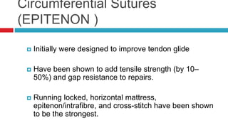
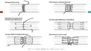
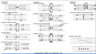
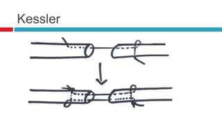
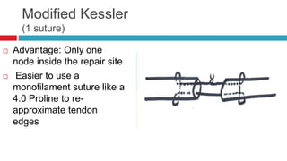
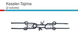
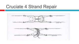
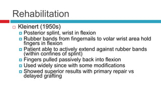
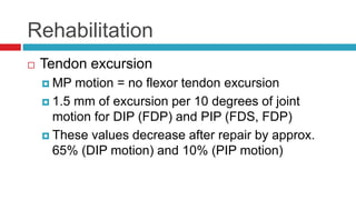
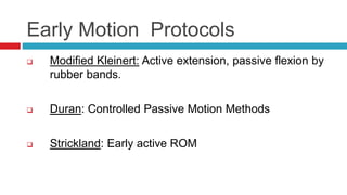
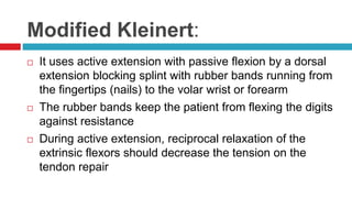
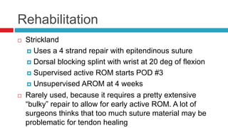
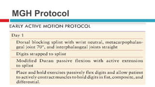
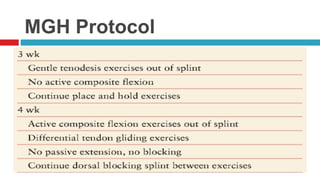
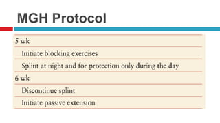
Sqn Ldr Deepak Kumar, Resident
Surgery
FLEXOR TENDON
INJURIES
Introduction
 FTI: considered one of the most challenging problems
for hand surgeons
 Suturing of a divided tendon usually results in:
 Some thickening enlarged area cannot pass the
constricting pulley and motion is prevented
 Bunnell coined the term "no man's land" to emphasize
the difficulties associated with injuries in this area of the
digital sheath
Introduction
 Restoration of satisfactory digital function after flexor
tendon lacerations remains one of the most challenging
problems in hand surgery
 Prior to the 1960’s tendons lacerated in “no man’s land”
were not repaired in favor of delayed grafting
 Kleinert and Verdan (1960’s) showed superior results
with primary repair leading to general acceptance of this
approach
Introduction
 In the past 25 years more scientifically sound research
has advanced our understanding of flexor tendon
structure, nutrition, healing, biomechanics, response to
stress, repair techniques
 Many studies have examined passive and active motion
protocols
Tendon Morphology
 70% collagen (Type I)
 Extracellular components
 Elastin
 Mucopolysaccharides (enhance
water-binding capability)
 Endotenon – around collagen bundles
 Epitenon – covers surface of tendon
 Paratenon – visceral/parietal adventitia
surrounding tendons in hand
 Synovial like fluid environment
Anatomy
 Flexor tendon system consists of intrinsic and extrinsic
components
 Extrinsics:
 FDP: flexing the DIP joint
 FDS: Flexing the PIP Joint
 FPL: Flexing the IP joint of the thumb
 Intrinsics:
 Lumbricals: Flex the MCP joints and Extend the IP
joints
Anatomy
 Extrinsic flexors
 Superficial group
PT, FCR, FCU,
PL
Arise from medial
epicondyle, MCL,
coronoid process
Anatomy
 Extrinsic Flexors
 Intermediate group
 FDS
 Arises from medial
epicondyle, UCL,
coronoid process
 Usually have
independent
musculotendinous
origins
Anatomy
 Extrinsic flexors
 Deep group
 FPL – originates from
entire medial third of
volar radius
 FDP – originates on
proximal two thirds of
the
ulna, often has common
musculotendinous
Anatomy
 FDP inserts on base
of distal phalanx
 FDS inserts on sides
of middle phalanx
 FPL inserts on
proximal portion of
the distal phalanx
Anatomy
 Intrinsics:
 Lumbricals:
Flex the
MCP joints
and Extend
the IP joints
Carpal Tunnel
 The tendons of the nine digital
flexors enter the proximal aspect
of the carpal tunnel in a fairly
constant relationship
 The most superficial tendons
are the FDS tendons to the long
and ring fingers
 Immediately beneath them are
the FDS tendons to the index
and little fingers
 In the deepest layer are four
tendons of the FDP and the FPL
Fibro-Osseous Sheath
 FDP and FDS tendons fibrous
sheaths on the palmar aspect of
the digits
 Extent: Ant to MCPJ to the distal
phalanges
 Hold the tendons to the bony
plane and prevent the tendons
from bowing when the digits are
flexed
 The tendons are surrounded by
a synovial sheath
Fibro-Osseous Sheath
 Allows smooth gliding of the tendon
 Facilitates nutrition to the tendon by
synovialdiffusion
 Tendons are enclosed within this sheath and
was defined as “No Man’s Land”, because of
the generally worse outcome associated with
this repair
Camper’s Chiasma
 In each finger, the FDS tendon
enters the A1 pulley and divides
into two equal halves that rotate
laterally and then dorsally
 The two slips rejoin deep to the
FDP tendon over the distal aspect
of the proximal phalanx and the
palmar plate of the PIP joint at
Camper's chiasma
 Insert as two separate slips on the
volar aspect of the middle phalanx.
Pulley system
 Synovial sheath is reinforced by a system of
fibrous pulleys
 5 annular pulleys (A) and 3 Cruciform
pulleys (C)
 A1: 8-10 mm over MCPJ
 A2: 18-20mm over proximal phalanx
 A3: 2-4 mm over PIPJ
 A4: 10-12mm over middle phalanx
 A5: 2-4 mm over DIPJ
Pulley system
 C1, C2, C3 proximal to A3,
A4, A5
 Allow shortening of the pulley
system in flexion
 A2 and A4 are considered
most important.
 Their disruption leads to
bowstringing, reduced
mechanical efficiency and
decreased flexion
Biomechanics
 Efficiency of Flexor system : degree to which tendon
excursion and muscle contraction translates into joint
motion
 Governed by the integrity of pulley system and resistence
to glide
 Pulley decreases the arm length at each joint leading to
motion
 A2 and A4 are considered most important. Their disruption
leads to bowstringing, reduced mechanical efficiency and
decreased flexion
Tendon Nutrition
 Vascular
 Longitudinal vessels
 Enter in palm
 Enter at proximal synovial fold
 Segmental branches from digital arteries
 Long and short vinculae
 Vessels at osseous insertions
 Synovial fluid diffusion
 Imbibition (pumping mechanism)
Vincular System
 Flexor tendon receives
blood supply within the
tendon sheath
 Each tendon is supplied
by
 short Vinculum
(Vinculum Breve)
 long Vinculum
(Vinculum Longus)
Vincular System
 VBP arises from distal
transverse digital artery at
DIP
 VBS & VLP from Central
Transverse digital artery at
PIP
 VLS arises just distal to
MCP from proximal
transverse digital artery
Zones of Flexor Tendon Injury
 Zone I: Between insertion of FDP and
FDS
 Zone II: From insertion of FDS to A1
Pulley
 Zone III: Between A1 pulley and distal
limit of carpal tunnel
 Zone IV: Within the carpal tunnel
 Zone V: Between the entrance of
Carpal tunnel and musculotendinous
junction
 Thumb zones:
 I: Distal to IPJ
 II: from A1 to IPJ
Tendon healing
 2 forms:
 Intrinsic healing : occurs without direct blood flow to
the tendons
 Extrinsic healing: occurs by proliferation of fibroblast
from peripheral epitendon; adhesions occur because
of this process and limit tendon gliding within the
synovial sheath
Tendon healing
 Intrinsic Tendon healing occurs in
three phases:
Inflammation
Active repair
Remodeling
Tendon healing
 Inflammatory phase (0-5 days)
 Epitenon proliferation within 1 cm of the repair site
 Fibronectin within the repair site: Chemotactic for
fibroblasts
 Macrophage and inflammatory cells accumulate to
debride non viable tissue
 Strength of repair in reliant on the strength of
suture itself
Tendon healing
 Active repair phase(5-28days):
 Epitenon fibroblasts secrete type I collagen to unite
the tendon ends
 The repair is biomechanically weakest from 10-15
days
 Remodeling phase(>28 days):
 Collagen in the repair site remodels and continues to
strenghten
Tendon Adhesion
 Healing that is largely based on intrinsic cellular activity
will result in fewer, less dense adhesions
 Factors influencing adhesion formation
 Traumatic injuries/reparative surgery
 Ischemia: disruption of vinculae
 Immobilization
 Gapping at repair site
 Injury/resection of flexor sheath component
Diagnosis of Flexor Injury
 Posture of Hand/ Normal cascade
 Passive tenodesis test
 Forearm compression test
 Independent testing of FDS & FDP
 Imaging
Normal Flexion Cascade
Principles of Evaluation:
 Full ROM of each tendon against resistance should be
assessed and compared with the uninjured side
 Important to test resistance because up to 90 % of a
tendon can be lacerated with preservation of ROM without
resistance
 Pain along the course of the tendon during resistance
testing suggests a partial laceration even if the strength
appears adequate
Clinical Examination
 FDP is tested by flexing the DIP against resistance
while the MP and PIP are held in extension
 FDS is tested by flexing the PIP against resistance
while the remaining fingers are held
 Sensation and ROM should be tested before
anesthesia applied
FDS: Clinical Exam
FDP: Clinical Exam
Imaging:
In case of closed tendon rupture:
 Ultrasonography : Not reliable in patial tears
 Computed Tomography
 MRI : Gold standard
Flexor Tendon Repair
TIMING
Temporally classified into
 Primary repair (within 12 hours)
 Delayed primary repair (within 10 to 14 days or
before the skin wound has healed)
 Secondary repair (2 to 4 weeks)
 Late secondary repair (after 4 weeks
Flexor Tendon Repair
 Best - done within hours and second best after 10 days.
worst results - done between 4 and 7 days.
 Adhesion formation and tendon gliding are adversely
affected when repairs were done after 7 days.
 Delay several days if wound infected.
General Principles
 Atraumatic technique:
 To maximize the healing potential and to avoid adhesions
 The tendon and surrounding tissues must be handled as
delicately as possible and kept moist
 Precise instruments are needed.
 Must be done under loupe magnification
 Early motion increases tendon strength, decreases adhesions,
increases excursion, and improves nutrition through the pumping of
synovial fluid.
 If both FDS and FDP are lacerated in zone II, both should be
Incisions
- Depend on
- Direction of initial laceration
- Need to expose other injured
structures
- Surgical preference
 Factors
 Avoid crossing joints at 90 deg
 Preference - Existing lacerations
 Need to expose other
Tendon Retrieval
 Retraction often limited to A1/A2 pullet region by vinculae
 If lacerated proximal to vinculae or if vinculae disrupted, tendon
ends may retract into plam
 If proximal stumps have retracted into the palm the correct
orientation of FDS and FDP must be re-established (such that FDP
lies volar to Camper’s Chiasm
 Avoid trauma to synovial sheath lining
 Forcep/hemostat/skin hook if proximal stump visible
 Proximal to distal milking
 Suction catheter
ZONE 1 INJURIES: Jersey Finger
 Involves only FDP
 If the distal stump is too small to allow the placement of
adequate sutures an osteo-periosteal flap is raised at
the base of the distal phalanx
Tendon Advancement
•An unlocked Tajima-
type suture of 3-0
polypropylene is
placed in the proximal
tendon stump
•The suture is brought
out through the nail
bed and a tie-over
button is used
Jersey Finger
Leddy Classification
 Type I: Retraction into palm
 Type II: Retraction to PIP level
 Type III: Bony avulsion (tendon
attached)
 Type IV: fracture and avulsion of
Tendon Advancement
 Previously advocated for zone 1 repairs, as moving
the repair site out of the sheath was felt to decrease
adhesion formation
 Disadvantages
 Shortening of flexor system
 Contracture
 Quadriga effect
Quadriga Effect
 If FDP tendon advanced too distally( > 1cm)
 Entire muscle bells gets pulled distally. Creates tension
in the other “yoked” tendons of FDP.
 Tendon excursion of FDP of other digits is limited
Zone 2 Injuries
 Zone 2: Deep and superficial
flexor gliding inside tendon
sheets
 Traditionally “No man’s land”:
Stiffness after repair
Injury: Tendons Retract
Repair Techniques
 Ideal
 Gap resistant
 Strong enough to tolerate forces generated by early
controlled active motion protocols
 10-50% decrease in repair strength from day 5-21 post repair
in immobilized tendons
 Minimized (possibly eliminated) through application of early
motion stress
 Uncomplicated
 Minimal bulk
 Minimal interference with tendon vascularity
Core Sutures
 Current literature supports several conclusions
regarding core sutures
 Strength proportional to number of strands
 Locking loops increase strength but may collapse and lead
to gapping
 Knots should be outside repair site
 Increased suture callibre = increases strength
 Braided 3-0 or 4-0 probably best suture material
 Dorsally placed suture stronger and biomechanically
advantageous
 Equal tension across all strands
SUTURE MATERIAL
 Non-absorbable
 Most prefer a synthetic braided 3.0 or 4.0 suture,
usually of polyester material
 However, monofilament sutures like nylon and wire are
also used (e.g. Proline)
 Additional running, circumferential 5-0 or 6-0 nylon is
used ofte
Dorsal vs Volar
 Dorsal placement avoided due to tendon vascular
anatomy, but now diffusion felt to be primary source of
nutrition during healing
 Biomechanical advantage and increased tensile
strength found during finger flexion(less work of
flexion) with dorsal sutures
 Increase work of flexion with volar sutures
Circumferential Sutures
(EPITENON )
 Initially were designed to improve tendon glide
 Have been shown to add tensile strength (by 10–
50%) and gap resistance to repairs.
 Running locked, horizontal mattress,
epitenon/intrafibre, and cross-stitch have been shown
to be the strongest.
Kessler
Modified Kessler
(1 suture)
 Advantage: Only one
node inside the repair site
 Easier to use a
monofilament suture like a
4.0 Proline to re-
approximate tendon
edges
Kessler-Tajima
(2 sutures)
Cruciate 4 Strand Repair
Sheath repair
 Advantages
 Barrier to extrinsic adhesion formation
 More rapid return of synovial nutrition
 Disadvantages
 Technically difficult
 Increased foreign material at repair site
 May narrow sheath and restrict glide
NO CLEAR ADVANTAGE ESTABLISHED
Partial laceration
 No repair if 60 % of the tendon intact
 Potential complications:
 Triggering
 Tendon entrapment
 Evaluation for the risk of triggering; debride if necessary
 Dorsal block splinting for 6 to 8 weeks
Complications
 Stiffness
 Re-rupture
 Tenolysis may be required in an estimated
18% to 25% of patients
 No earlier than 3 months after repair
 If no ROM improvement for 1-2 months
Zone 3 Injuries
 Lumbrical muscle bellies usually are not
sutured because this can increase the tension
of these muscles and result in a “lumbrical
plus” finger (paradoxical proximal
interphalangeal extension on attempted active
finger flexion).
POST-OP
REHABILITATION
Post –op Rehabilitation
 For flexor tendon injuries is divided into four groups on
the basis of the exercises instituted during the first 3 to 4
weeks aftertendon repair:
(1) Immobilization
(2) Early passive mobilization
(3) Early active extension and passive flexion
(4) Early controlled active flexion
Immobilisation:
Rarely is there an indication for immobilization after flexortendon
surgery
 Exceptions
1) children or
2) adults unable to cooperate with hand therapy,
3) unstable bone repair, and
4) concerns about the effect of tension on
microneurovascular repairs.
Immobilisation:
 Splint - wrist in neutral or slight flexion, the
MCP joints are significantly flexed, and the
interphalangeal joints are only slightly
flexed or extended.
Rehabilitation
 Mayer (1916)
 The operation should never be undertaken unless
the surgeon himself can perform effective
postoperative care
 Early motion should be instituted at the right time
 graded exercises should be used with corrective
splints
Rehabilitation
 Bunnel (1918)
 Postoperative immobilization
 Active motion beginning at 3 wks postop
 Suboptimal results by today’s standards
 Improved suture material/technique as well as
postoperative rehabilitation protocols
Rehabilitation
 Kleinert (1950s)
 Posterior splint, wrist in flexion
 Rubber bands from fingernails to volar wrist area hold
fingers in flexion
 Patient able to actively extend against rubber bands
(within confines of splint)
 Fingers pulled passively back into flexion
 Used widely since with some modifications
 Showed superior results with primary repair vs
delayed grafting
Rehabilitation
 Tendon excursion
 MP motion = no flexor tendon excursion
 1.5 mm of excursion per 10 degrees of joint
motion for DIP (FDP) and PIP (FDS, FDP)
 These values decrease after repair by approx.
65% (DIP motion) and 10% (PIP motion)
Early Motion Protocols
 Modified Kleinert: Active extension, passive flexion by
rubber bands.
 Duran: Controlled Passive Motion Methods
 Strickland: Early active ROM
Modified Kleinert:
 It uses active extension with passive flexion by a dorsal
extension blocking splint with rubber bands running from
the fingertips (nails) to the volar wrist or forearm
 The rubber bands keep the patient from flexing the digits
against resistance
 During active extension, reciprocal relaxation of the
extrinsic flexors should decrease the tension on the
tendon repair
Modified Kleinert
Modified Kleinert
 After 3 to 4 weeks, the dorsal splint is removed but the
rubber bands are maintained
 Wrist range-of-motion exercises are started and finger
exercises continued
Duran Protocol
 Dorsal Splint in 20 deg wrist flexion
 No rubber bands
 Passive flexion
 Designed in response to notion 3-5mm of
tendon gliding sufficient to prevent restrictive
adhesions
Duran protocol
Rehabilitation
 Strickland
 Uses a 4 strand repair with epitendinous suture
 Dorsal blocking splint with wrist at 20 deg of flexion
 Supervised active ROM starts POD #3
 Unsupervised AROM at 4 weeks
 Rarely used, because it requires a pretty extensive
“bulky” repair to allow for early active ROM. A lot of
surgeons thinks that too much suture material may be
problematic for tendon healing
MGH Protocol
MGH Protocol
MGH Protocol
MGH Protocol
Children
 Usually not able to reliably participate in
rehabilitation programs
 No benefit to early mobilization in patients
under 16 years
 Immobilization >4 wks may lead to poorer
outcomes
COMPLICATIONS
Complications
 Short term:
 Infection
 Injury to neurovascular structures or pulley system
 Abnormal scarring
 Long term:
 Adhesion
 Rupture
 Joint contracture
 Triggering
Complications
 Adhesions
 Most common complication despite early motion
protocols
 Tenolysis when patients progressive gain in digital
motion has plateaued, usually 3- 6 months after repair
Complications
 Tendon rupture
 Noted by the patient at “popping” in the hand
 7-10 days postop when tensile strength is weakest
 MRI may help in diagnosis
 Flexion contracture
 FDP advancement more than 1 cm may lead to
flexion contracture and weakened hand grip because
of quadrigia effect
THANK YOU

Flexor tendon injuries

  • 1.
    Sqn Ldr DeepakKumar, Resident Surgery FLEXOR TENDON INJURIES
  • 2.
    Introduction  FTI: consideredone of the most challenging problems for hand surgeons  Suturing of a divided tendon usually results in:  Some thickening enlarged area cannot pass the constricting pulley and motion is prevented  Bunnell coined the term "no man's land" to emphasize the difficulties associated with injuries in this area of the digital sheath
  • 3.
    Introduction  Restoration ofsatisfactory digital function after flexor tendon lacerations remains one of the most challenging problems in hand surgery  Prior to the 1960’s tendons lacerated in “no man’s land” were not repaired in favor of delayed grafting  Kleinert and Verdan (1960’s) showed superior results with primary repair leading to general acceptance of this approach
  • 4.
    Introduction  In thepast 25 years more scientifically sound research has advanced our understanding of flexor tendon structure, nutrition, healing, biomechanics, response to stress, repair techniques  Many studies have examined passive and active motion protocols
  • 5.
    Tendon Morphology  70%collagen (Type I)  Extracellular components  Elastin  Mucopolysaccharides (enhance water-binding capability)  Endotenon – around collagen bundles  Epitenon – covers surface of tendon  Paratenon – visceral/parietal adventitia surrounding tendons in hand  Synovial like fluid environment
  • 6.
    Anatomy  Flexor tendonsystem consists of intrinsic and extrinsic components  Extrinsics:  FDP: flexing the DIP joint  FDS: Flexing the PIP Joint  FPL: Flexing the IP joint of the thumb  Intrinsics:  Lumbricals: Flex the MCP joints and Extend the IP joints
  • 7.
    Anatomy  Extrinsic flexors Superficial group PT, FCR, FCU, PL Arise from medial epicondyle, MCL, coronoid process
  • 8.
    Anatomy  Extrinsic Flexors Intermediate group  FDS  Arises from medial epicondyle, UCL, coronoid process  Usually have independent musculotendinous origins
  • 9.
    Anatomy  Extrinsic flexors Deep group  FPL – originates from entire medial third of volar radius  FDP – originates on proximal two thirds of the ulna, often has common musculotendinous
  • 10.
    Anatomy  FDP insertson base of distal phalanx  FDS inserts on sides of middle phalanx  FPL inserts on proximal portion of the distal phalanx
  • 11.
    Anatomy  Intrinsics:  Lumbricals: Flexthe MCP joints and Extend the IP joints
  • 12.
    Carpal Tunnel  Thetendons of the nine digital flexors enter the proximal aspect of the carpal tunnel in a fairly constant relationship  The most superficial tendons are the FDS tendons to the long and ring fingers  Immediately beneath them are the FDS tendons to the index and little fingers  In the deepest layer are four tendons of the FDP and the FPL
  • 13.
    Fibro-Osseous Sheath  FDPand FDS tendons fibrous sheaths on the palmar aspect of the digits  Extent: Ant to MCPJ to the distal phalanges  Hold the tendons to the bony plane and prevent the tendons from bowing when the digits are flexed  The tendons are surrounded by a synovial sheath
  • 14.
    Fibro-Osseous Sheath  Allowssmooth gliding of the tendon  Facilitates nutrition to the tendon by synovialdiffusion  Tendons are enclosed within this sheath and was defined as “No Man’s Land”, because of the generally worse outcome associated with this repair
  • 15.
    Camper’s Chiasma  Ineach finger, the FDS tendon enters the A1 pulley and divides into two equal halves that rotate laterally and then dorsally  The two slips rejoin deep to the FDP tendon over the distal aspect of the proximal phalanx and the palmar plate of the PIP joint at Camper's chiasma  Insert as two separate slips on the volar aspect of the middle phalanx.
  • 16.
    Pulley system  Synovialsheath is reinforced by a system of fibrous pulleys  5 annular pulleys (A) and 3 Cruciform pulleys (C)  A1: 8-10 mm over MCPJ  A2: 18-20mm over proximal phalanx  A3: 2-4 mm over PIPJ  A4: 10-12mm over middle phalanx  A5: 2-4 mm over DIPJ
  • 17.
    Pulley system  C1,C2, C3 proximal to A3, A4, A5  Allow shortening of the pulley system in flexion  A2 and A4 are considered most important.  Their disruption leads to bowstringing, reduced mechanical efficiency and decreased flexion
  • 18.
    Biomechanics  Efficiency ofFlexor system : degree to which tendon excursion and muscle contraction translates into joint motion  Governed by the integrity of pulley system and resistence to glide  Pulley decreases the arm length at each joint leading to motion  A2 and A4 are considered most important. Their disruption leads to bowstringing, reduced mechanical efficiency and decreased flexion
  • 19.
    Tendon Nutrition  Vascular Longitudinal vessels  Enter in palm  Enter at proximal synovial fold  Segmental branches from digital arteries  Long and short vinculae  Vessels at osseous insertions  Synovial fluid diffusion  Imbibition (pumping mechanism)
  • 20.
    Vincular System  Flexortendon receives blood supply within the tendon sheath  Each tendon is supplied by  short Vinculum (Vinculum Breve)  long Vinculum (Vinculum Longus)
  • 21.
    Vincular System  VBParises from distal transverse digital artery at DIP  VBS & VLP from Central Transverse digital artery at PIP  VLS arises just distal to MCP from proximal transverse digital artery
  • 22.
    Zones of FlexorTendon Injury  Zone I: Between insertion of FDP and FDS  Zone II: From insertion of FDS to A1 Pulley  Zone III: Between A1 pulley and distal limit of carpal tunnel  Zone IV: Within the carpal tunnel  Zone V: Between the entrance of Carpal tunnel and musculotendinous junction  Thumb zones:  I: Distal to IPJ  II: from A1 to IPJ
  • 24.
    Tendon healing  2forms:  Intrinsic healing : occurs without direct blood flow to the tendons  Extrinsic healing: occurs by proliferation of fibroblast from peripheral epitendon; adhesions occur because of this process and limit tendon gliding within the synovial sheath
  • 25.
    Tendon healing  IntrinsicTendon healing occurs in three phases: Inflammation Active repair Remodeling
  • 26.
    Tendon healing  Inflammatoryphase (0-5 days)  Epitenon proliferation within 1 cm of the repair site  Fibronectin within the repair site: Chemotactic for fibroblasts  Macrophage and inflammatory cells accumulate to debride non viable tissue  Strength of repair in reliant on the strength of suture itself
  • 27.
    Tendon healing  Activerepair phase(5-28days):  Epitenon fibroblasts secrete type I collagen to unite the tendon ends  The repair is biomechanically weakest from 10-15 days  Remodeling phase(>28 days):  Collagen in the repair site remodels and continues to strenghten
  • 28.
    Tendon Adhesion  Healingthat is largely based on intrinsic cellular activity will result in fewer, less dense adhesions  Factors influencing adhesion formation  Traumatic injuries/reparative surgery  Ischemia: disruption of vinculae  Immobilization  Gapping at repair site  Injury/resection of flexor sheath component
  • 29.
    Diagnosis of FlexorInjury  Posture of Hand/ Normal cascade  Passive tenodesis test  Forearm compression test  Independent testing of FDS & FDP  Imaging
  • 30.
  • 31.
    Principles of Evaluation: Full ROM of each tendon against resistance should be assessed and compared with the uninjured side  Important to test resistance because up to 90 % of a tendon can be lacerated with preservation of ROM without resistance  Pain along the course of the tendon during resistance testing suggests a partial laceration even if the strength appears adequate
  • 32.
    Clinical Examination  FDPis tested by flexing the DIP against resistance while the MP and PIP are held in extension  FDS is tested by flexing the PIP against resistance while the remaining fingers are held  Sensation and ROM should be tested before anesthesia applied
  • 33.
  • 34.
  • 35.
    Imaging: In case ofclosed tendon rupture:  Ultrasonography : Not reliable in patial tears  Computed Tomography  MRI : Gold standard
  • 36.
    Flexor Tendon Repair TIMING Temporallyclassified into  Primary repair (within 12 hours)  Delayed primary repair (within 10 to 14 days or before the skin wound has healed)  Secondary repair (2 to 4 weeks)  Late secondary repair (after 4 weeks
  • 37.
    Flexor Tendon Repair Best - done within hours and second best after 10 days. worst results - done between 4 and 7 days.  Adhesion formation and tendon gliding are adversely affected when repairs were done after 7 days.  Delay several days if wound infected.
  • 38.
    General Principles  Atraumatictechnique:  To maximize the healing potential and to avoid adhesions  The tendon and surrounding tissues must be handled as delicately as possible and kept moist  Precise instruments are needed.  Must be done under loupe magnification  Early motion increases tendon strength, decreases adhesions, increases excursion, and improves nutrition through the pumping of synovial fluid.  If both FDS and FDP are lacerated in zone II, both should be
  • 39.
    Incisions - Depend on -Direction of initial laceration - Need to expose other injured structures - Surgical preference  Factors  Avoid crossing joints at 90 deg  Preference - Existing lacerations  Need to expose other
  • 41.
    Tendon Retrieval  Retractionoften limited to A1/A2 pullet region by vinculae  If lacerated proximal to vinculae or if vinculae disrupted, tendon ends may retract into plam  If proximal stumps have retracted into the palm the correct orientation of FDS and FDP must be re-established (such that FDP lies volar to Camper’s Chiasm  Avoid trauma to synovial sheath lining  Forcep/hemostat/skin hook if proximal stump visible  Proximal to distal milking  Suction catheter
  • 42.
    ZONE 1 INJURIES:Jersey Finger  Involves only FDP  If the distal stump is too small to allow the placement of adequate sutures an osteo-periosteal flap is raised at the base of the distal phalanx
  • 43.
    Tendon Advancement •An unlockedTajima- type suture of 3-0 polypropylene is placed in the proximal tendon stump •The suture is brought out through the nail bed and a tie-over button is used
  • 44.
  • 45.
    Leddy Classification  TypeI: Retraction into palm  Type II: Retraction to PIP level  Type III: Bony avulsion (tendon attached)  Type IV: fracture and avulsion of
  • 46.
    Tendon Advancement  Previouslyadvocated for zone 1 repairs, as moving the repair site out of the sheath was felt to decrease adhesion formation  Disadvantages  Shortening of flexor system  Contracture  Quadriga effect
  • 47.
    Quadriga Effect  IfFDP tendon advanced too distally( > 1cm)  Entire muscle bells gets pulled distally. Creates tension in the other “yoked” tendons of FDP.  Tendon excursion of FDP of other digits is limited
  • 48.
    Zone 2 Injuries Zone 2: Deep and superficial flexor gliding inside tendon sheets  Traditionally “No man’s land”: Stiffness after repair
  • 49.
  • 50.
    Repair Techniques  Ideal Gap resistant  Strong enough to tolerate forces generated by early controlled active motion protocols  10-50% decrease in repair strength from day 5-21 post repair in immobilized tendons  Minimized (possibly eliminated) through application of early motion stress  Uncomplicated  Minimal bulk  Minimal interference with tendon vascularity
  • 51.
    Core Sutures  Currentliterature supports several conclusions regarding core sutures  Strength proportional to number of strands  Locking loops increase strength but may collapse and lead to gapping  Knots should be outside repair site  Increased suture callibre = increases strength  Braided 3-0 or 4-0 probably best suture material  Dorsally placed suture stronger and biomechanically advantageous  Equal tension across all strands
  • 52.
    SUTURE MATERIAL  Non-absorbable Most prefer a synthetic braided 3.0 or 4.0 suture, usually of polyester material  However, monofilament sutures like nylon and wire are also used (e.g. Proline)  Additional running, circumferential 5-0 or 6-0 nylon is used ofte
  • 53.
    Dorsal vs Volar Dorsal placement avoided due to tendon vascular anatomy, but now diffusion felt to be primary source of nutrition during healing  Biomechanical advantage and increased tensile strength found during finger flexion(less work of flexion) with dorsal sutures  Increase work of flexion with volar sutures
  • 54.
    Circumferential Sutures (EPITENON ) Initially were designed to improve tendon glide  Have been shown to add tensile strength (by 10– 50%) and gap resistance to repairs.  Running locked, horizontal mattress, epitenon/intrafibre, and cross-stitch have been shown to be the strongest.
  • 57.
  • 58.
    Modified Kessler (1 suture) Advantage: Only one node inside the repair site  Easier to use a monofilament suture like a 4.0 Proline to re- approximate tendon edges
  • 59.
  • 60.
  • 61.
    Sheath repair  Advantages Barrier to extrinsic adhesion formation  More rapid return of synovial nutrition  Disadvantages  Technically difficult  Increased foreign material at repair site  May narrow sheath and restrict glide NO CLEAR ADVANTAGE ESTABLISHED
  • 62.
    Partial laceration  Norepair if 60 % of the tendon intact  Potential complications:  Triggering  Tendon entrapment  Evaluation for the risk of triggering; debride if necessary  Dorsal block splinting for 6 to 8 weeks
  • 63.
    Complications  Stiffness  Re-rupture Tenolysis may be required in an estimated 18% to 25% of patients  No earlier than 3 months after repair  If no ROM improvement for 1-2 months
  • 64.
    Zone 3 Injuries Lumbrical muscle bellies usually are not sutured because this can increase the tension of these muscles and result in a “lumbrical plus” finger (paradoxical proximal interphalangeal extension on attempted active finger flexion).
  • 65.
  • 66.
    Post –op Rehabilitation For flexor tendon injuries is divided into four groups on the basis of the exercises instituted during the first 3 to 4 weeks aftertendon repair: (1) Immobilization (2) Early passive mobilization (3) Early active extension and passive flexion (4) Early controlled active flexion
  • 67.
    Immobilisation: Rarely is therean indication for immobilization after flexortendon surgery  Exceptions 1) children or 2) adults unable to cooperate with hand therapy, 3) unstable bone repair, and 4) concerns about the effect of tension on microneurovascular repairs.
  • 68.
    Immobilisation:  Splint -wrist in neutral or slight flexion, the MCP joints are significantly flexed, and the interphalangeal joints are only slightly flexed or extended.
  • 69.
    Rehabilitation  Mayer (1916) The operation should never be undertaken unless the surgeon himself can perform effective postoperative care  Early motion should be instituted at the right time  graded exercises should be used with corrective splints
  • 70.
    Rehabilitation  Bunnel (1918) Postoperative immobilization  Active motion beginning at 3 wks postop  Suboptimal results by today’s standards  Improved suture material/technique as well as postoperative rehabilitation protocols
  • 71.
    Rehabilitation  Kleinert (1950s) Posterior splint, wrist in flexion  Rubber bands from fingernails to volar wrist area hold fingers in flexion  Patient able to actively extend against rubber bands (within confines of splint)  Fingers pulled passively back into flexion  Used widely since with some modifications  Showed superior results with primary repair vs delayed grafting
  • 72.
    Rehabilitation  Tendon excursion MP motion = no flexor tendon excursion  1.5 mm of excursion per 10 degrees of joint motion for DIP (FDP) and PIP (FDS, FDP)  These values decrease after repair by approx. 65% (DIP motion) and 10% (PIP motion)
  • 73.
    Early Motion Protocols Modified Kleinert: Active extension, passive flexion by rubber bands.  Duran: Controlled Passive Motion Methods  Strickland: Early active ROM
  • 74.
    Modified Kleinert:  Ituses active extension with passive flexion by a dorsal extension blocking splint with rubber bands running from the fingertips (nails) to the volar wrist or forearm  The rubber bands keep the patient from flexing the digits against resistance  During active extension, reciprocal relaxation of the extrinsic flexors should decrease the tension on the tendon repair
  • 75.
  • 76.
    Modified Kleinert  After3 to 4 weeks, the dorsal splint is removed but the rubber bands are maintained  Wrist range-of-motion exercises are started and finger exercises continued
  • 77.
    Duran Protocol  DorsalSplint in 20 deg wrist flexion  No rubber bands  Passive flexion  Designed in response to notion 3-5mm of tendon gliding sufficient to prevent restrictive adhesions
  • 78.
  • 79.
    Rehabilitation  Strickland  Usesa 4 strand repair with epitendinous suture  Dorsal blocking splint with wrist at 20 deg of flexion  Supervised active ROM starts POD #3  Unsupervised AROM at 4 weeks  Rarely used, because it requires a pretty extensive “bulky” repair to allow for early active ROM. A lot of surgeons thinks that too much suture material may be problematic for tendon healing
  • 80.
  • 81.
  • 82.
  • 83.
  • 84.
    Children  Usually notable to reliably participate in rehabilitation programs  No benefit to early mobilization in patients under 16 years  Immobilization >4 wks may lead to poorer outcomes
  • 85.
  • 86.
    Complications  Short term: Infection  Injury to neurovascular structures or pulley system  Abnormal scarring  Long term:  Adhesion  Rupture  Joint contracture  Triggering
  • 87.
    Complications  Adhesions  Mostcommon complication despite early motion protocols  Tenolysis when patients progressive gain in digital motion has plateaued, usually 3- 6 months after repair
  • 88.
    Complications  Tendon rupture Noted by the patient at “popping” in the hand  7-10 days postop when tensile strength is weakest  MRI may help in diagnosis  Flexion contracture  FDP advancement more than 1 cm may lead to flexion contracture and weakened hand grip because of quadrigia effect
  • 89.