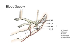
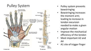
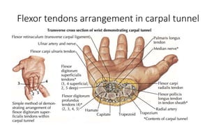
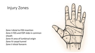
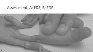
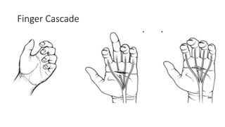
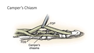
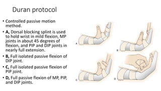
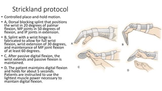
Flexor Tendons
Injuries
By
Mohamed Mohamed Salah Eldeeb
Basic Science
Blood Supply
V: Vinculum
L: Longus
B: Brevis
P: Profundus
S: Superficialis
Pulley System • Pulley system prevents
bowstringing
• Bowstringing increases
the moment arm;
leading to increase in
tendon excursion
needed to make a given
angular motion
• Improve the mechanical
efficiency of the tendon
• Most important A2 and
A4
• A1 site of trigger finger
Flexor tendons arrangement in carpal tunnel
Injury Zones
Zone I distal to FDS insertion
Zone II FDS and FDP slide in common
sheath
Zone III area of lumbircal origin
Zone IV carpal tunnel
Zone V distal forearm
Tendon
healing
Two forms:
• Intrinsic healing: by the activity of fibroblast
derived from the tendon.
• Extrinsic healing: by activity of fibroblast
derived from the epitendon and haematoma
of injury.
• Extrinsic healing results in adhesions
formation and limit the gliding of the tendon
through the sheath.
Biological
fact
• Traumatized tendon ends tend to soften
during the first 2 weeks post-surgery, thus
reducing the holding power of the suture.
• The innately weak and slow biological
healing creates a “no gain” lag period in
tendon strength during the first 3 to 4 weeks
after surgery.
• We should ensure that the strength of
surgical repairs is greater than the forces
generated during unresisted active finger
flexion plus a certain safety margin.
DIAGNOSIS
Assessment: A; FDS, B; FDP
Finger Cascade
Flexor Tendon
Repair Techniques
Incisions
Avoid crossing
perpendicular on the
creases.
Tendon
Retrieval
• Avoid trauma to synovial sheath lining
• Forcep/hemostat/skin hook if proximal stump
visible
• Proximal to distal milking, reverse Esmarch
• Suture catheter to proximal tendons in palm
and deliver distally
• Retraction often limited to A1/A2 pulley region
by vinculae If lacerated proximal to vinculae or if
vinculae disrupted, tendon ends may retract
into palm If proximal stumps have retracted into
the palm the correct orientation of FDS and FDP
must be re-established (such that FDP lies volar
to Camper's Chiasm)
Camper’s Chiasm
Tendon
Advancement
• Previously advocated for zone 1 repairs, as
moving the repair site out of the sheath was
felt to decrease adhesion formation –
• Disadvantages Shortening of flexor system
• Contracture
• Quadregia effect
Quadregia effect
General
principles for
tendon
repair
• To provide repair strength sufficient to
permit motion in the first 6 weeks
• Good end to end coaptations, no gap
• Adequate tensile strength to resist gapping
during early 6wks
• Frictionless repair, minimal bulk
• Non-traumatizing
Items of
tendon
repair
• Suture material
• Core or epitendinous repair, and number of
strands
• Knot out or in
• Locking vs grasping
• Repair of both tendons or only one
• Repair techniques
Suture material
• Non-absorbable, braided or monofilament
• Comparison: nylon, a monofilament suture
• Prolene, a monofilament polypropylene suture
• Ethibond, a braided polyester suture
• Stainless steel (braided)
• Fibrewire, a polyethylene-based braided suture with reported
superior strength characteristics.
Core or
epitendinous,
crossing
strands
• Core suture is mechanically better
• Epitendinous just to smooth the repair site
with little mechanical advantages
• The tensile strengrth of repair is related to
the number of crossing strands
• Four strands or more should be chosen, two
crossing strands less effiecient
• Suture calibre related to the size of the
tendon
Examples of four or more strands
Knot in or
out
• The knot being either within or away
from the repair site has not been
shown to have an independent effect
on tensile strength.
• Greater quantity of suture within the
repair site may increase repair site
bulk.
• External knot distant to the repair site
may also adversely affect tendon
gliding within the flexor tendon sheath
by increased friction or knot trapping
between tendon and sheath.
Locking vs Grasping loop
• The loop of the core
suture that is
positioned to “lock”
rather than “grasp”
the tendon stumps
shows greater
strength.
• Similarly, increasing
the number of locks
or grasps increases
tensile strength
Repair both
flexors or
one
• For better functional results repair of both is the
role.
• If the tendon ends are severely injured, unclean,
or ragged, or if there is insufficient tendon for
repair, excision of the FDS tendon and isolated
repair of the FDP tendon may be the best
alternative.
• Excision of one slip of the FDS tendon may be
necessary to diminish the bulk of the repair and
facilitate gliding through the tendon sheath.
• In cases where only repair of the FDS tendon is
possible, FDP tenodesis to the middle phalanx or
DIP joint fusion may be necessary.
Repair techniques
• Tendon to bone.
• In FDP avulsion
End to end
Post Operative Care
Rehabilitation
Goals
• Promote intrinsic tendon healing and
minimize extrinsic healing and adhesion
formation to improve gliding.
• Early post operative tendon mobilization
biologically affect the scar formation at the
repair site.
• The collagen is laid down longitudinally
parellel to axial force.
• Load at failure for mobilized tendon is
twice that for immobilized tendon at two
weeks post-operative.
Immobilization
Programme
• Indicated in:
• Children and adults who are difficult or
untrustful to comply with the
mobilization programme.
• Associated injuries or co-morbidities
precluding mobilization programme
compliance.
Kleinert splint
• Dorsal splint with wrist in flexion.
• Finger tied by rubber band in
flexion.
• Active extension is done.
• Finger return back in flexion by pull
of the rubber band.
Duran protocol
• Controlled passive motion
method.
• A, Dorsal blocking splint is used
to hold wrist in mild flexion, MP
joints in about 45 degrees of
flexion, and PIP and DIP joints in
nearly full extension.
• B, Full isolated passive flexion of
DIP joint.
• C, Full isolated passive flexion of
PIP joint.
• D, Full passive flexion of MP, PIP,
and DIP joints.
Strickland protocol
• Controlled place-and-hold motion.
• A, Dorsal blocking splint that positions
the wrist in 20 degrees of palmar
flexion, MP joints in 50 degrees of
flexion, and IP joints in extension.
• B, Splint with a wrist hinge is
fabricated to allow for full wrist
flexion, wrist extension of 30 degrees,
and maintenance of MP joint flexion
of at least 60 degrees.
• C, After passive digital flexion, the
wrist extends and passive flexion is
maintained.
• D, The patient maintains digital flexion
and holds for about 5 seconds.
Patients are instructed to use the
lightest muscle power necessary to
maintain digital flexion.
Thank You

Flexor tendon injuries

  • 1.
  • 2.
  • 3.
    Blood Supply V: Vinculum L:Longus B: Brevis P: Profundus S: Superficialis
  • 4.
    Pulley System •Pulley system prevents bowstringing • Bowstringing increases the moment arm; leading to increase in tendon excursion needed to make a given angular motion • Improve the mechanical efficiency of the tendon • Most important A2 and A4 • A1 site of trigger finger
  • 5.
  • 6.
    Injury Zones Zone Idistal to FDS insertion Zone II FDS and FDP slide in common sheath Zone III area of lumbircal origin Zone IV carpal tunnel Zone V distal forearm
  • 7.
    Tendon healing Two forms: • Intrinsichealing: by the activity of fibroblast derived from the tendon. • Extrinsic healing: by activity of fibroblast derived from the epitendon and haematoma of injury. • Extrinsic healing results in adhesions formation and limit the gliding of the tendon through the sheath.
  • 8.
    Biological fact • Traumatized tendonends tend to soften during the first 2 weeks post-surgery, thus reducing the holding power of the suture. • The innately weak and slow biological healing creates a “no gain” lag period in tendon strength during the first 3 to 4 weeks after surgery. • We should ensure that the strength of surgical repairs is greater than the forces generated during unresisted active finger flexion plus a certain safety margin.
  • 9.
  • 10.
  • 11.
  • 12.
  • 13.
  • 14.
    Tendon Retrieval • Avoid traumato synovial sheath lining • Forcep/hemostat/skin hook if proximal stump visible • Proximal to distal milking, reverse Esmarch • Suture catheter to proximal tendons in palm and deliver distally • Retraction often limited to A1/A2 pulley region by vinculae If lacerated proximal to vinculae or if vinculae disrupted, tendon ends may retract into palm If proximal stumps have retracted into the palm the correct orientation of FDS and FDP must be re-established (such that FDP lies volar to Camper's Chiasm)
  • 15.
  • 16.
    Tendon Advancement • Previously advocatedfor zone 1 repairs, as moving the repair site out of the sheath was felt to decrease adhesion formation – • Disadvantages Shortening of flexor system • Contracture • Quadregia effect
  • 17.
  • 18.
    General principles for tendon repair • Toprovide repair strength sufficient to permit motion in the first 6 weeks • Good end to end coaptations, no gap • Adequate tensile strength to resist gapping during early 6wks • Frictionless repair, minimal bulk • Non-traumatizing
  • 19.
    Items of tendon repair • Suturematerial • Core or epitendinous repair, and number of strands • Knot out or in • Locking vs grasping • Repair of both tendons or only one • Repair techniques
  • 20.
    Suture material • Non-absorbable,braided or monofilament • Comparison: nylon, a monofilament suture • Prolene, a monofilament polypropylene suture • Ethibond, a braided polyester suture • Stainless steel (braided) • Fibrewire, a polyethylene-based braided suture with reported superior strength characteristics.
  • 21.
    Core or epitendinous, crossing strands • Coresuture is mechanically better • Epitendinous just to smooth the repair site with little mechanical advantages • The tensile strengrth of repair is related to the number of crossing strands • Four strands or more should be chosen, two crossing strands less effiecient • Suture calibre related to the size of the tendon
  • 22.
    Examples of fouror more strands
  • 23.
    Knot in or out •The knot being either within or away from the repair site has not been shown to have an independent effect on tensile strength. • Greater quantity of suture within the repair site may increase repair site bulk. • External knot distant to the repair site may also adversely affect tendon gliding within the flexor tendon sheath by increased friction or knot trapping between tendon and sheath.
  • 24.
    Locking vs Graspingloop • The loop of the core suture that is positioned to “lock” rather than “grasp” the tendon stumps shows greater strength. • Similarly, increasing the number of locks or grasps increases tensile strength
  • 25.
    Repair both flexors or one •For better functional results repair of both is the role. • If the tendon ends are severely injured, unclean, or ragged, or if there is insufficient tendon for repair, excision of the FDS tendon and isolated repair of the FDP tendon may be the best alternative. • Excision of one slip of the FDS tendon may be necessary to diminish the bulk of the repair and facilitate gliding through the tendon sheath. • In cases where only repair of the FDS tendon is possible, FDP tenodesis to the middle phalanx or DIP joint fusion may be necessary.
  • 26.
    Repair techniques • Tendonto bone. • In FDP avulsion
  • 27.
  • 28.
  • 29.
    Rehabilitation Goals • Promote intrinsictendon healing and minimize extrinsic healing and adhesion formation to improve gliding. • Early post operative tendon mobilization biologically affect the scar formation at the repair site. • The collagen is laid down longitudinally parellel to axial force. • Load at failure for mobilized tendon is twice that for immobilized tendon at two weeks post-operative.
  • 30.
    Immobilization Programme • Indicated in: •Children and adults who are difficult or untrustful to comply with the mobilization programme. • Associated injuries or co-morbidities precluding mobilization programme compliance.
  • 31.
    Kleinert splint • Dorsalsplint with wrist in flexion. • Finger tied by rubber band in flexion. • Active extension is done. • Finger return back in flexion by pull of the rubber band.
  • 32.
    Duran protocol • Controlledpassive motion method. • A, Dorsal blocking splint is used to hold wrist in mild flexion, MP joints in about 45 degrees of flexion, and PIP and DIP joints in nearly full extension. • B, Full isolated passive flexion of DIP joint. • C, Full isolated passive flexion of PIP joint. • D, Full passive flexion of MP, PIP, and DIP joints.
  • 33.
    Strickland protocol • Controlledplace-and-hold motion. • A, Dorsal blocking splint that positions the wrist in 20 degrees of palmar flexion, MP joints in 50 degrees of flexion, and IP joints in extension. • B, Splint with a wrist hinge is fabricated to allow for full wrist flexion, wrist extension of 30 degrees, and maintenance of MP joint flexion of at least 60 degrees. • C, After passive digital flexion, the wrist extends and passive flexion is maintained. • D, The patient maintains digital flexion and holds for about 5 seconds. Patients are instructed to use the lightest muscle power necessary to maintain digital flexion.
  • 34.