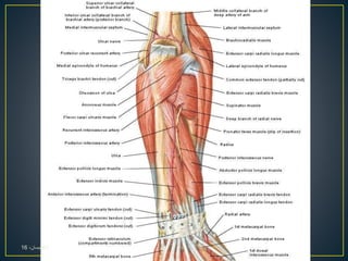
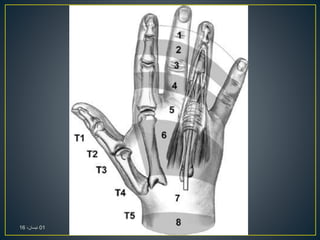
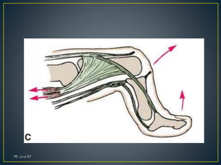
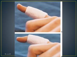
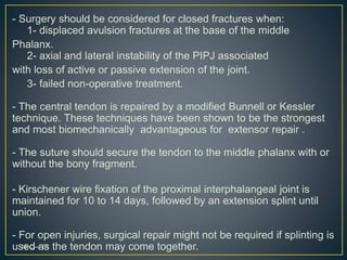
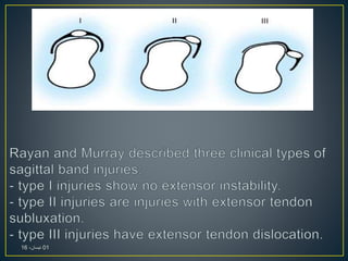
Ismaeel Al-Saadi Hospital
Orthopedic Surgery Department
The forearm consists of many compartment:
- Anterior: contains flexor muscles.
- lateral: considered as part of posterior compartment which
consists of (Brachioradialis and extensor carpi radialis
Longus)
- Posterior: consists of two groups of muscles (superficial
an deep group)
01،‫نيسان‬16
01،‫نيسان‬16
01،‫نيسان‬16
Notice:
- The four tendons of the extensor digitorum emerge from
under the extensor retinaculum and fan out over the
dorsum of the hand.
- Strong oblique fibrous bands connect the tendons to the
little, ring, and middle fingers.
- The tendon to the index finger is joined on its medial side
by the tendon of the extensor indicis, which is the same for
little finger.
01،‫نيسان‬16
- On the posterior surface of each finger, the extensor
tendon joins the fascial expansion called the (extensor
Expansion).
- Near the proximal interphalangeal joint, the extensor
expansion splits into three parts:
- Central part, which is inserted into the base of the
middle phalanx.
- Two lateral parts, which converge to be inserted into
the base of the distal phalanx.
01،‫نيسان‬16
01،‫نيسان‬16
01،‫نيسان‬16
01،‫نيسان‬16
01،‫نيسان‬16
01،‫نيسان‬16
01،‫نيسان‬16
It’s a thickening of deep fascia that stretches across the
back of the wrist and holds the long extensor tendons in
position, It converts the grooves on the posterior surface of
the distal ends of the radius and ulna into six separate
tunnels ( 6 Compartments) lined with a synovial sheath.
It’s attached medially to the pisiform bone and the hook of
the hamate and laterally to the distal end of the radius.
Extensor Retinaculum
01،‫نيسان‬16
The 6 Compartments contents are (from lateral to medial):
1- abductor pollicis longus , extensor pollicis brevis
and to the medial, there is the radial artery.
2- extensor carpi radialis longus and brevis.
3- extensor pollicis longus.
4- extensor digitorum and extensor indicis above it.
5- extensor digiti minimi.
6- extensor carpi ulnaris.
01،‫نيسان‬16
01،‫نيسان‬16
01،‫نيسان‬16
- Extensor tendon injuries are more frequent than flexor
tendon injuries. and are very common (61%) as they
are not protected as well as the flexor tendons due to:
- their superficial location
- and lack of overlying subcutaneous tissue.
- Extensors are particularly difficult for surgeons because of:
- their reduced size compared with the flexors
- and their lack of collagen-bundle linkage, which reduces the
grip strength available for the suture material.
01،‫نيسان‬16
- Extensor digiti minimi is responsible for extension of the fifth digit. It
can be tested by asking the patient to lie their hand flat on a surface
and hyperextend the fifth digit.
- The extensor digitorum tendons extend digits two through five. The
hand can be laid flat again on a surface and the tendon of each
finger tested by having the patient hyperextend the digits against
resistance.
- Extension of the second finger is also performed by extensor
indicis, which can be tested in a similar fashion to extensor
digiti minimi.
- Extension of the first digit is controlled by two muscles, extensor
pollicis longus and brevis.
01،‫نيسان‬16
- Extension of the thumb at the interphalangeal joint and the
metacarpophalangeal joint can be tested separately against
Resistance
- When checking the long extensor tendon of the thumb, the
examiner must stabilize the metacarpophalangeal joint and
must test carefully for active extension of the interphalangeal
joint and active retropulsion of the thumb toward the dorsum of
the hand, because an intact short thumb extensor can actively
extend the thumb.
- Radiographs are needed to assess any associated
fracture as they will need to be fixed before tendon and
nerve injuries during surgery
01،‫نيسان‬16
01،‫نيسان‬16
01،‫نيسان‬16
- Kleinert and Verdan wrote a classification system for
extensor tendon lacerations according to the eight zones of
the hand, wrist and forearm which has been widely accepted.
- The type of injury, surgical approach, potential deformity
varies according to the zone.
- Zone I refers to the area from the DIP joint to the fingertip;
zone II encompasses the middle phalanx;
zone III refers to the PIP joint;
zone IV is over the proximal phalanx;
zone V refers to the MP joint;
zone VI encompasses the metacarpal and
zone VII is over the wrist
zone VIII refers to the forearm.01،‫نيسان‬16
01،‫نيسان‬16
Extensor Tendon Suture Methods:
– For more distal injuries (zones 1-4) the tendon is flat:
Figure of 8 repair or mattress suture repair
–As the caliber thickens (zones 5-8):
Core suture method
01،‫نيسان‬16
- Rockwell et al Explained that treatment of tendon injuries is
dependent on the location and type of injury. Repair should
take place very soon after the injury especially within the first 2
weeks.
- Extensor tendon repair techniques are not as complicated
as flexor tendon repairs, this is due to the:
- extensor tendon being smaller with a relatively flat
cross section.
- its collagen is longitudinally orientated with little or
no cross-linking.
01،‫نيسان‬16
- Exploration of the wound will often occur with the patient
awake (local or regional anaesthetic),
- but more complex injuries would be explored whilst the
patient is asleep (general anaesthetic).
- The surgeon will apply a tourniquet to the limb to prevent
bleeding, which makes the operation much easier.
- It is likely that the wound will be made larger by the
surgeon to enable a thorough inspection of the structures in
the vicinity as well as to retrieve tendon ends that tend to
spring apart when cut.
- The hand/wrist is almost always protected by a splint to
prevent the tendon repair being over-stretched
01،‫نيسان‬16
01،‫نيسان‬16
01،‫نيسان‬16
- Zone I injury often referred to as mallet finger is when
there is disruption to the extensor tendon over the distal
interphalangeal joint causing a flexion deformity of the distal
interphalangeal joint.
- It’s caused by Forceful flexion of the distal interphalangeal
joint in an extended digit, results in rupture of the extensor
tendon or avulsion from its insertion at the distal phalanx.
- When left untreated, hyperextension of the proximal
interphalangeal joint may develop due to the retraction of the
central band causing a swan neck deformity.
01،‫نيسان‬16
01،‫نيسان‬16
01،‫نيسان‬16
Mallet fingers are classified into 4 types:
Type 1: Closed with or without avulsion fracture
Type 2: Laceration at or proximal to the distal interphalangeal
joint with loss of tendon continuity
Type 3: Deep abrasion with loss of skin, subcutaneous
cover, and tendon substance
Type 4: A- Transepiphyseal plate fracture in children.
B- hyperflexion injury with fracture of the articular
surface of 20 to 50 percent.
C- hyperextension injury with fracture of the articular
surface usually greater than 50 percent and with
early or late palmar subluxation of the distal phalanx.
01،‫نيسان‬16
- Closed mallet fingers, which is type I fractures should be
treated with an immobilisation splint in extension or slight
hyperextension for 6 to 8 weeks and at night only for 1 additional
week.
- After 8 weeks the fingers should be examined again and if active
extension is present splinting can be reduced to high-risk times such
as sleeping, manual work or athletic performance.
- Type II required simple suture through the tendon alone or a roll
type suture (tenodermodesis technique) incorporating the tendon
and skin in the same suture and then splinting for 6-8 weeks.
- Type III fractures include loss of tendon substance which requires
immediate soft tissue coverage and primary grafting or
reconstruction with free tendon graft.
01،‫نيسان‬16
- Type IV-A are best treated with closed reduction followed by
splinting and are the most likely fracture in children.
- Type IV-B is usually treated by splinting for 6 weeks with 2 weeks
of night splinting yields good results.
- Type IV-C with palmar subluxation of the distal phalanx is surgically
managed with open reduction and internal fixation using a Kirschner
wire and sometimes a pull-out wire or suture.
A splint for 6 weeks is then used after which the Kirschner wire is
removed and motion started.
01،‫نيسان‬16
- Repair the extensor tendon with 4-0 monofilament nylon or 4-0
monofilament wire as a pull-out roll stitch.
- Close the skin with interrupted 5-0 nylon. As an alternative,
use 4-0 nylon as a dermotenodermal suture.
- The sutures are removed at 10 to 14 days.
The Kirschner wire is removed after 4 to 6 weeks.
01،‫نيسان‬16
01،‫نيسان‬16
01،‫نيسان‬16
01،‫نيسان‬16
01،‫نيسان‬16
- Zone II injures or middle phalanx injuries are usually as a
result of laceration or crush injuries rather than avulsion like
zone I.
- If there is extensor lag on examination then exploration and
repair is needed, whereas if there active extension with only a
degree of weakness than splinting can be used for 3-4 weeks.
01،‫نيسان‬16
- According to experimental work by Newport et al. and
Newport and Williams, the Kleinert modification of the Bunnell
suture and the modified Kessler sutures were stronger than a
figure-of-eight or mattress suture for repair of extensor tendons
in zone II.
- Injuries greater than 50% of the tendon should be repaired
with a fashion-of eight suture or similar fashion.
01،‫نيسان‬16
01،‫نيسان‬16
01،‫نيسان‬16
01،‫نيسان‬16
- Known as Boutonniere Deformity is caused by disruption
of the central slip at the proximal interphalangeal joint.
- The boutonniere deformity usually occurs 10-14 days
after the first injury.
- Closed deformities require splinting for 4-6 weeks of the
PIP joint in extension with the DIP joint and wrist joints left
free.
01،‫نيسان‬16
01،‫نيسان‬16
01،‫نيسان‬16
01،‫نيسان‬16
- Surgery should be considered for closed fractures when:
1- displaced avulsion fractures at the base of the middle
Phalanx.
2- axial and lateral instability of the PIPJ associated
with loss of active or passive extension of the joint.
3- failed non-operative treatment.
- The central tendon is repaired by a modified Bunnell or Kessler
technique. These techniques have been shown to be the strongest
and most biomechanically advantageous for extensor repair .
- The suture should secure the tendon to the middle phalanx with or
without the bony fragment.
- Kirschener wire fixation of the proximal interphalangeal joint is
maintained for 10 to 14 days, followed by an extension splint until
union.
- For open injuries, surgical repair might not be required if splinting is
used as the tendon may come together.01،‫نيسان‬16
- known as proximal phalanx injuries, usually involve the broad
extensor mechanism, it’s diagnosed usually by inspection.
- Splinting the PIPJ in extension for 3-4 weeks without repair as
shown to have the same outcome as repairing it with nonabsorbale
sutures.
- However, if the laceration is complete, surgical primary repair
should be performed followed by 6 weeks of splinting in extension.
- In the first 3 weeks volar positioning should be used with passive
extension allowed.
At week 4 gentle active extension is introduced but no passive
flexion at this time.
In the last two weeks active flexion is introduced and graded
resisted exercises are implemented.01،‫نيسان‬16
01،‫نيسان‬16
- Injuries in zone V are nearly always open.
- It should be repaired with the modified Bunnell repair
technique because this technique has been shown to be the
best repair method.
- The saggital bands should be repaired in a simple or
mattress fashion to prevent lateral migration of the extensor
digtorum communis tendon and subsequent
metacarpophalangeal extension loss.
- Splinting of the wrist in 30-45 degrees of extension and the
metacarpophalangeal joint in 20-30 degrees of flexion is
performed with the proximal interphalangeal joint free.
01،‫نيسان‬16
01،‫نيسان‬16
- It occurs most commonly in the long finger.
- The dislocation usually occurs as a result of a tear in the
proximal radial portion of the shroud ligament (sagittal bands)
and the more proximal fascia, as the finger is suddenly extended
against a force, as in a flicking or thumping motion.
- If seen within the first few days, this dislocation can be treated
effectively with splinting of the metacarpophalangeal joint and
wrist in extension for about 3 to 4 weeks, followed by 3 to 4
weeks of removable splinting or buddy taping to the adjacent
finger on the radial side in the case of ulnar displacement.
01،‫نيسان‬16
01،‫نيسان‬16
01،‫نيسان‬16
01،‫نيسان‬16
- The tendons in this area are close to the thin subcutaneous
tissue. Injuries in this zone are situated in the dorsal hand and may
not always result in loss of the extension at the
metacarpophalangeal joint.
- Surgical repair is needed with stronger core type sutures and then
splinting should be placed in extension for 4-6 weeks.
- If the extensor digitorum communis is involved, all fingers should
be splinted but if the proprius tendon is solely involved, only the
affected finger need be splinted with the wrist.
- As the tendons in this area are larger, stronger core suture should
be used.
01،‫نيسان‬16
01،‫نيسان‬16
- In the dorsal forearm, many tendons are likely to be
lacerated, including the muscletendious junction and tendon
bellies.
- Thumb and wrist extension should be repaired first.
- Multiple figure of eight sutures should be used to repair
the muscle bellies.
- Static immobilisation of the wrist in 45 degrees of extension
and metacarpophalangeal joints in 15-20 degrees should be
maintained for 4-5 weeks
01،‫نيسان‬16
- Mallet injuries are uncommon in the thumb because the
terminal extensor tendon is thicker on the thumb.
- Open injuries most surgeons would recommend primary
repair with splinting for 6 weeks. For closed injuries
splinting for 6 weeks (8 weeks: Campbells) without surgical
repair is a suitable alternative but surgical repair is also used.
- Extensor pollicis brevis is rare to be solely lacerated, so
its repair is debatable because extension of the
metacarpophalangeal joint is possible with an intact extensor
pollicis longus.
01،‫نيسان‬16
- Splinting is usually for 3 to 4 weeks, with the thumb
metacarpophalangeal joint in full extension and wrist in 40
degrees of extension with slight radial deviation.
- For injuries in zone VI and VII the abductor pollicis longus
retracts when divided and therefore requires to be released for
successful repair.
- Splinting is then needed for 4-5 weeks with the wrist in radial
deviation and the thumb in maximal abduction.
01،‫نيسان‬16
- The ultimate aim of any rehabilitation is to obtain healing
with minimal gapping and prevent adhesions.
- complications include:
- tendon rupture
- adhesion formation requiring tenolysis.
- extension lad
- loss of flexion and decreased grip strength
- deformities
- infection
- Early mobilisation rehabilitation programes can be in
two categories:
(1) early active mobilisation
(2) early controlled mobilization using a dynamic splint which
has been found to decrease adhesions and subsequent
contractures.
01،‫نيسان‬16
01،‫نيسان‬16

Extensor tendon injuries

  • 1.
  • 2.
    The forearm consistsof many compartment: - Anterior: contains flexor muscles. - lateral: considered as part of posterior compartment which consists of (Brachioradialis and extensor carpi radialis Longus) - Posterior: consists of two groups of muscles (superficial an deep group) 01،‫نيسان‬16
  • 3.
  • 4.
  • 5.
    Notice: - The fourtendons of the extensor digitorum emerge from under the extensor retinaculum and fan out over the dorsum of the hand. - Strong oblique fibrous bands connect the tendons to the little, ring, and middle fingers. - The tendon to the index finger is joined on its medial side by the tendon of the extensor indicis, which is the same for little finger. 01،‫نيسان‬16
  • 6.
    - On theposterior surface of each finger, the extensor tendon joins the fascial expansion called the (extensor Expansion). - Near the proximal interphalangeal joint, the extensor expansion splits into three parts: - Central part, which is inserted into the base of the middle phalanx. - Two lateral parts, which converge to be inserted into the base of the distal phalanx. 01،‫نيسان‬16
  • 7.
  • 8.
  • 9.
  • 10.
  • 11.
  • 12.
  • 13.
    It’s a thickeningof deep fascia that stretches across the back of the wrist and holds the long extensor tendons in position, It converts the grooves on the posterior surface of the distal ends of the radius and ulna into six separate tunnels ( 6 Compartments) lined with a synovial sheath. It’s attached medially to the pisiform bone and the hook of the hamate and laterally to the distal end of the radius. Extensor Retinaculum 01،‫نيسان‬16
  • 14.
    The 6 Compartmentscontents are (from lateral to medial): 1- abductor pollicis longus , extensor pollicis brevis and to the medial, there is the radial artery. 2- extensor carpi radialis longus and brevis. 3- extensor pollicis longus. 4- extensor digitorum and extensor indicis above it. 5- extensor digiti minimi. 6- extensor carpi ulnaris. 01،‫نيسان‬16
  • 15.
  • 16.
  • 17.
    - Extensor tendoninjuries are more frequent than flexor tendon injuries. and are very common (61%) as they are not protected as well as the flexor tendons due to: - their superficial location - and lack of overlying subcutaneous tissue. - Extensors are particularly difficult for surgeons because of: - their reduced size compared with the flexors - and their lack of collagen-bundle linkage, which reduces the grip strength available for the suture material. 01،‫نيسان‬16
  • 18.
    - Extensor digitiminimi is responsible for extension of the fifth digit. It can be tested by asking the patient to lie their hand flat on a surface and hyperextend the fifth digit. - The extensor digitorum tendons extend digits two through five. The hand can be laid flat again on a surface and the tendon of each finger tested by having the patient hyperextend the digits against resistance. - Extension of the second finger is also performed by extensor indicis, which can be tested in a similar fashion to extensor digiti minimi. - Extension of the first digit is controlled by two muscles, extensor pollicis longus and brevis. 01،‫نيسان‬16
  • 19.
    - Extension ofthe thumb at the interphalangeal joint and the metacarpophalangeal joint can be tested separately against Resistance - When checking the long extensor tendon of the thumb, the examiner must stabilize the metacarpophalangeal joint and must test carefully for active extension of the interphalangeal joint and active retropulsion of the thumb toward the dorsum of the hand, because an intact short thumb extensor can actively extend the thumb. - Radiographs are needed to assess any associated fracture as they will need to be fixed before tendon and nerve injuries during surgery 01،‫نيسان‬16
  • 20.
  • 21.
  • 22.
    - Kleinert andVerdan wrote a classification system for extensor tendon lacerations according to the eight zones of the hand, wrist and forearm which has been widely accepted. - The type of injury, surgical approach, potential deformity varies according to the zone. - Zone I refers to the area from the DIP joint to the fingertip; zone II encompasses the middle phalanx; zone III refers to the PIP joint; zone IV is over the proximal phalanx; zone V refers to the MP joint; zone VI encompasses the metacarpal and zone VII is over the wrist zone VIII refers to the forearm.01،‫نيسان‬16
  • 23.
  • 24.
    Extensor Tendon SutureMethods: – For more distal injuries (zones 1-4) the tendon is flat: Figure of 8 repair or mattress suture repair –As the caliber thickens (zones 5-8): Core suture method 01،‫نيسان‬16
  • 25.
    - Rockwell etal Explained that treatment of tendon injuries is dependent on the location and type of injury. Repair should take place very soon after the injury especially within the first 2 weeks. - Extensor tendon repair techniques are not as complicated as flexor tendon repairs, this is due to the: - extensor tendon being smaller with a relatively flat cross section. - its collagen is longitudinally orientated with little or no cross-linking. 01،‫نيسان‬16
  • 26.
    - Exploration ofthe wound will often occur with the patient awake (local or regional anaesthetic), - but more complex injuries would be explored whilst the patient is asleep (general anaesthetic). - The surgeon will apply a tourniquet to the limb to prevent bleeding, which makes the operation much easier. - It is likely that the wound will be made larger by the surgeon to enable a thorough inspection of the structures in the vicinity as well as to retrieve tendon ends that tend to spring apart when cut. - The hand/wrist is almost always protected by a splint to prevent the tendon repair being over-stretched 01،‫نيسان‬16
  • 27.
  • 28.
  • 29.
    - Zone Iinjury often referred to as mallet finger is when there is disruption to the extensor tendon over the distal interphalangeal joint causing a flexion deformity of the distal interphalangeal joint. - It’s caused by Forceful flexion of the distal interphalangeal joint in an extended digit, results in rupture of the extensor tendon or avulsion from its insertion at the distal phalanx. - When left untreated, hyperextension of the proximal interphalangeal joint may develop due to the retraction of the central band causing a swan neck deformity. 01،‫نيسان‬16
  • 30.
  • 31.
  • 32.
    Mallet fingers areclassified into 4 types: Type 1: Closed with or without avulsion fracture Type 2: Laceration at or proximal to the distal interphalangeal joint with loss of tendon continuity Type 3: Deep abrasion with loss of skin, subcutaneous cover, and tendon substance Type 4: A- Transepiphyseal plate fracture in children. B- hyperflexion injury with fracture of the articular surface of 20 to 50 percent. C- hyperextension injury with fracture of the articular surface usually greater than 50 percent and with early or late palmar subluxation of the distal phalanx. 01،‫نيسان‬16
  • 33.
    - Closed malletfingers, which is type I fractures should be treated with an immobilisation splint in extension or slight hyperextension for 6 to 8 weeks and at night only for 1 additional week. - After 8 weeks the fingers should be examined again and if active extension is present splinting can be reduced to high-risk times such as sleeping, manual work or athletic performance. - Type II required simple suture through the tendon alone or a roll type suture (tenodermodesis technique) incorporating the tendon and skin in the same suture and then splinting for 6-8 weeks. - Type III fractures include loss of tendon substance which requires immediate soft tissue coverage and primary grafting or reconstruction with free tendon graft. 01،‫نيسان‬16
  • 34.
    - Type IV-Aare best treated with closed reduction followed by splinting and are the most likely fracture in children. - Type IV-B is usually treated by splinting for 6 weeks with 2 weeks of night splinting yields good results. - Type IV-C with palmar subluxation of the distal phalanx is surgically managed with open reduction and internal fixation using a Kirschner wire and sometimes a pull-out wire or suture. A splint for 6 weeks is then used after which the Kirschner wire is removed and motion started. 01،‫نيسان‬16
  • 35.
    - Repair theextensor tendon with 4-0 monofilament nylon or 4-0 monofilament wire as a pull-out roll stitch. - Close the skin with interrupted 5-0 nylon. As an alternative, use 4-0 nylon as a dermotenodermal suture. - The sutures are removed at 10 to 14 days. The Kirschner wire is removed after 4 to 6 weeks. 01،‫نيسان‬16
  • 36.
  • 37.
  • 38.
  • 39.
  • 40.
    - Zone IIinjures or middle phalanx injuries are usually as a result of laceration or crush injuries rather than avulsion like zone I. - If there is extensor lag on examination then exploration and repair is needed, whereas if there active extension with only a degree of weakness than splinting can be used for 3-4 weeks. 01،‫نيسان‬16
  • 41.
    - According toexperimental work by Newport et al. and Newport and Williams, the Kleinert modification of the Bunnell suture and the modified Kessler sutures were stronger than a figure-of-eight or mattress suture for repair of extensor tendons in zone II. - Injuries greater than 50% of the tendon should be repaired with a fashion-of eight suture or similar fashion. 01،‫نيسان‬16
  • 42.
  • 43.
  • 44.
  • 45.
    - Known asBoutonniere Deformity is caused by disruption of the central slip at the proximal interphalangeal joint. - The boutonniere deformity usually occurs 10-14 days after the first injury. - Closed deformities require splinting for 4-6 weeks of the PIP joint in extension with the DIP joint and wrist joints left free. 01،‫نيسان‬16
  • 46.
  • 47.
  • 48.
  • 49.
    - Surgery shouldbe considered for closed fractures when: 1- displaced avulsion fractures at the base of the middle Phalanx. 2- axial and lateral instability of the PIPJ associated with loss of active or passive extension of the joint. 3- failed non-operative treatment. - The central tendon is repaired by a modified Bunnell or Kessler technique. These techniques have been shown to be the strongest and most biomechanically advantageous for extensor repair . - The suture should secure the tendon to the middle phalanx with or without the bony fragment. - Kirschener wire fixation of the proximal interphalangeal joint is maintained for 10 to 14 days, followed by an extension splint until union. - For open injuries, surgical repair might not be required if splinting is used as the tendon may come together.01،‫نيسان‬16
  • 50.
    - known asproximal phalanx injuries, usually involve the broad extensor mechanism, it’s diagnosed usually by inspection. - Splinting the PIPJ in extension for 3-4 weeks without repair as shown to have the same outcome as repairing it with nonabsorbale sutures. - However, if the laceration is complete, surgical primary repair should be performed followed by 6 weeks of splinting in extension. - In the first 3 weeks volar positioning should be used with passive extension allowed. At week 4 gentle active extension is introduced but no passive flexion at this time. In the last two weeks active flexion is introduced and graded resisted exercises are implemented.01،‫نيسان‬16
  • 51.
  • 52.
    - Injuries inzone V are nearly always open. - It should be repaired with the modified Bunnell repair technique because this technique has been shown to be the best repair method. - The saggital bands should be repaired in a simple or mattress fashion to prevent lateral migration of the extensor digtorum communis tendon and subsequent metacarpophalangeal extension loss. - Splinting of the wrist in 30-45 degrees of extension and the metacarpophalangeal joint in 20-30 degrees of flexion is performed with the proximal interphalangeal joint free. 01،‫نيسان‬16
  • 53.
  • 54.
    - It occursmost commonly in the long finger. - The dislocation usually occurs as a result of a tear in the proximal radial portion of the shroud ligament (sagittal bands) and the more proximal fascia, as the finger is suddenly extended against a force, as in a flicking or thumping motion. - If seen within the first few days, this dislocation can be treated effectively with splinting of the metacarpophalangeal joint and wrist in extension for about 3 to 4 weeks, followed by 3 to 4 weeks of removable splinting or buddy taping to the adjacent finger on the radial side in the case of ulnar displacement. 01،‫نيسان‬16
  • 55.
  • 56.
  • 57.
  • 58.
    - The tendonsin this area are close to the thin subcutaneous tissue. Injuries in this zone are situated in the dorsal hand and may not always result in loss of the extension at the metacarpophalangeal joint. - Surgical repair is needed with stronger core type sutures and then splinting should be placed in extension for 4-6 weeks. - If the extensor digitorum communis is involved, all fingers should be splinted but if the proprius tendon is solely involved, only the affected finger need be splinted with the wrist. - As the tendons in this area are larger, stronger core suture should be used. 01،‫نيسان‬16
  • 59.
  • 60.
    - In thedorsal forearm, many tendons are likely to be lacerated, including the muscletendious junction and tendon bellies. - Thumb and wrist extension should be repaired first. - Multiple figure of eight sutures should be used to repair the muscle bellies. - Static immobilisation of the wrist in 45 degrees of extension and metacarpophalangeal joints in 15-20 degrees should be maintained for 4-5 weeks 01،‫نيسان‬16
  • 61.
    - Mallet injuriesare uncommon in the thumb because the terminal extensor tendon is thicker on the thumb. - Open injuries most surgeons would recommend primary repair with splinting for 6 weeks. For closed injuries splinting for 6 weeks (8 weeks: Campbells) without surgical repair is a suitable alternative but surgical repair is also used. - Extensor pollicis brevis is rare to be solely lacerated, so its repair is debatable because extension of the metacarpophalangeal joint is possible with an intact extensor pollicis longus. 01،‫نيسان‬16
  • 62.
    - Splinting isusually for 3 to 4 weeks, with the thumb metacarpophalangeal joint in full extension and wrist in 40 degrees of extension with slight radial deviation. - For injuries in zone VI and VII the abductor pollicis longus retracts when divided and therefore requires to be released for successful repair. - Splinting is then needed for 4-5 weeks with the wrist in radial deviation and the thumb in maximal abduction. 01،‫نيسان‬16
  • 63.
    - The ultimateaim of any rehabilitation is to obtain healing with minimal gapping and prevent adhesions. - complications include: - tendon rupture - adhesion formation requiring tenolysis. - extension lad - loss of flexion and decreased grip strength - deformities - infection - Early mobilisation rehabilitation programes can be in two categories: (1) early active mobilisation (2) early controlled mobilization using a dynamic splint which has been found to decrease adhesions and subsequent contractures. 01،‫نيسان‬16
  • 64.