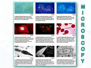
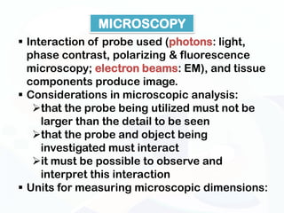
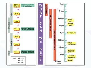
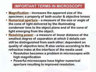
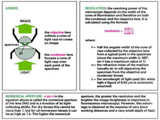
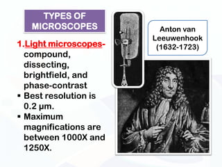
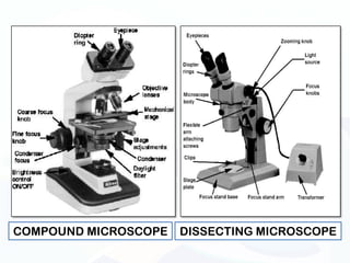
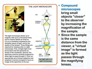
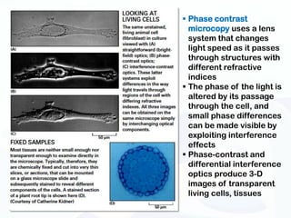
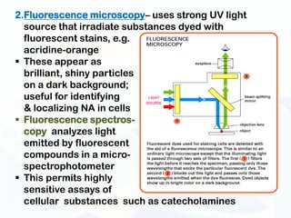
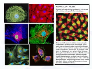
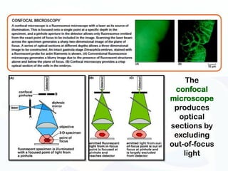
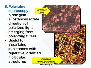
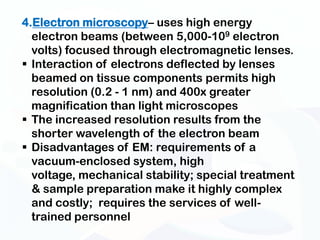
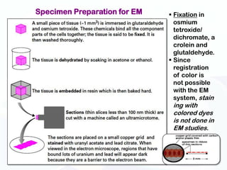
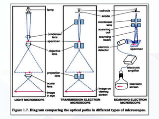
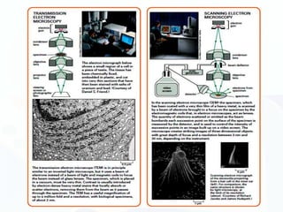
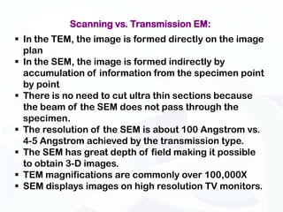
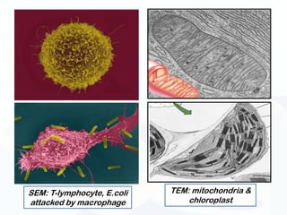
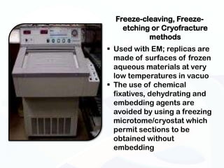
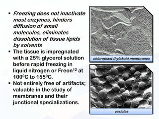
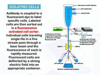
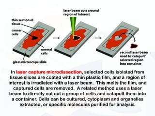
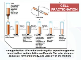
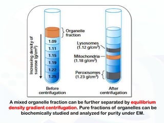
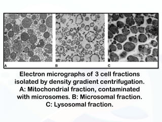
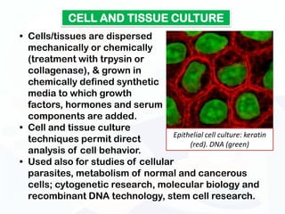
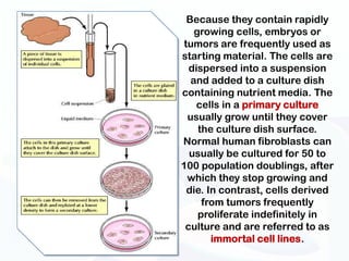
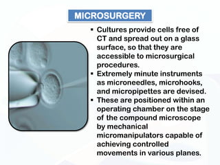
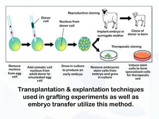
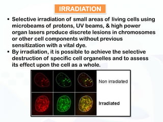
Department of Natural Sciences
   University of St. La Salle
INVESTIGATION OF CELLS AND THEIR PARTS
EXPERIMENTAL SUBJECTS OF CELL
         BIOLOGISTS
MICROSCOPY
 Interaction of probe used (photons: light,
  phase contrast, polarizing & fluorescence
  microscopy; electron beams: EM), and tissue
  components produce image.
 Considerations in microscopic analysis:
   that the probe being utilized must not be
     larger than the detail to be seen
   that the probe and object being
     investigated must interact
   it must be possible to observe and
     interpret this interaction
 Units for measuring microscopic dimensions:
W
H
A
T

C
A
N

W
E

S
E
E
?
IMPORTANT TERMS IN MICROSCOPY
 Magnification – increases the apparent size of the
  specimen; a property of both ocular & objective lenses
 Numerical aperture – a measure of the size or angle of
  the cone of light delivered by the illuminating
  condenser lens to the object plane and of the cone of
  light emerging from the object.
 Resolving power – a measure of linear distance of the
  smallest degree of separation at which 2 details can
  still be distinguished from each other; dependent on
  quality of objective lens; R also varies according to the
  refractive index at the interface of the media used
     Resolution becomes a problem in microscopes with
      high magnification
     Powerful microscopes have higher numerical
      aperture resulting to improved resolution.
TYPES OF
   MICROSCOPES           Anton van
                       Leeuwenhook
1.Light microscopes-    (1632-1723)
  compound,
  dissecting,
  brightfield, and
  phase-contrast
 Best resolution is
  0.2 µm.
 Maximum
  magnifications are
  between 1000X and
  1250X.
COMPOUND MICROSCOPE   DISSECTING MICROSCOPE
 Compound
  microscopes
  bring small
  objects "closer"
  to the observer
  by increasing the
  magnification of
  the sample.
 Since the sample
  is the same
  distance from the
  viewer, a "virtual
  image" is formed
  as the light
  passes through
  the magnifying
  lenses.
 Phase contrast
  microcopy uses a lens
  system that changes
  light speed as it passes
  through structures with
  different refractive
  indices
 The phase of the light is
  altered by its passage
  through the cell, and
  small phase differences
  can be made visible by
  exploiting interference
  effects
 Phase-contrast and
  differential interference
  optics produce 3-D
  images of transparent
  living cells, tissues
2.Fluorescence microscopy– uses strong UV light
  source that irradiate substances dyed with
  fluorescent stains, e.g.
  acridine-orange
 These appear as
  brilliant, shiny particles
  on a dark background;
  useful for identifying
  & localizing NA in cells
 Fluorescence spectros-
  copy analyzes light
  emitted by fluorescent
  compounds in a micro-
  spectrophotometer
 This permits highly
  sensitive assays of
  cellular substances such as catecholamines
The
 confocal
microscope
 produces
  optical
sections by
 excluding
out-of-focus
    light
3. Polarizing
                      Compact
   microscopy–          bone
   birefrigent
   substances rotate
   direction of
   polarized light
   emerging from
   polarizing filters
 Useful for
   visualizing
   substances with
   repetitive, oriented
   molecular              Collagen
   structures         fibers, polarizing
                           microscopy
4.Electron microscopy– uses high energy
  electron beams (between 5,000-109 electron
  volts) focused through electromagnetic lenses.
 Interaction of electrons deflected by lenses
  beamed on tissue components permits high
  resolution (0.2 - 1 nm) and 400x greater
  magnification than light microscopes
 The increased resolution results from the
  shorter wavelength of the electron beam
 Disadvantages of EM: requirements of a
  vacuum-enclosed system, high
  voltage, mechanical stability; special treatment
  & sample preparation make it highly complex
  and costly; requires the services of well-
  trained personnel
Specimen Preparation for EM    Fixation in
                                osmium
                                tetroxide/
                                dichromate, a
                                crolein and
                                glutaldehyde.
                               Since
                                registration
                                of color is
                                not possible
                                with the EM
                                system, stain
                               ing with
                               colored dyes
                               is not done in
                               EM studies.
Scanning vs. Transmission EM:
 In the TEM, the image is formed directly on the image
  plan
 In the SEM, the image is formed indirectly by
  accumulation of information from the specimen point
  by point
 There is no need to cut ultra thin sections because
  the beam of the SEM does not pass through the
  specimen.
 The resolution of the SEM is about 100 Angstrom vs.
  4-5 Angstrom achieved by the transmission type.
 The SEM has great depth of field making it possible
  to obtain 3-D images.
 TEM magnifications are commonly over 100,000X
 SEM displays images on high resolution TV monitors.
SEM: T-lymphocyte, E.coli   TEM: mitochondria &
attacked by macrophage          chloroplast
Freeze-cleaving, Freeze-
    etching or Cryofracture
           methods
 Used with EM; replicas are
  made of surfaces of frozen
  aqueous materials at very
  low temperatures in vacuo
 The use of chemical
  fixatives, dehydrating and
  embedding agents are
  avoided by using a freezing
  microtome/cryostat which
  permit sections to be
  obtained without
  embedding
 Freezing does not inactivate
  most enzymes, hinders
  diffusion of small
  molecules, eliminates
  dissolution of tissue lipids
  by solvents
 The tissue is impregnated
  with a 25% glycerol solution      chloroplast thylakoid membranes
  before rapid freezing in
  liquid nitrogen or Freon12 at
  1000C to 1550C.
 Not entirely free of artifacts;
  valuable in the study of
  membranes and their
  junctional specializations.
                                               vesicles
ISOLATING CELLS
Antibody is coupled to a
 fluorescent dye to label
  specific cells. Labeled
cells are then sorted out
     in a fluorescence
   activated cell sorter.
Individual cells traveling
     single file in a fine
  stream pass through a
    laser beam and the
 fluorescence of each is
     rapidly measured.
   Fluorescent cells are
   deflected by a strong
   electric field into an
  appropriate container.
In laser capture microdissection, selected cells isolated from
 tissue slices are coated with a thin plastic film, and a region of
interest is irradiated with a laser beam. This melts the film, and
    captured cells are removed. A related method uses a laser
 beam to directly cut out a group of cells and catapult them into
   a container. Cells can be cultured, cytoplasm and organelles
       extracted, or specific molecules purified for analysis.
CELL
                                         FRACTIONATION


Homogenization/ differential centrifugation separate organelles
 based on their sedimentation coefficients. The latter depends
   on its size, form and density, and viscosity of the medium.
A mixed organelle fraction can be further separated by equilibrium
density gradient centrifugation. Pure fractions of organelles can be
     biochemically studied and analyzed for purity under EM.
Electron micrographs of 3 cell fractions
isolated by density gradient centrifugation.
  A: Mitochondrial fraction, contaminated
 with microsomes. B: Microsomal fraction.
           C: Lysosomal fraction.
CELL AND TISSUE CULTURE
• Cells/tissues are dispersed
  mechanically or chemically
  (treatment with trpysin or
  collagenase), & grown in
  chemically defined synthetic
  media to which growth
  factors, hormones and serum
  components are added.
• Cell and tissue culture
                                   Epithelial cell culture: keratin
  techniques permit direct              (red). DNA (green)
  analysis of cell behavior.
• Used also for studies of cellular
  parasites, metabolism of normal and cancerous
  cells; cytogenetic research, molecular biology and
  recombinant DNA technology, stem cell research.
Because they contain rapidly
   growing cells, embryos or
tumors are frequently used as
starting material. The cells are
  dispersed into a suspension
  and added to a culture dish
containing nutrient media. The
    cells in a primary culture
 usually grow until they cover
    the culture dish surface.
Normal human fibroblasts can
  usually be cultured for 50 to
100 population doublings, after
 which they stop growing and
 die. In contrast, cells derived
     from tumors frequently
    proliferate indefinitely in
 culture and are referred to as
       immortal cell lines.
MICROSURGERY
   Cultures provide cells free of
    CT and spread out on a glass
    surface, so that they are
    accessible to microsurgical
    procedures.
   Extremely minute instruments
    as microneedles, microhooks,
    and micropipettes are devised.
   These are positioned within an
    operating chamber on the stage
    of the compound microscope
    by mechanical
    micromanipulators capable of
    achieving controlled
    movements in various planes.
Reproductive cloning
                   Donor
                    cell      Nucleus from
                              donor cell
                                                     Implant embryo in      Clone of
                                                     surrogate mother     donor is born


                                                              Therapeutic cloning


 Remove                        Grow in culture                             Induce stem
           Add somatic cell                      Remove embryonic          cells to form
 nucleus    nucleus from       to produce an       stem cells from       specialized cells
from egg    adult donor to      early embryo      embryo and grow         for therapeutic
   cell    enucleated egg                             in culture                use
                 cell


     Transplantation & explantation techniques
       used in grafting experiments as well as
        embryo transfer utilize this method.
IRRADIATION
 Selective irradiation of small areas of living cells using
  microbeams of protons, UV beams, & high power
  organ lasers produce discrete lesions in chromosomes
  or other cell components without previous
  sensitization with a vital dye.
 By irradiation, it is possible to achieve the selective
  destruction of specific cell organelles and to assess
  its effect upon the cell as a whole.
Investigating cells

Investigating cells

  • 1.
    Department of NaturalSciences University of St. La Salle
  • 2.
    INVESTIGATION OF CELLSAND THEIR PARTS
  • 3.
    EXPERIMENTAL SUBJECTS OFCELL BIOLOGISTS
  • 6.
    MICROSCOPY  Interaction ofprobe used (photons: light, phase contrast, polarizing & fluorescence microscopy; electron beams: EM), and tissue components produce image.  Considerations in microscopic analysis: that the probe being utilized must not be larger than the detail to be seen that the probe and object being investigated must interact it must be possible to observe and interpret this interaction  Units for measuring microscopic dimensions:
  • 7.
  • 8.
    IMPORTANT TERMS INMICROSCOPY  Magnification – increases the apparent size of the specimen; a property of both ocular & objective lenses  Numerical aperture – a measure of the size or angle of the cone of light delivered by the illuminating condenser lens to the object plane and of the cone of light emerging from the object.  Resolving power – a measure of linear distance of the smallest degree of separation at which 2 details can still be distinguished from each other; dependent on quality of objective lens; R also varies according to the refractive index at the interface of the media used  Resolution becomes a problem in microscopes with high magnification  Powerful microscopes have higher numerical aperture resulting to improved resolution.
  • 10.
    TYPES OF MICROSCOPES Anton van Leeuwenhook 1.Light microscopes- (1632-1723) compound, dissecting, brightfield, and phase-contrast  Best resolution is 0.2 µm.  Maximum magnifications are between 1000X and 1250X.
  • 11.
    COMPOUND MICROSCOPE DISSECTING MICROSCOPE
  • 12.
     Compound microscopes bring small objects "closer" to the observer by increasing the magnification of the sample.  Since the sample is the same distance from the viewer, a "virtual image" is formed as the light passes through the magnifying lenses.
  • 13.
     Phase contrast microcopy uses a lens system that changes light speed as it passes through structures with different refractive indices  The phase of the light is altered by its passage through the cell, and small phase differences can be made visible by exploiting interference effects  Phase-contrast and differential interference optics produce 3-D images of transparent living cells, tissues
  • 14.
    2.Fluorescence microscopy– usesstrong UV light source that irradiate substances dyed with fluorescent stains, e.g. acridine-orange  These appear as brilliant, shiny particles on a dark background; useful for identifying & localizing NA in cells  Fluorescence spectros- copy analyzes light emitted by fluorescent compounds in a micro- spectrophotometer  This permits highly sensitive assays of cellular substances such as catecholamines
  • 16.
    The confocal microscope produces optical sections by excluding out-of-focus light
  • 17.
    3. Polarizing Compact microscopy– bone birefrigent substances rotate direction of polarized light emerging from polarizing filters  Useful for visualizing substances with repetitive, oriented molecular Collagen structures fibers, polarizing microscopy
  • 18.
    4.Electron microscopy– useshigh energy electron beams (between 5,000-109 electron volts) focused through electromagnetic lenses.  Interaction of electrons deflected by lenses beamed on tissue components permits high resolution (0.2 - 1 nm) and 400x greater magnification than light microscopes  The increased resolution results from the shorter wavelength of the electron beam  Disadvantages of EM: requirements of a vacuum-enclosed system, high voltage, mechanical stability; special treatment & sample preparation make it highly complex and costly; requires the services of well- trained personnel
  • 19.
    Specimen Preparation forEM  Fixation in osmium tetroxide/ dichromate, a crolein and glutaldehyde.  Since registration of color is not possible with the EM system, stain ing with colored dyes is not done in EM studies.
  • 22.
    Scanning vs. TransmissionEM:  In the TEM, the image is formed directly on the image plan  In the SEM, the image is formed indirectly by accumulation of information from the specimen point by point  There is no need to cut ultra thin sections because the beam of the SEM does not pass through the specimen.  The resolution of the SEM is about 100 Angstrom vs. 4-5 Angstrom achieved by the transmission type.  The SEM has great depth of field making it possible to obtain 3-D images.  TEM magnifications are commonly over 100,000X  SEM displays images on high resolution TV monitors.
  • 23.
    SEM: T-lymphocyte, E.coli TEM: mitochondria & attacked by macrophage chloroplast
  • 24.
    Freeze-cleaving, Freeze- etching or Cryofracture methods  Used with EM; replicas are made of surfaces of frozen aqueous materials at very low temperatures in vacuo  The use of chemical fixatives, dehydrating and embedding agents are avoided by using a freezing microtome/cryostat which permit sections to be obtained without embedding
  • 25.
     Freezing doesnot inactivate most enzymes, hinders diffusion of small molecules, eliminates dissolution of tissue lipids by solvents  The tissue is impregnated with a 25% glycerol solution chloroplast thylakoid membranes before rapid freezing in liquid nitrogen or Freon12 at 1000C to 1550C.  Not entirely free of artifacts; valuable in the study of membranes and their junctional specializations. vesicles
  • 26.
    ISOLATING CELLS Antibody iscoupled to a fluorescent dye to label specific cells. Labeled cells are then sorted out in a fluorescence activated cell sorter. Individual cells traveling single file in a fine stream pass through a laser beam and the fluorescence of each is rapidly measured. Fluorescent cells are deflected by a strong electric field into an appropriate container.
  • 27.
    In laser capturemicrodissection, selected cells isolated from tissue slices are coated with a thin plastic film, and a region of interest is irradiated with a laser beam. This melts the film, and captured cells are removed. A related method uses a laser beam to directly cut out a group of cells and catapult them into a container. Cells can be cultured, cytoplasm and organelles extracted, or specific molecules purified for analysis.
  • 28.
    CELL FRACTIONATION Homogenization/ differential centrifugation separate organelles based on their sedimentation coefficients. The latter depends on its size, form and density, and viscosity of the medium.
  • 29.
    A mixed organellefraction can be further separated by equilibrium density gradient centrifugation. Pure fractions of organelles can be biochemically studied and analyzed for purity under EM.
  • 30.
    Electron micrographs of3 cell fractions isolated by density gradient centrifugation. A: Mitochondrial fraction, contaminated with microsomes. B: Microsomal fraction. C: Lysosomal fraction.
  • 31.
    CELL AND TISSUECULTURE • Cells/tissues are dispersed mechanically or chemically (treatment with trpysin or collagenase), & grown in chemically defined synthetic media to which growth factors, hormones and serum components are added. • Cell and tissue culture Epithelial cell culture: keratin techniques permit direct (red). DNA (green) analysis of cell behavior. • Used also for studies of cellular parasites, metabolism of normal and cancerous cells; cytogenetic research, molecular biology and recombinant DNA technology, stem cell research.
  • 32.
    Because they containrapidly growing cells, embryos or tumors are frequently used as starting material. The cells are dispersed into a suspension and added to a culture dish containing nutrient media. The cells in a primary culture usually grow until they cover the culture dish surface. Normal human fibroblasts can usually be cultured for 50 to 100 population doublings, after which they stop growing and die. In contrast, cells derived from tumors frequently proliferate indefinitely in culture and are referred to as immortal cell lines.
  • 33.
    MICROSURGERY Cultures provide cells free of CT and spread out on a glass surface, so that they are accessible to microsurgical procedures.  Extremely minute instruments as microneedles, microhooks, and micropipettes are devised.  These are positioned within an operating chamber on the stage of the compound microscope by mechanical micromanipulators capable of achieving controlled movements in various planes.
  • 34.
    Reproductive cloning Donor cell Nucleus from donor cell Implant embryo in Clone of surrogate mother donor is born Therapeutic cloning Remove Grow in culture Induce stem Add somatic cell Remove embryonic cells to form nucleus nucleus from to produce an stem cells from specialized cells from egg adult donor to early embryo embryo and grow for therapeutic cell enucleated egg in culture use cell Transplantation & explantation techniques used in grafting experiments as well as embryo transfer utilize this method.
  • 35.
    IRRADIATION  Selective irradiationof small areas of living cells using microbeams of protons, UV beams, & high power organ lasers produce discrete lesions in chromosomes or other cell components without previous sensitization with a vital dye.  By irradiation, it is possible to achieve the selective destruction of specific cell organelles and to assess its effect upon the cell as a whole.