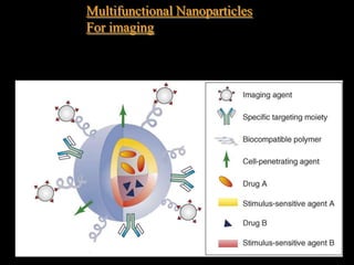
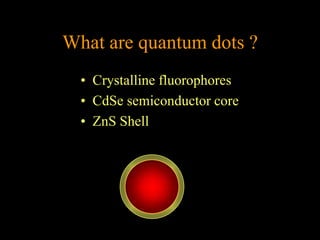
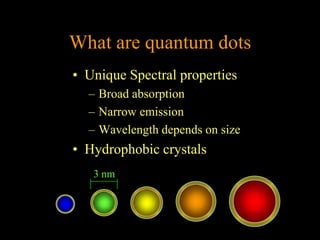
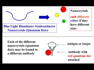
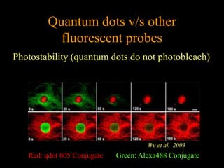
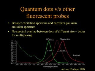
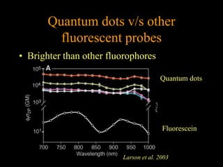
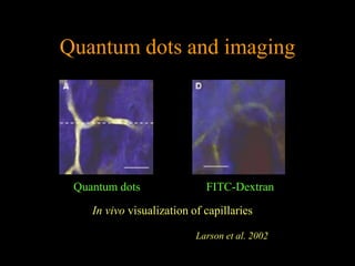
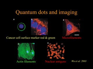
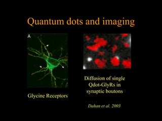
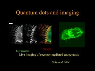
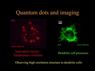
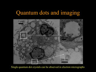
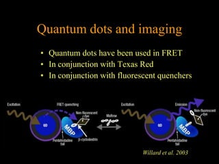
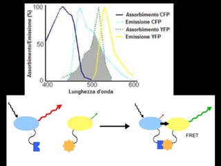
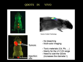
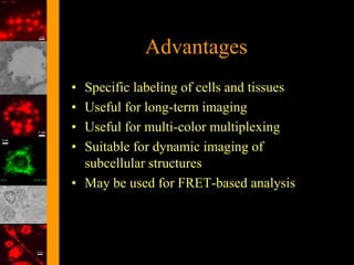
Imaging in vitro
Quantum dots as tools
 for cellular imaging
What are quantum dots ?
  • Crystalline fluorophores
  • CdSe semiconductor core
  • ZnS Shell
What are quantum dots
• Unique Spectral properties
  – Broad absorption
  – Narrow emission
  – Wavelength depends on size
• Hydrophobic crystals
   3 nm
Making hydrophobic quantum
      dots bio-compatible
• Various methods for making them
  water-soluble
  – Derivatizing surface with
    mercaptoacetic acid
  – Encapsulating in phospholipid
    micelles or liposomes
  – Coating them with amine-modified
    polyacrylic acid
Conjugating quantum dots to
         biomolecules
                              Avidin

• Avidin or protein-G with
  positively charged tail
  conjugated to negatively
  charged DHLA coat of
  quantum dots

                  protein G
Quantum dots v/s other
           fluorescent probes
Photostability (quantum dots do not photobleach)


                                      Wu et al. 2003
   Red: qdot 605 Conjugate   Green: Alexa488 Conjugate
Quantum dots v/s other
           fluorescent probes
• Broader excitation spectrum and narrower gaussian
  emission spectrum
• No spectral overlap between dots of different size – better
  for multiplexing


                                          Jaiswal & Simon 2004
Quantum dots v/s other
         fluorescent probes
• Brighter than other fluorophores

                                       Quantum dots


                                        Fluorescein


                         Larson et al. 2003
Quantum dots and imaging


 Quantum dots                FITC-Dextran

    In vivo visualization of capillaries

                           Larson et al. 2002
Quantum dots and imaging


Cancer cell surface marker red & green     Microfilaments


       Actin filaments        Nuclear antigens   Wu et al. 2003
Quantum dots and imaging


                    Diffusion of single
                      Qdot-GlyRs in
                     synaptic boutons
Glycine Receptors
                     Dahan et al. 2003
Quantum dots and imaging


                   EGF-QD
EGF receptor
  Live imaging of receptor mediated endocytosis

                            Lidke et al. 2004
Quantum dots and imaging


 Individual vesicles -             Dendritic cell processes
Temperature variations

 Observing high resolution structure in dendritic cells
Quantum dots and imaging
      1 m


                                             200 nm


                                             200 nm


Single quantum dot crystals can be observed in electron micrographs
Quantum dots and imaging
• Quantum dots have been used in FRET
• In conjunction with Texas Red
• In conjunction with fluorescent quenchers


                             Willard et al. 2003
QDOTS   IN   VIVO
Advantages
• Specific labeling of cells and tissues
• Useful for long-term imaging
• Useful for multi-color multiplexing
• Suitable for dynamic imaging of
  subcellular structures
• May be used for FRET-based analysis
Disadvantages
• Colloidal polymer-coated quantum dots can
  aggregate irreversibly
• Toxic in vivo
• Quantum dots are bulkier than many organic
  fluorophores
   – Accessibility issues
   – Mobility issues
• Cannot diffuse through cell membrane
   – Use of invasive techniques may change physiology
Imaging in vivo
X-raydense,
fluorescent,
metallic,
or magnetic cores
Multifunctional Nanoparticles
For imaging


                                25
X-ray CT

CT is ubiquitous in the clinical setting as. The increasing use and
development of micro-CT and hybrid systems that with
PET, MRI.
The most investigated NPs in this field are gold NPs, since they
have large absorption coefficients against the x-ray source used
for CT imaging and may increase the signal-to-noise ratio of the
technique.
To date, different types of gold NPs have been tested in a
preclinical setting as contrast agents for molecular imaging:
nanospheres, nanocages, nanorods and nanoshells. Gold NPs
formulations as an injectable imaging agent have been utilized to
study the distribution in rodent brain ex vivo
nanocage
                              nanorod


                              nanosphere
   nanoshell
               Size 4-40 nm
MRI
several nanotechnological approaches have been devised, based on the idea of
carrying a substantial payload of Gd chelates. Examples include liposomes
micelles dendrimers fullerenes. However, this approach has not yet achieved
clinical applications.

To this end, magnetic NPs (MNPs) are of considerable interest because they
may behave either as contrast agents or carriers for drug delivery. Among these,
the most promising and developed NP system is represented
bysuperparamagnetic iron oxide agents
PET
The strategy utilized is consisting in incorporating
PET emitters within the components of the NP, or
entrapping them within the core. Oku et al (2011)
employed PET to image brain cancer using positron-
emitting labelled liposomes in rats. Plotkin et al
(2006) used PET radioisotopes for targeting the intra-
tumourally injected magnetic NPs in patients with
glioblastoma.
Magnetic Nanoparticles


                         31
Types of Magnets
• Ferromagnetic materials: the magnetic moments of
  neighboring atoms align resulting in a net magnetic moment.

• Paramagnetic materials are randomly oriented due to Brownian
   motion, except in the presence of external magnetic field.


                                                                 32
Superparamagnetic
• Combination of paramagnetic and ferromagnetic
  properties
   – Made of nano-sized ferrous
   magnetic particles, but affected by Brownian Motion.
• They will align in the presence of an external magnetic
  field.
The most promising and developed NP system is
represented by superparamagnetic iron oxide
agents, consisting of a magnetite (Fe3O4) and/or
maghemite (Fe2O3) crystalline core surrounded by a
low molecular weight carbohydrate (usually dextran
or carboxydextran) or polymer coat.
. Iron oxide NPs can be classified according to their
core structure, such as Monocrystalline (MION;
10–30 nm diameter), or according to their size as
ultra-small superparamagnetic (USPIO) (20–50 nm
diameter), superparamagnetic (SPIO) (60–250 nm).
Dextran Coated Magnetite Nanoparticles
• Synthesis of polysaccharide covered superparamagnetic
  oxide colloids
   –   For MRI imaging


• FDA max size for injectables = 220 nm.

• Smaller sizes (<100 nm) have longer plasma half-life.
   –   Blood clearance by Reticuloendothelial system (RES)
   –   Liver and Spleen


• Without coating, opsonin proteins deposit on               Magnetite
  and mark for removal by RES
Formation of Nanoparticles
• Solution of Dextran and Ferric hexahydrate (acidic
  solution)
   –     Less Dextran Larger Particles
• Drip in Ammonium hydroxide (basic) at ~2oC
• Stirred at 75oC for 75 min.
• Purified by washing and
  ultra-centrifugation
• Resulting Size ~ 10-20 nm
• Plasma half-life: 200 min


                                                       37
Variation of Formation
• Change Coating Material
  –      Various other starches, Sulfated Dextran (for
      functionalization)


• Crosslinking coating material
  –      Increases plasma half-life
  –      Same Particle Size


                                                    38
Magnetite Cationic Liposomes (MCL)
      Fe3O4

• Why Cationic?
  –    Interaction between + liposome and – cell
  –    membrane results in 10x uptake.
Formation of MCL
• magnetite NP dispersed in distilled water
• N-( -trimethyl-amminoacetyl)-didodecyl-D-
  glutamate chloride (TMAG)
  Dilauroylphosphatidylcholine (DLPC)
  Dioleoylphosphatidyl-ethanolamine (DOPE)
  added to dispersion at ratio of 1:2:2
• Stirred and sonicated for 15 min
• pH raised to 7.4 by NaCl and Na phosphate
  buffered and then sonicated
Uses of Nano Magnets
• Hyperthermia
   – An oscillating magnetic field on nanomagnets result
    in local heating by (1) hysteresis, (2) frictional losses
    (3) Neel or Brown relaxation
Cancer Treatment


       • Heating due to magnetic field results in two
         possibilities
             Death due to overheating
             Increase in heat shock
             proteins result in
             anti-cancer immunity.
                                                                                44
Ito A., Honda H., Kobayashi T. Cancer Immunol Immunother Res 2006 55; 320-328
Delivery Magnetic nanoparticles
     • Magnetite nanoparticles
       encapsulated in liposomes
        – (1) Antibody conjugated
          (AML)
        – (2) Positive Surface Charge
          (MCL)
     • Sprague-Dawley rats injected with
       two human tumors.
        – Liposomes injected into 1
        tumor (black) and applied
        Alternating Magnetic Field


Ito A., Honda H., Kobayashi T. Cancer Immunol Immunother Res 2006 55; 320-328   45
Effect of Hyperthermia


Treated
Tumor
                                         Before Treatment
Untreated
Tumor

                                Rectum
• Non-local heating in body is the
  result of eddy-currents
   – The currents resulting from the      After Treatment
     magnetic field produce heat
Uses of Nano Magnets

• MRI imaging.
Iron oxide agents shorten T2 and T2* relaxation times on T2-
and T2*-weighted MRI images, creating low signal or
negative contrast. They can also be detected by MRI with
T1, off resonance, and steady-state free precession sequences
Uses of Nano Magnets

• External Magnetic field for nanoparticle delivery
   – Magnetic nanoparticles loaded with
   drug can be directed to diseased site for
   Drug Delivery or MRI imaging.
Magnetic Drug Delivery System


• Using Magnetic Nanoparticles for Drug
  Delivery
  • Widder & others developed method in late 1970s
  • Drug loaded magnetic nanoparticles introduced through IV or IA
    injection and directed with External Magnets
  • Requires smaller dosage because of targeting, resulting in fewer side
    effects
                                                                       51
Magnetic Nanoparticles/Carriers
                                                    M
                                                                      M
• Magnetite Core                               M


• Starch Polymer Coating                   M
                                                        Magnetite
                                                          Core
                                                                          M

     • Bioavailable
                                                    Starch Polymer
     • Phosphate in coating for functionalization
                                                                     M
• Chemo Drug attached to Coating                    M

     • Mitoxantrone
• Drug Delivered to Rabbit with Carcinoma


                                                                     52
Results of Drug Delivery


• External magnetic
  field (dark)
     • deliver more
       nanoparticles to tumor
• No magnetic field
  (white)
     • most nanoparticles in
       non tumor regions

                                    53
Magnetic nanoparticles in medicine
They consist of a metal or metallic oxide core, encapsulated in
an inorganic or a polymeric coating, that renders the particles
biocompatible, stable, and may serve as a support for
biomolecules.


• Drug or therapeutic radionuclide is bound to a magnetic
NP, introduced in the body, and then concentrated in the target
area by means of a magnetic field.
• Depending on the application, the particles release the drug or
give rise to a local effect (hyperthermia).
• Drug release can proceed by simple diffusion or take place
through mechanisms requiring enzymatic activity or changes in
physiological conditions (pH, osmolality, temperature, etc…).
Multifunctional Magnetic Nanoparticles


•   Magnetic nanocrystals as ultrasensitive MR contrast agents: MnFe2O4
•   Anticancer drugs as chemotherapeutic agents: doxorubicin, DOX
•   Amphiphilic block copolymers as stabilizers: PLGA-PEG
•   Antibodies to target cancer cells: anti-HER antibody (HER, herceptin)
    conjugated by carboxyl group on the surface of the MMPNs
                                                                                55
                                   Yang, etal. Angew. Chem. 2007, 119, 8992 –8995.
Magnetic nanoparticles
 The application of magnetic nanoparticles in cancer therapy is
 one of the most successful biomedical exploitations of
 nanotechnology.
 The efficacy of the particles in the treatment depends upon the
 specific targeting capacity of the nanoparticles to the cancer cells.
 Efficient, surface-engineered magnetic nanoparticles open up new
 possibilities for their therapeutic potential.


… effective conjugation of folic acid on the surface of
superparamagnetic iron oxide nanoparticles (SPION) enables their
high intracellular uptake by cancer cells.
Such magnetic-folate conjugate nanoparticles are stable for a long
time over a wide biological pH range: additionally, such particles
show remarkably low phagocytosis as verified with peritoneal
macrophages.
Conclusions
• Nanomagnets can be made bioavailable by
  liposomal encapsulation with targeting
• Nanoparticles smaller than 20 nm can be useful
  for local heat generation
• Intracellular hyperthermia kills the cancer cell
  and releases heat shock proteins. These are used
  to target and kill other cancer cells.
• Results in reduction in growth of tumor size
• Nanomagnets can be used for MR Imaging in
  vivo                                          57
MICROBUBBLES
• Used with ultrasound echocardiography and
  magnetic resonance imaging (MRI)
• Diagnostic imaging - Traces blood flow and
  outlines images


• Drug Delivery and Cancer Therapy
• Small (1-7 m) bubbles of air (CO2, Helium) or
  high molecular weight gases (perfluorocarbon).
• Enveloped by a shell (proteins, fatty acid esters).
• Exist - For a limited time only! 4 minutes-24
  hours; gases diffuse into liquid medium after
  use.
• Size varies according to Ideal Gas Law
  (PV=nRT) and thickness of shell.
ultrasound
• Ultrasound uses high frequency sound
  waves to image internal structures
• The wave reflects off different density
  liquids and tissues at different rates and
  magnitudes
• It is harmless, but not very accurate
Ultrasound and Microbubbles
• Air in microbubbles in the blood stream
  have almost 0 density and have a distinct
  reflection in ultrasound
• The bubbles must be able to fit through all
  capillaries and remain stable
Shell


Air or High Molecular
Weight Gases

                        1-   m
Preparation of microbubbles

  1. Water
  2. Fluorinated hydrocarbon
  3. Polymer solution
  4. Ethanol   palmitic   acid   solution   with
    Epikuron® 200
  5. Homogeneization for 10 min at 12000 rpm
O2 microbubbles coated with PAAs
  Cationic PAA                    PAA-cholesterol


  Diameter = 549.5 ± 94.7 nm   Diameter = 491.4 ± 38.2 nm
  PZ      = 8.54±1.21          PZ      = 6.22±1.17
  pH      = 3.28               pH      = 6.50
Application of microbubble technology for ultrasound
imaging of the heart

7 imaging

  • 1.
  • 2.
    Quantum dots astools for cellular imaging
  • 3.
    What are quantumdots ? • Crystalline fluorophores • CdSe semiconductor core • ZnS Shell
  • 4.
    What are quantumdots • Unique Spectral properties – Broad absorption – Narrow emission – Wavelength depends on size • Hydrophobic crystals 3 nm
  • 5.
    Making hydrophobic quantum dots bio-compatible • Various methods for making them water-soluble – Derivatizing surface with mercaptoacetic acid – Encapsulating in phospholipid micelles or liposomes – Coating them with amine-modified polyacrylic acid
  • 6.
    Conjugating quantum dotsto biomolecules Avidin • Avidin or protein-G with positively charged tail conjugated to negatively charged DHLA coat of quantum dots protein G
  • 8.
    Quantum dots v/sother fluorescent probes Photostability (quantum dots do not photobleach) Wu et al. 2003 Red: qdot 605 Conjugate Green: Alexa488 Conjugate
  • 9.
    Quantum dots v/sother fluorescent probes • Broader excitation spectrum and narrower gaussian emission spectrum • No spectral overlap between dots of different size – better for multiplexing Jaiswal & Simon 2004
  • 10.
    Quantum dots v/sother fluorescent probes • Brighter than other fluorophores Quantum dots Fluorescein Larson et al. 2003
  • 11.
    Quantum dots andimaging Quantum dots FITC-Dextran In vivo visualization of capillaries Larson et al. 2002
  • 12.
    Quantum dots andimaging Cancer cell surface marker red & green Microfilaments Actin filaments Nuclear antigens Wu et al. 2003
  • 13.
    Quantum dots andimaging Diffusion of single Qdot-GlyRs in synaptic boutons Glycine Receptors Dahan et al. 2003
  • 14.
    Quantum dots andimaging EGF-QD EGF receptor Live imaging of receptor mediated endocytosis Lidke et al. 2004
  • 15.
    Quantum dots andimaging Individual vesicles - Dendritic cell processes Temperature variations Observing high resolution structure in dendritic cells
  • 16.
    Quantum dots andimaging 1 m 200 nm 200 nm Single quantum dot crystals can be observed in electron micrographs
  • 17.
    Quantum dots andimaging • Quantum dots have been used in FRET • In conjunction with Texas Red • In conjunction with fluorescent quenchers Willard et al. 2003
  • 19.
    QDOTS IN VIVO
  • 20.
    Advantages • Specific labelingof cells and tissues • Useful for long-term imaging • Useful for multi-color multiplexing • Suitable for dynamic imaging of subcellular structures • May be used for FRET-based analysis
  • 21.
    Disadvantages • Colloidal polymer-coatedquantum dots can aggregate irreversibly • Toxic in vivo • Quantum dots are bulkier than many organic fluorophores – Accessibility issues – Mobility issues • Cannot diffuse through cell membrane – Use of invasive techniques may change physiology
  • 22.
  • 24.
  • 25.
  • 27.
    X-ray CT CT isubiquitous in the clinical setting as. The increasing use and development of micro-CT and hybrid systems that with PET, MRI. The most investigated NPs in this field are gold NPs, since they have large absorption coefficients against the x-ray source used for CT imaging and may increase the signal-to-noise ratio of the technique. To date, different types of gold NPs have been tested in a preclinical setting as contrast agents for molecular imaging: nanospheres, nanocages, nanorods and nanoshells. Gold NPs formulations as an injectable imaging agent have been utilized to study the distribution in rodent brain ex vivo
  • 28.
    nanocage nanorod nanosphere nanoshell Size 4-40 nm
  • 29.
    MRI several nanotechnological approacheshave been devised, based on the idea of carrying a substantial payload of Gd chelates. Examples include liposomes micelles dendrimers fullerenes. However, this approach has not yet achieved clinical applications. To this end, magnetic NPs (MNPs) are of considerable interest because they may behave either as contrast agents or carriers for drug delivery. Among these, the most promising and developed NP system is represented bysuperparamagnetic iron oxide agents
  • 30.
    PET The strategy utilizedis consisting in incorporating PET emitters within the components of the NP, or entrapping them within the core. Oku et al (2011) employed PET to image brain cancer using positron- emitting labelled liposomes in rats. Plotkin et al (2006) used PET radioisotopes for targeting the intra- tumourally injected magnetic NPs in patients with glioblastoma.
  • 31.
  • 32.
    Types of Magnets •Ferromagnetic materials: the magnetic moments of neighboring atoms align resulting in a net magnetic moment. • Paramagnetic materials are randomly oriented due to Brownian motion, except in the presence of external magnetic field. 32
  • 33.
    Superparamagnetic • Combination ofparamagnetic and ferromagnetic properties – Made of nano-sized ferrous magnetic particles, but affected by Brownian Motion. • They will align in the presence of an external magnetic field.
  • 34.
    The most promisingand developed NP system is represented by superparamagnetic iron oxide agents, consisting of a magnetite (Fe3O4) and/or maghemite (Fe2O3) crystalline core surrounded by a low molecular weight carbohydrate (usually dextran or carboxydextran) or polymer coat. . Iron oxide NPs can be classified according to their core structure, such as Monocrystalline (MION; 10–30 nm diameter), or according to their size as ultra-small superparamagnetic (USPIO) (20–50 nm diameter), superparamagnetic (SPIO) (60–250 nm).
  • 36.
    Dextran Coated MagnetiteNanoparticles • Synthesis of polysaccharide covered superparamagnetic oxide colloids – For MRI imaging • FDA max size for injectables = 220 nm. • Smaller sizes (<100 nm) have longer plasma half-life. – Blood clearance by Reticuloendothelial system (RES) – Liver and Spleen • Without coating, opsonin proteins deposit on Magnetite and mark for removal by RES
  • 37.
    Formation of Nanoparticles •Solution of Dextran and Ferric hexahydrate (acidic solution) – Less Dextran Larger Particles • Drip in Ammonium hydroxide (basic) at ~2oC • Stirred at 75oC for 75 min. • Purified by washing and ultra-centrifugation • Resulting Size ~ 10-20 nm • Plasma half-life: 200 min 37
  • 38.
    Variation of Formation •Change Coating Material – Various other starches, Sulfated Dextran (for functionalization) • Crosslinking coating material – Increases plasma half-life – Same Particle Size 38
  • 39.
    Magnetite Cationic Liposomes(MCL) Fe3O4 • Why Cationic? – Interaction between + liposome and – cell – membrane results in 10x uptake.
  • 40.
    Formation of MCL •magnetite NP dispersed in distilled water • N-( -trimethyl-amminoacetyl)-didodecyl-D- glutamate chloride (TMAG) Dilauroylphosphatidylcholine (DLPC) Dioleoylphosphatidyl-ethanolamine (DOPE) added to dispersion at ratio of 1:2:2 • Stirred and sonicated for 15 min • pH raised to 7.4 by NaCl and Na phosphate buffered and then sonicated
  • 41.
    Uses of NanoMagnets • Hyperthermia – An oscillating magnetic field on nanomagnets result in local heating by (1) hysteresis, (2) frictional losses (3) Neel or Brown relaxation
  • 44.
    Cancer Treatment • Heating due to magnetic field results in two possibilities Death due to overheating Increase in heat shock proteins result in anti-cancer immunity. 44 Ito A., Honda H., Kobayashi T. Cancer Immunol Immunother Res 2006 55; 320-328
  • 45.
    Delivery Magnetic nanoparticles • Magnetite nanoparticles encapsulated in liposomes – (1) Antibody conjugated (AML) – (2) Positive Surface Charge (MCL) • Sprague-Dawley rats injected with two human tumors. – Liposomes injected into 1 tumor (black) and applied Alternating Magnetic Field Ito A., Honda H., Kobayashi T. Cancer Immunol Immunother Res 2006 55; 320-328 45
  • 46.
    Effect of Hyperthermia Treated Tumor Before Treatment Untreated Tumor Rectum • Non-local heating in body is the result of eddy-currents – The currents resulting from the After Treatment magnetic field produce heat
  • 47.
    Uses of NanoMagnets • MRI imaging. Iron oxide agents shorten T2 and T2* relaxation times on T2- and T2*-weighted MRI images, creating low signal or negative contrast. They can also be detected by MRI with T1, off resonance, and steady-state free precession sequences
  • 50.
    Uses of NanoMagnets • External Magnetic field for nanoparticle delivery – Magnetic nanoparticles loaded with drug can be directed to diseased site for Drug Delivery or MRI imaging.
  • 51.
    Magnetic Drug DeliverySystem • Using Magnetic Nanoparticles for Drug Delivery • Widder & others developed method in late 1970s • Drug loaded magnetic nanoparticles introduced through IV or IA injection and directed with External Magnets • Requires smaller dosage because of targeting, resulting in fewer side effects 51
  • 52.
    Magnetic Nanoparticles/Carriers M M • Magnetite Core M • Starch Polymer Coating M Magnetite Core M • Bioavailable Starch Polymer • Phosphate in coating for functionalization M • Chemo Drug attached to Coating M • Mitoxantrone • Drug Delivered to Rabbit with Carcinoma 52
  • 53.
    Results of DrugDelivery • External magnetic field (dark) • deliver more nanoparticles to tumor • No magnetic field (white) • most nanoparticles in non tumor regions 53
  • 54.
    Magnetic nanoparticles inmedicine They consist of a metal or metallic oxide core, encapsulated in an inorganic or a polymeric coating, that renders the particles biocompatible, stable, and may serve as a support for biomolecules. • Drug or therapeutic radionuclide is bound to a magnetic NP, introduced in the body, and then concentrated in the target area by means of a magnetic field. • Depending on the application, the particles release the drug or give rise to a local effect (hyperthermia). • Drug release can proceed by simple diffusion or take place through mechanisms requiring enzymatic activity or changes in physiological conditions (pH, osmolality, temperature, etc…).
  • 55.
    Multifunctional Magnetic Nanoparticles • Magnetic nanocrystals as ultrasensitive MR contrast agents: MnFe2O4 • Anticancer drugs as chemotherapeutic agents: doxorubicin, DOX • Amphiphilic block copolymers as stabilizers: PLGA-PEG • Antibodies to target cancer cells: anti-HER antibody (HER, herceptin) conjugated by carboxyl group on the surface of the MMPNs 55 Yang, etal. Angew. Chem. 2007, 119, 8992 –8995.
  • 56.
    Magnetic nanoparticles Theapplication of magnetic nanoparticles in cancer therapy is one of the most successful biomedical exploitations of nanotechnology. The efficacy of the particles in the treatment depends upon the specific targeting capacity of the nanoparticles to the cancer cells. Efficient, surface-engineered magnetic nanoparticles open up new possibilities for their therapeutic potential. … effective conjugation of folic acid on the surface of superparamagnetic iron oxide nanoparticles (SPION) enables their high intracellular uptake by cancer cells. Such magnetic-folate conjugate nanoparticles are stable for a long time over a wide biological pH range: additionally, such particles show remarkably low phagocytosis as verified with peritoneal macrophages.
  • 57.
    Conclusions • Nanomagnets canbe made bioavailable by liposomal encapsulation with targeting • Nanoparticles smaller than 20 nm can be useful for local heat generation • Intracellular hyperthermia kills the cancer cell and releases heat shock proteins. These are used to target and kill other cancer cells. • Results in reduction in growth of tumor size • Nanomagnets can be used for MR Imaging in vivo 57
  • 58.
    MICROBUBBLES • Used withultrasound echocardiography and magnetic resonance imaging (MRI) • Diagnostic imaging - Traces blood flow and outlines images • Drug Delivery and Cancer Therapy
  • 59.
    • Small (1-7m) bubbles of air (CO2, Helium) or high molecular weight gases (perfluorocarbon). • Enveloped by a shell (proteins, fatty acid esters). • Exist - For a limited time only! 4 minutes-24 hours; gases diffuse into liquid medium after use. • Size varies according to Ideal Gas Law (PV=nRT) and thickness of shell.
  • 60.
    ultrasound • Ultrasound useshigh frequency sound waves to image internal structures • The wave reflects off different density liquids and tissues at different rates and magnitudes • It is harmless, but not very accurate
  • 61.
    Ultrasound and Microbubbles •Air in microbubbles in the blood stream have almost 0 density and have a distinct reflection in ultrasound • The bubbles must be able to fit through all capillaries and remain stable
  • 62.
    Shell Air or HighMolecular Weight Gases 1- m
  • 63.
    Preparation of microbubbles 1. Water 2. Fluorinated hydrocarbon 3. Polymer solution 4. Ethanol palmitic acid solution with Epikuron® 200 5. Homogeneization for 10 min at 12000 rpm
  • 64.
    O2 microbubbles coatedwith PAAs Cationic PAA PAA-cholesterol Diameter = 549.5 ± 94.7 nm Diameter = 491.4 ± 38.2 nm PZ = 8.54±1.21 PZ = 6.22±1.17 pH = 3.28 pH = 6.50
  • 65.
    Application of microbubbletechnology for ultrasound imaging of the heart