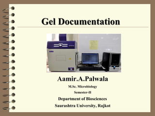
Gel Documentation

Aamir.A.Palwala
M.Sc. Microbiology

Semester-II

Department of Biosciences
Saurashtra University, Rajkot
INTRODUCTION
Gel documentation , also known as gel doc. system , gel image

system , or gel imager.

It is widely used in molecular biology laboratories for imaging and

documentation of nucleic acid and protein polyacrylamide or
agarose gels typically stained with ethidium bromide or flurophores
such as SYBR green.
Generally , a gel doc. is composed of ultraviolet ( UV ) light

transilluminator , a hood to shield external light sources and CCTV
camera for image capturing.
PRINCIPLE
Principle of fluorescence with fluorescent staining of

nucleic acids , a fluorescent substance that has bound to
nucleic acid is exited by ultraviolet irradiation and emits
fluorescent light.
Ethidium bromide binds specifically to nucleic acid and

the amount of bonding depends on the molecular weight
and concentration of the nucleic acid.
In other words , a band for a large amount will shine

brighter ; conversely , fluorescence will be weaker for a
band for a small amount.
PURPOSE AND ANALYSIS
Photography of stained gel.
Print out of photographic data.
Image data is displayed in

real time.
Image still displayed can be printed
out with a video printer or saved to a
compact flash media for future use.
COMPONENTS OF GEL
DOC. SYSTEM
Camera
Lenses

Filters

Overhead illuminator

Visible light
Adjustable stage

Darkroom

Tr ansilluminator
COMPONENTS OF GEL
DOC. SYSTEM
Camera : Ultraviolet camera ranging from 1.4m upto

8.3m

pixel.
Lenses : All lanses are computer controlled and motor

driven. Here lances automatically tracks upwards or
downwards movements of sample using auto focus.
Filters : There is an extensive range of emission filters

use for an array of applications.

Over head illuminator : White light , Ultra violet light ,

and LED lighting option

Visible light : For extending the transmitted light

application. Visible light convert screen for visible light
application.
COMPONENTS OF GEL
DOC. SYSTEM
Adjustable stage : This enables samples to be moved

closer to the camera.
Darkroom

: dark room is suitable for advanced
chemiluminescence, fluorescence and visible light
application. An electronic auto-door lock
with security
function prevents interruption to long exposures.
Transilluminator : It has a variable intensity setting and

has a safety cut-off.
QUANTITY ONE FROM
BIORED
It is a software which is required to operate gel doc. system.
It is user friendly version windows based option.
It has a various tool for image capturing and transformation.
There is a separate tool for identifying , detecting and drawing lense and bands.
Band attributes include band creation , GAUSS model bands , density of a

band , band boundries etc……
There is a separate function for storing standard data , standard curve for

known molecular weight and size.
A separate volume analysis tool for volume contour , free hand , rectangle ,

circle and array.
The software also has options for VNTR display , VNTR

calculation ,

differential display and colony counting applictions.
The software can generate reports for individual bands , all lenses , band types

, 1-dimension analysis , lane compaing , phylogenetic tree and other simmilarity
matrix options.
APPLICATION
1-D Electrophoresis Documentation
2-D Documentation
Protein Gel Documentation
Blotting technique analysis
Gel doc system

Gel doc system

  • 1.
  • 2.
    INTRODUCTION Gel documentation ,also known as gel doc. system , gel image system , or gel imager. It is widely used in molecular biology laboratories for imaging and documentation of nucleic acid and protein polyacrylamide or agarose gels typically stained with ethidium bromide or flurophores such as SYBR green. Generally , a gel doc. is composed of ultraviolet ( UV ) light transilluminator , a hood to shield external light sources and CCTV camera for image capturing.
  • 3.
    PRINCIPLE Principle of fluorescencewith fluorescent staining of nucleic acids , a fluorescent substance that has bound to nucleic acid is exited by ultraviolet irradiation and emits fluorescent light. Ethidium bromide binds specifically to nucleic acid and the amount of bonding depends on the molecular weight and concentration of the nucleic acid. In other words , a band for a large amount will shine brighter ; conversely , fluorescence will be weaker for a band for a small amount.
  • 4.
    PURPOSE AND ANALYSIS Photographyof stained gel. Print out of photographic data. Image data is displayed in real time. Image still displayed can be printed out with a video printer or saved to a compact flash media for future use.
  • 5.
    COMPONENTS OF GEL DOC.SYSTEM Camera Lenses Filters Overhead illuminator Visible light Adjustable stage Darkroom Tr ansilluminator
  • 6.
    COMPONENTS OF GEL DOC.SYSTEM Camera : Ultraviolet camera ranging from 1.4m upto 8.3m pixel. Lenses : All lanses are computer controlled and motor driven. Here lances automatically tracks upwards or downwards movements of sample using auto focus. Filters : There is an extensive range of emission filters use for an array of applications. Over head illuminator : White light , Ultra violet light , and LED lighting option Visible light : For extending the transmitted light application. Visible light convert screen for visible light application.
  • 7.
    COMPONENTS OF GEL DOC.SYSTEM Adjustable stage : This enables samples to be moved closer to the camera. Darkroom : dark room is suitable for advanced chemiluminescence, fluorescence and visible light application. An electronic auto-door lock with security function prevents interruption to long exposures. Transilluminator : It has a variable intensity setting and has a safety cut-off.
  • 8.
    QUANTITY ONE FROM BIORED Itis a software which is required to operate gel doc. system. It is user friendly version windows based option. It has a various tool for image capturing and transformation. There is a separate tool for identifying , detecting and drawing lense and bands. Band attributes include band creation , GAUSS model bands , density of a band , band boundries etc…… There is a separate function for storing standard data , standard curve for known molecular weight and size. A separate volume analysis tool for volume contour , free hand , rectangle , circle and array. The software also has options for VNTR display , VNTR calculation , differential display and colony counting applictions. The software can generate reports for individual bands , all lenses , band types , 1-dimension analysis , lane compaing , phylogenetic tree and other simmilarity matrix options.
  • 9.
    APPLICATION 1-D Electrophoresis Documentation 2-DDocumentation Protein Gel Documentation Blotting technique analysis