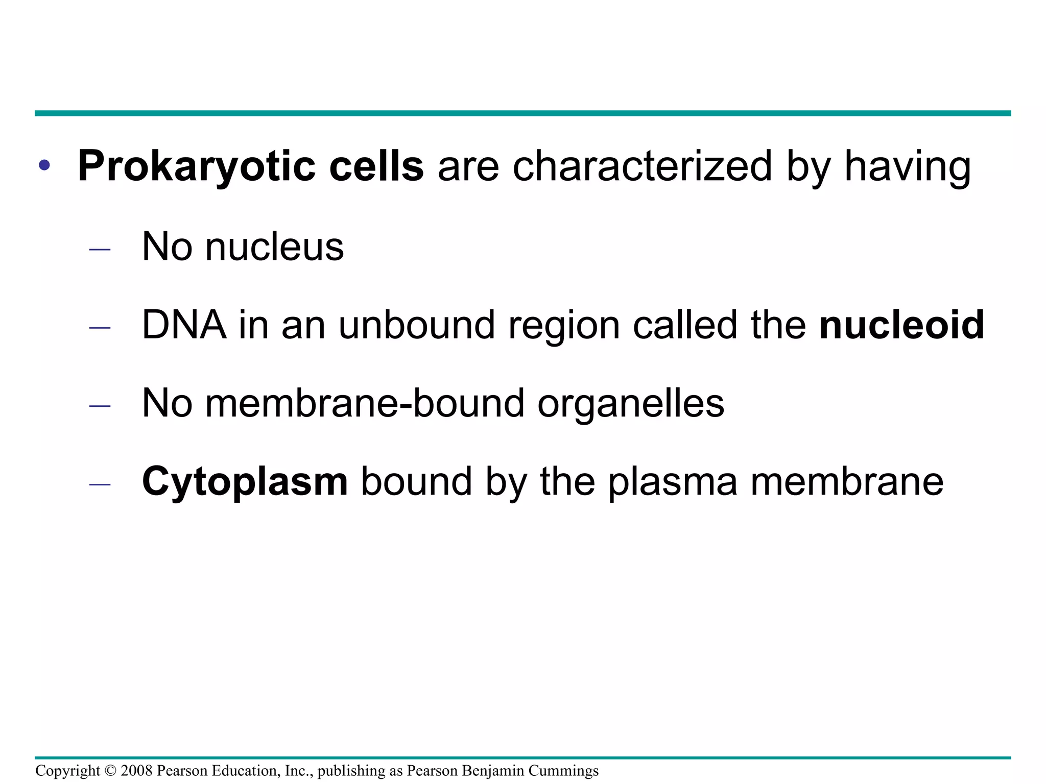
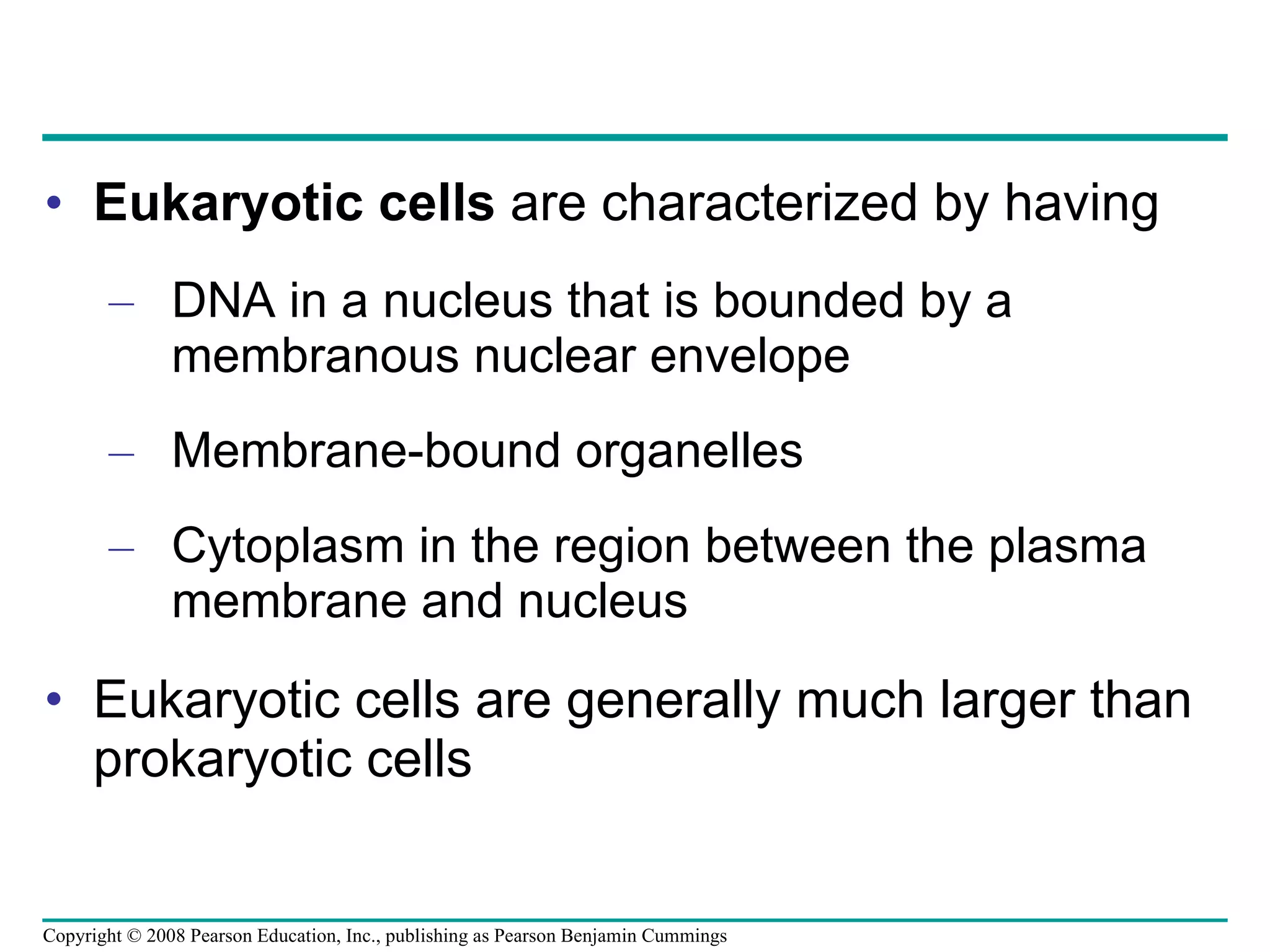
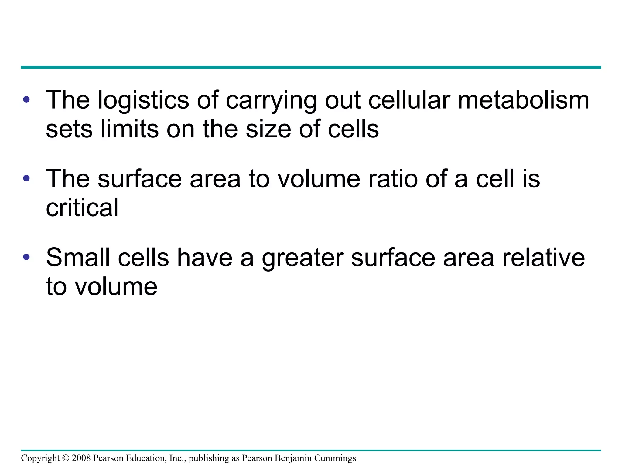
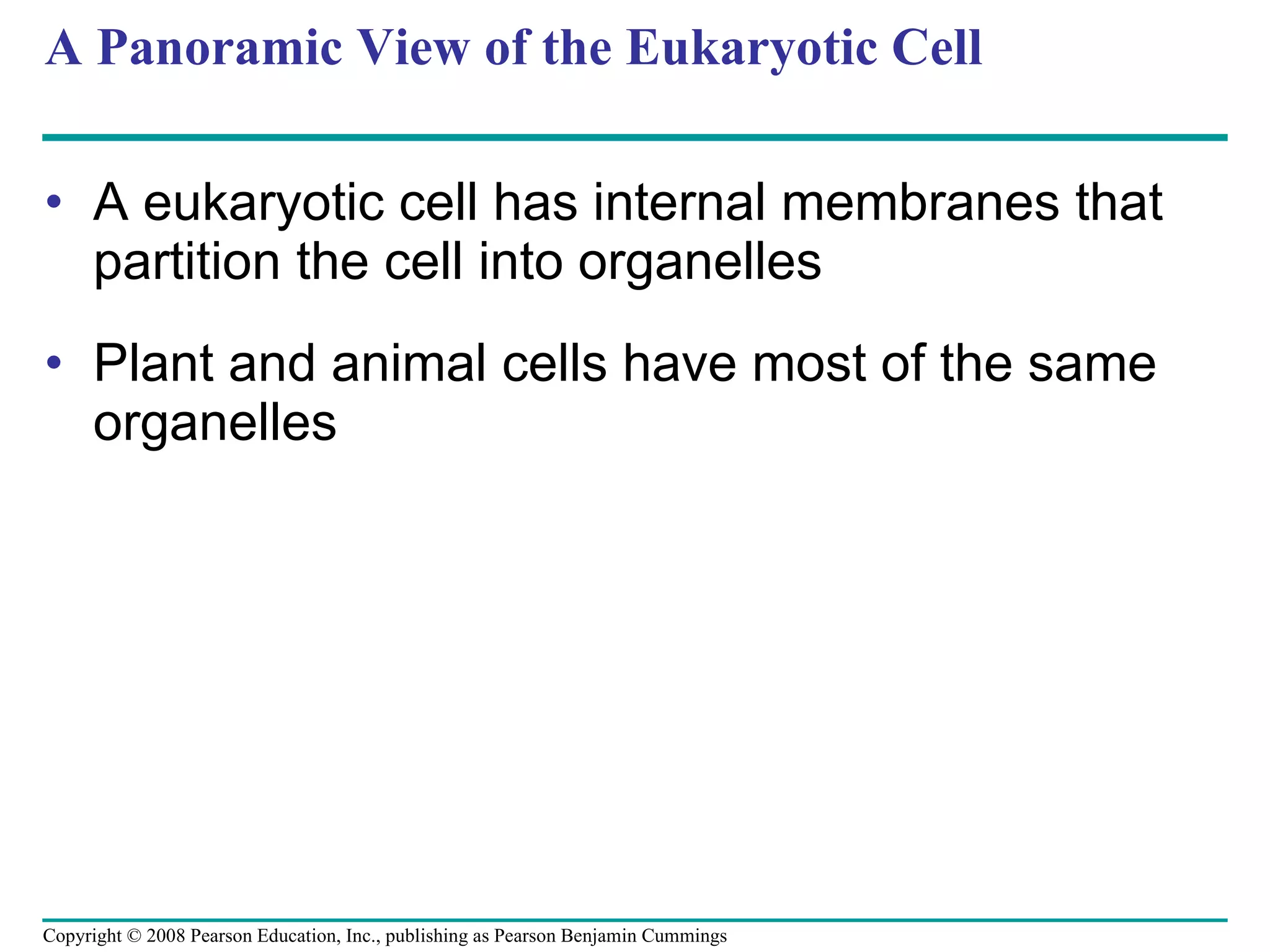
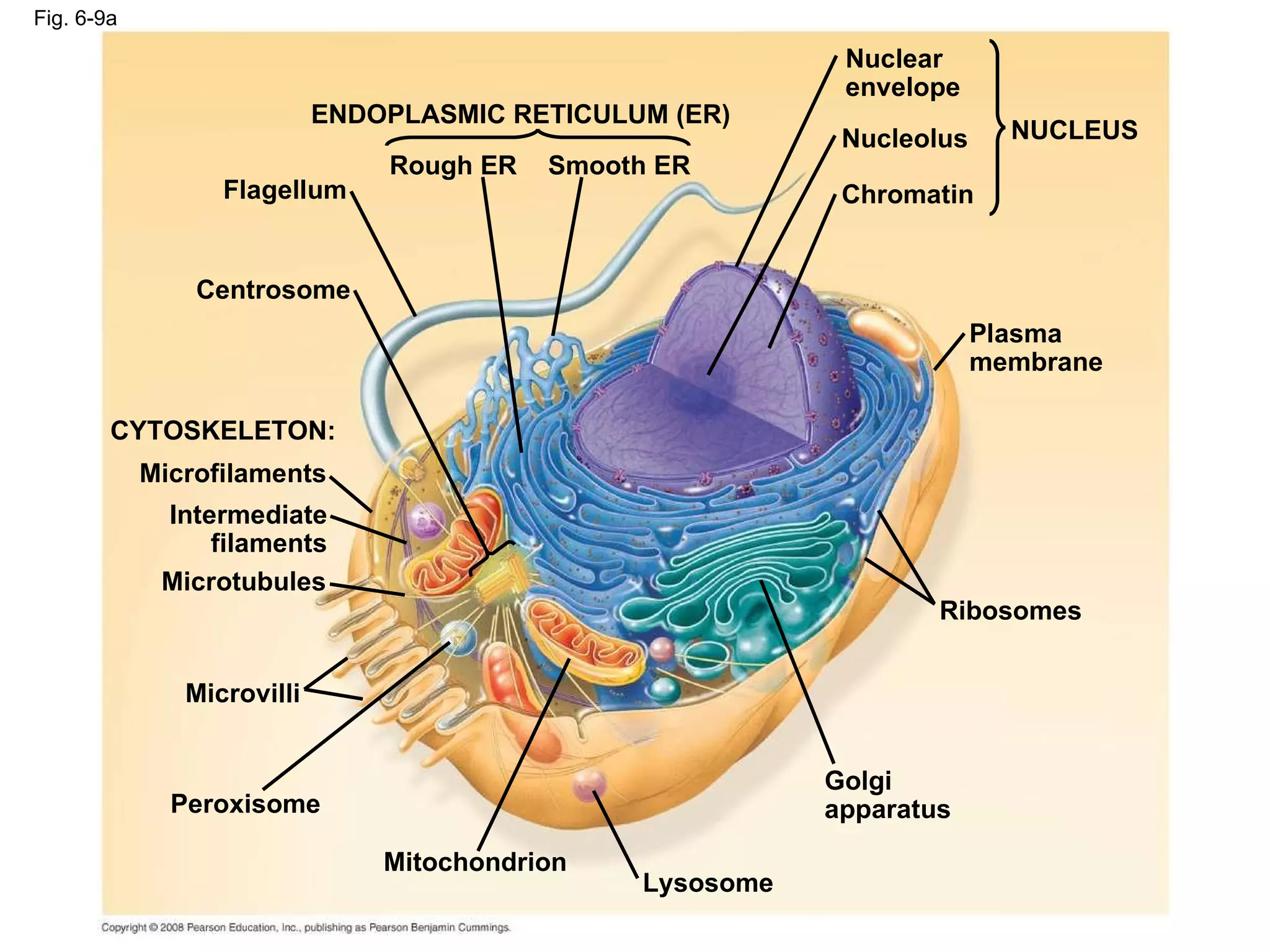
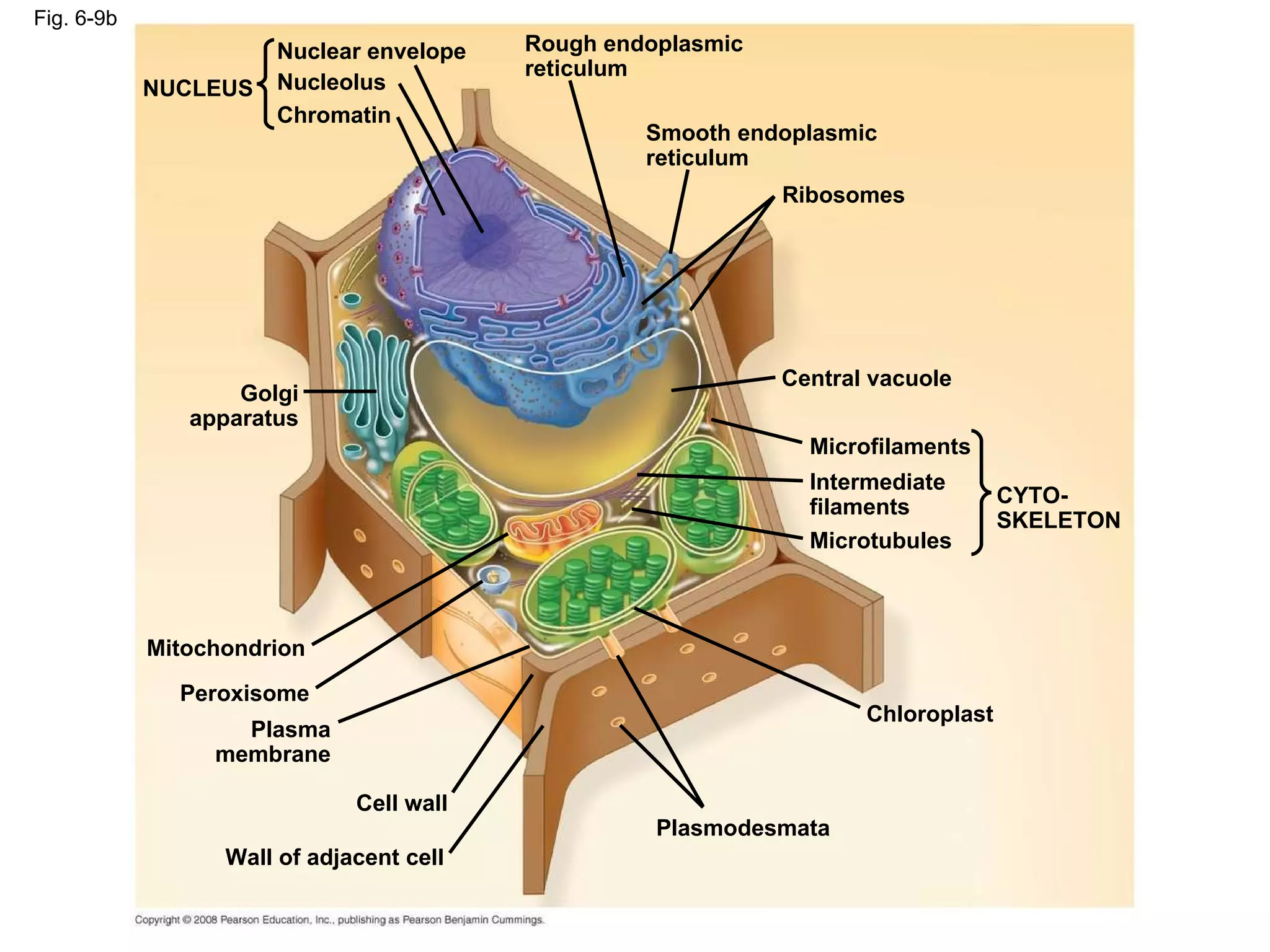
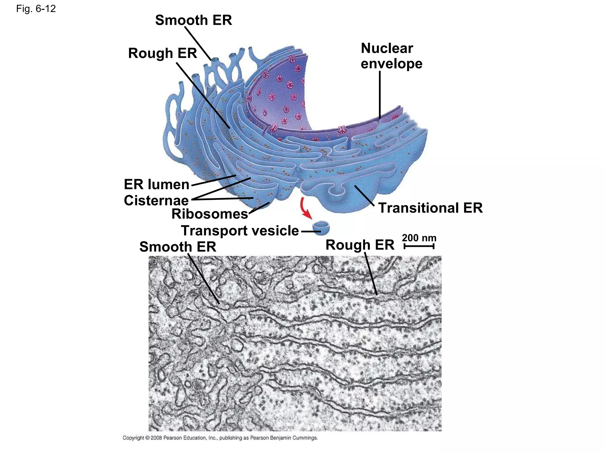
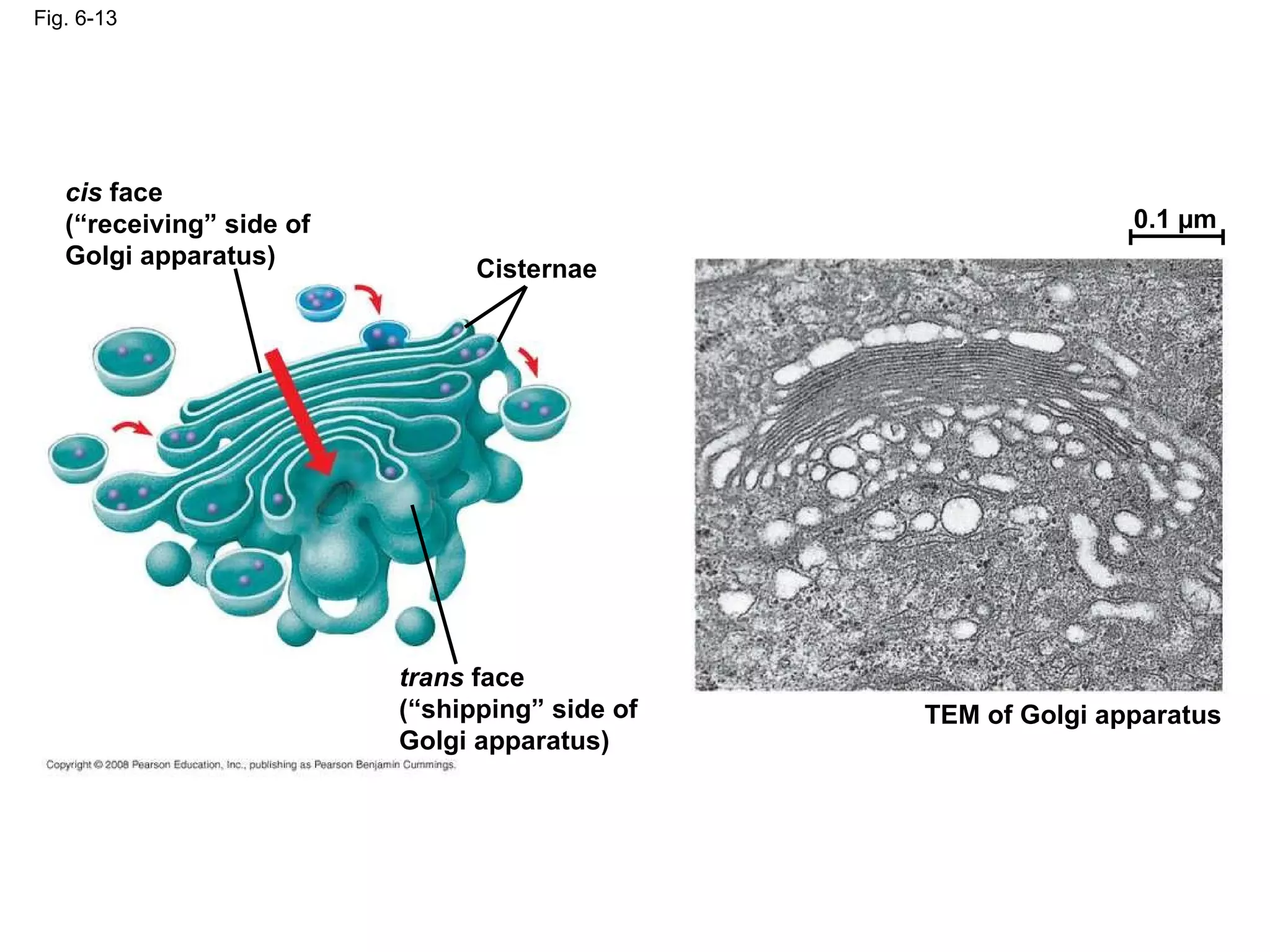
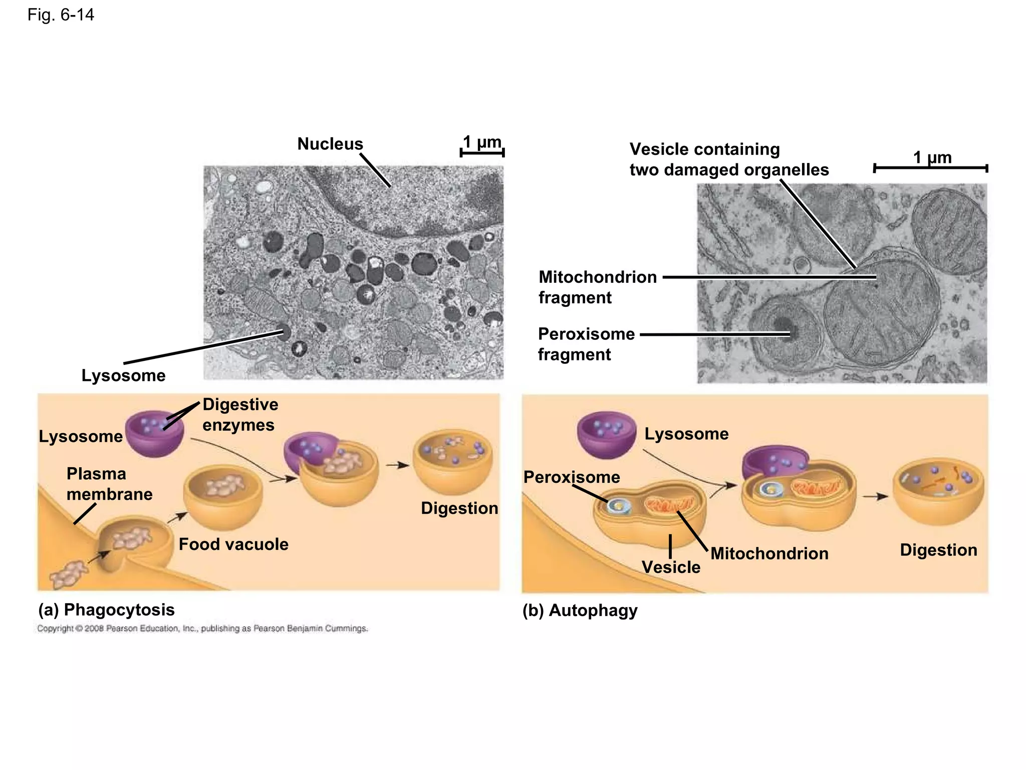
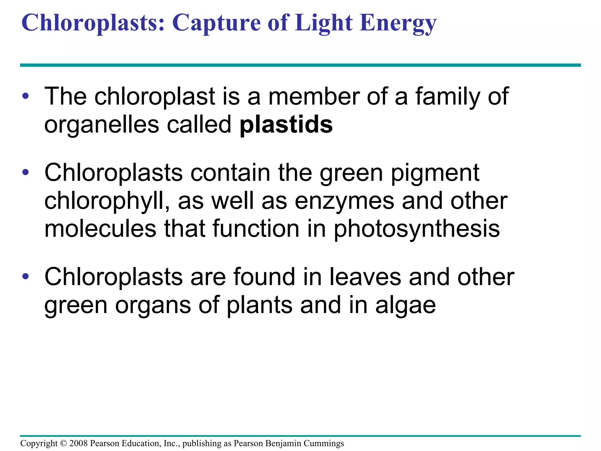
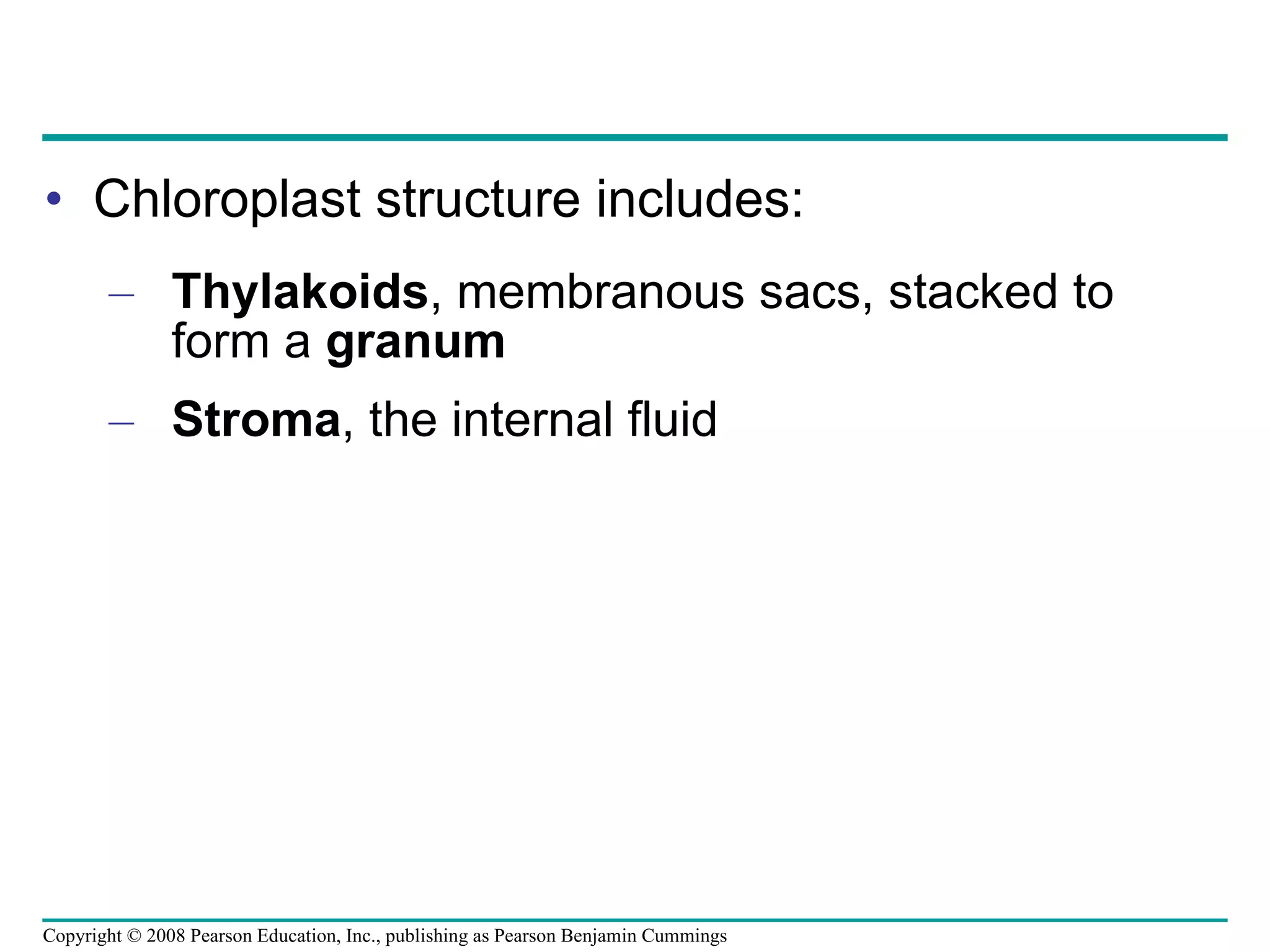
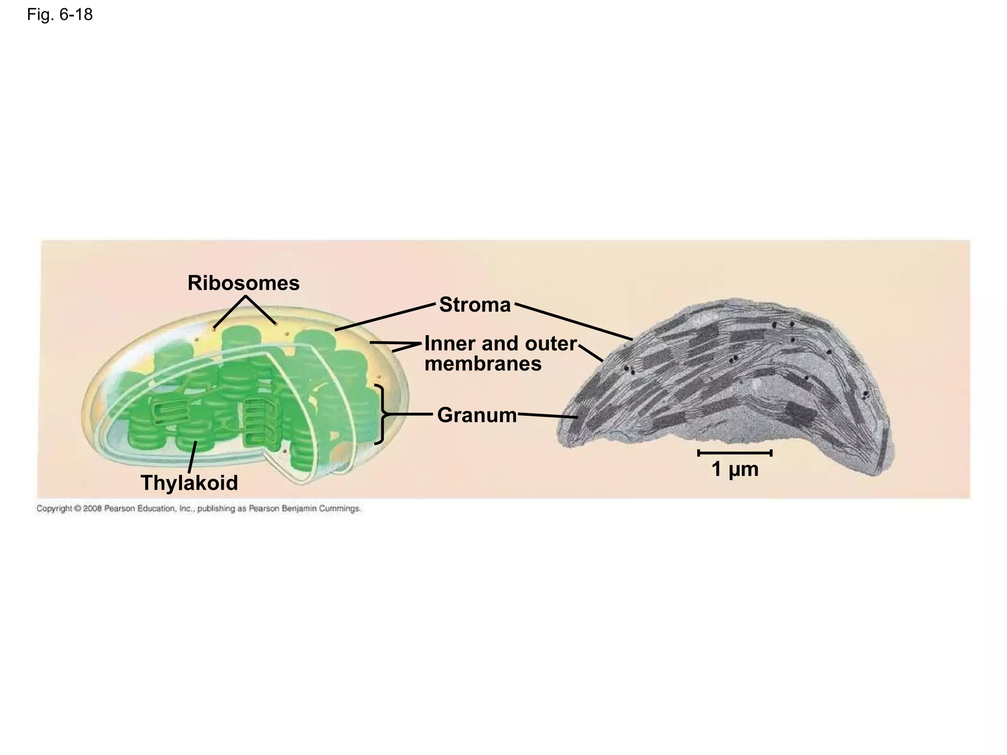
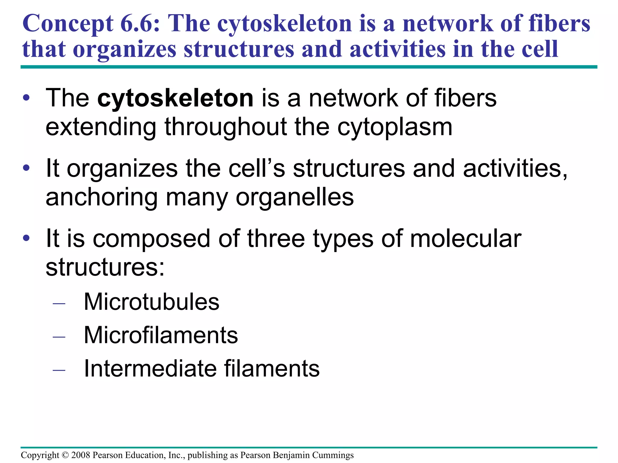
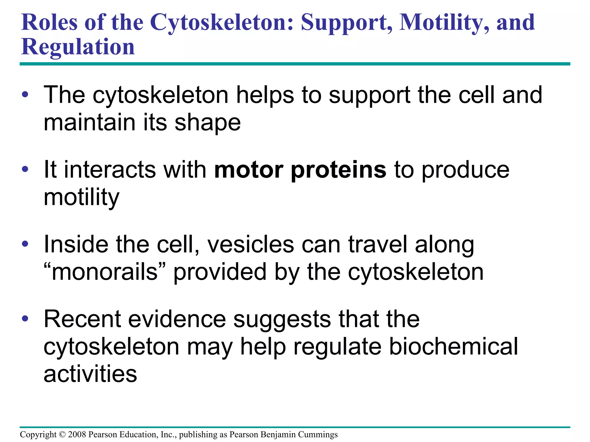
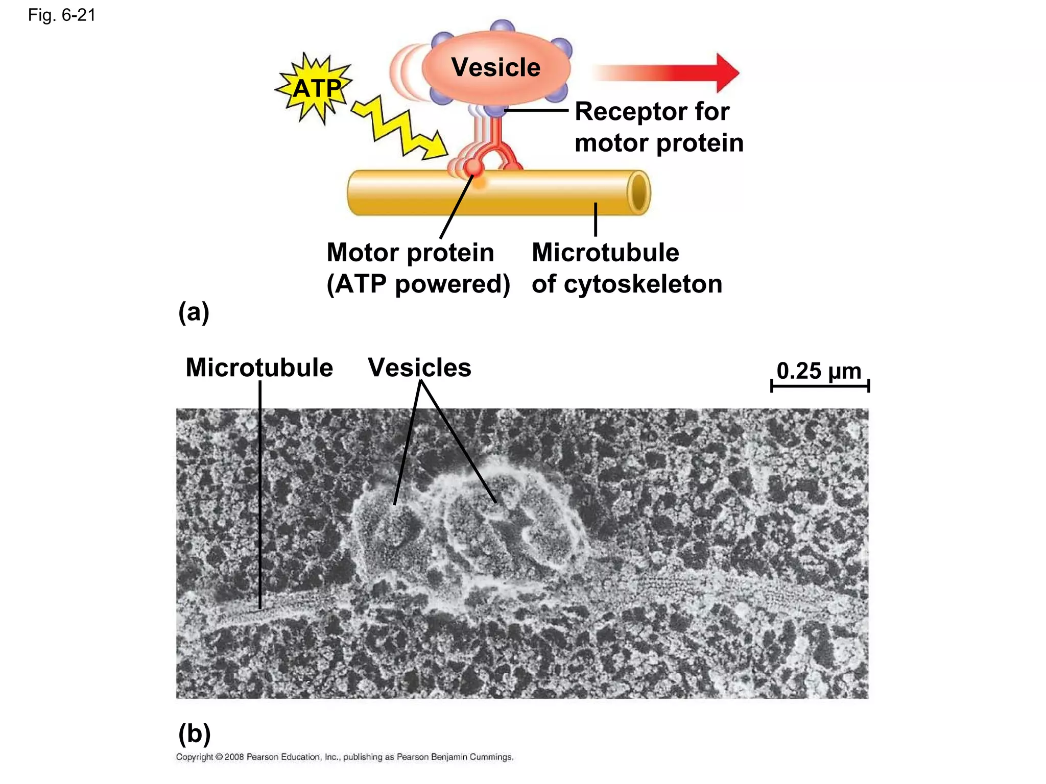
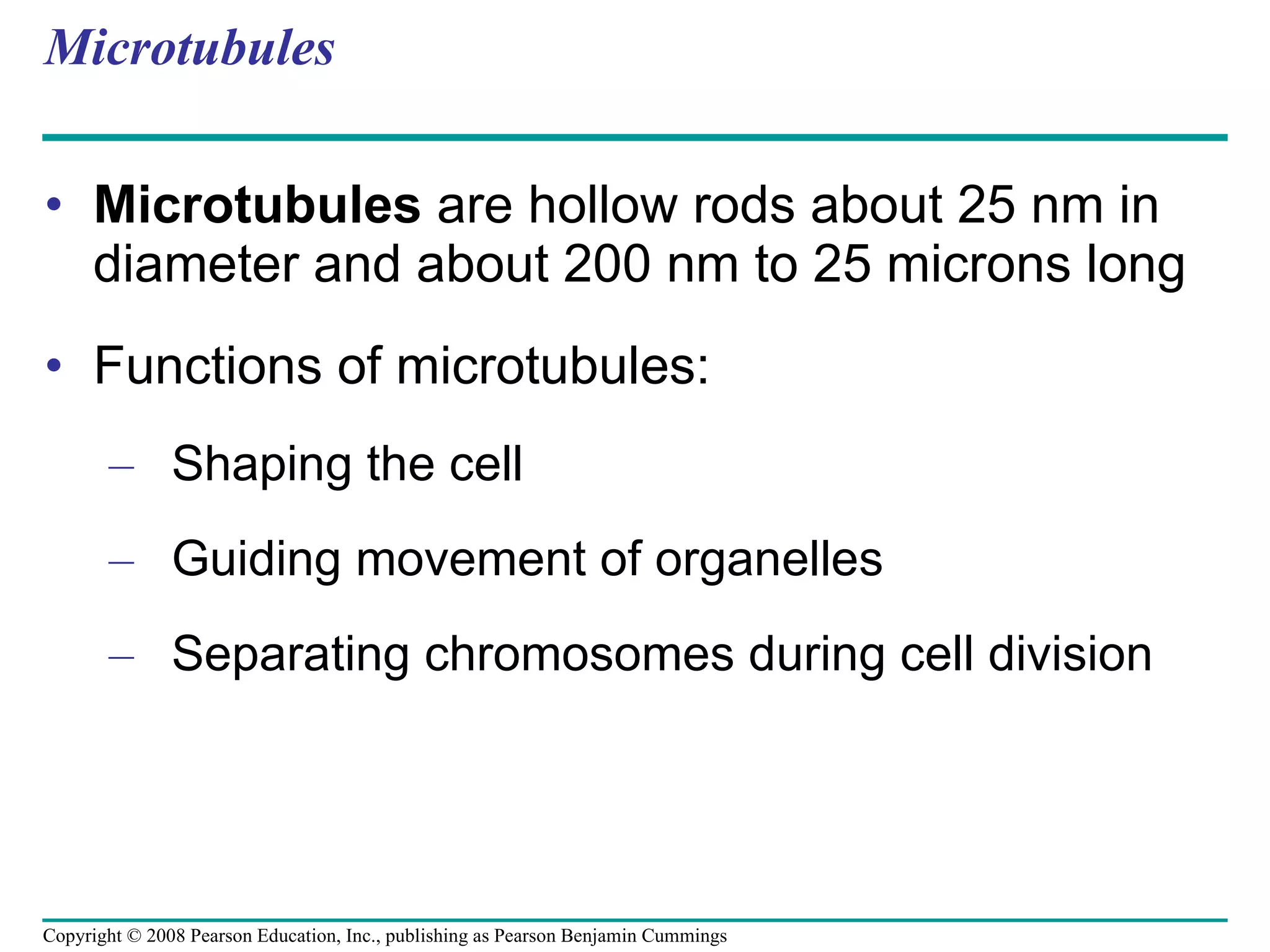
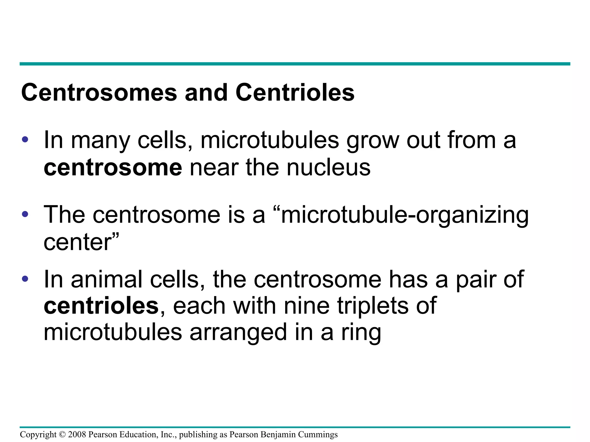
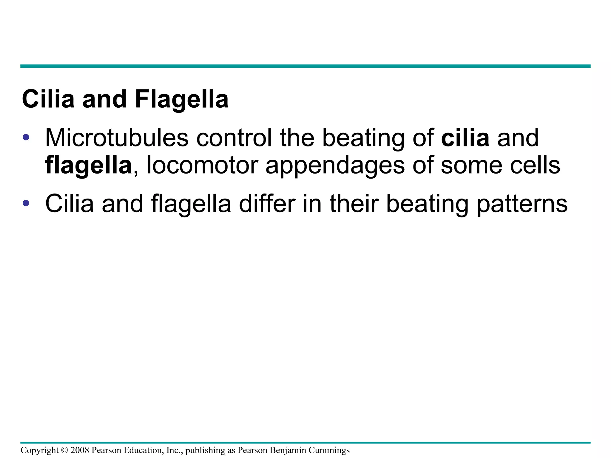
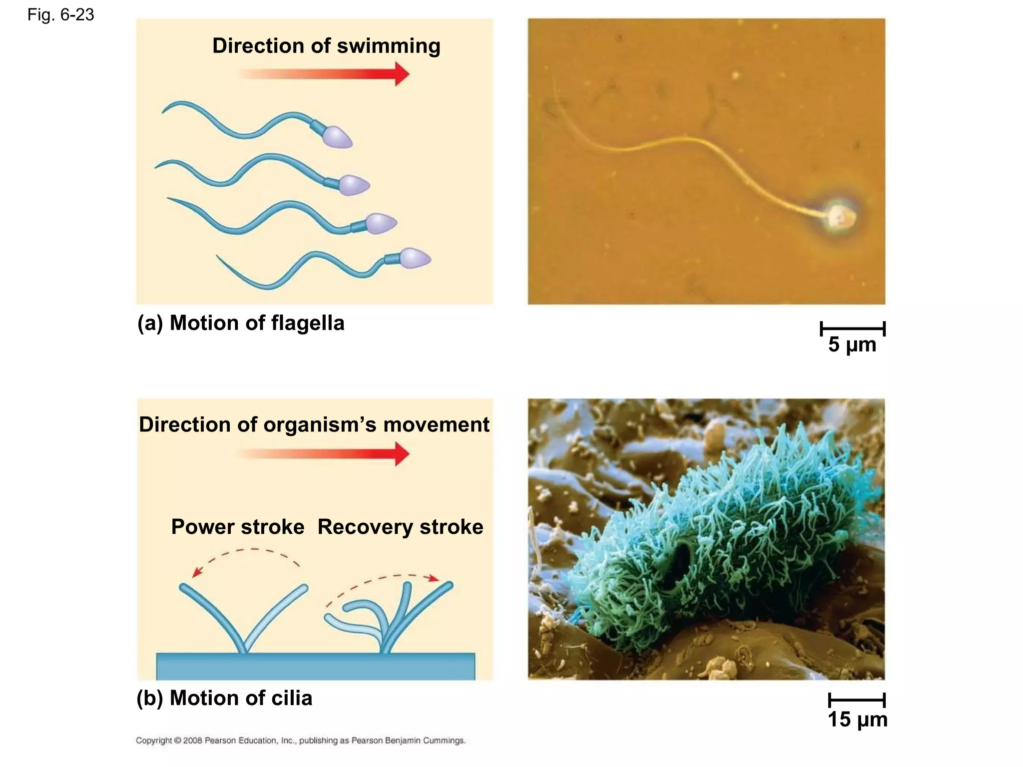
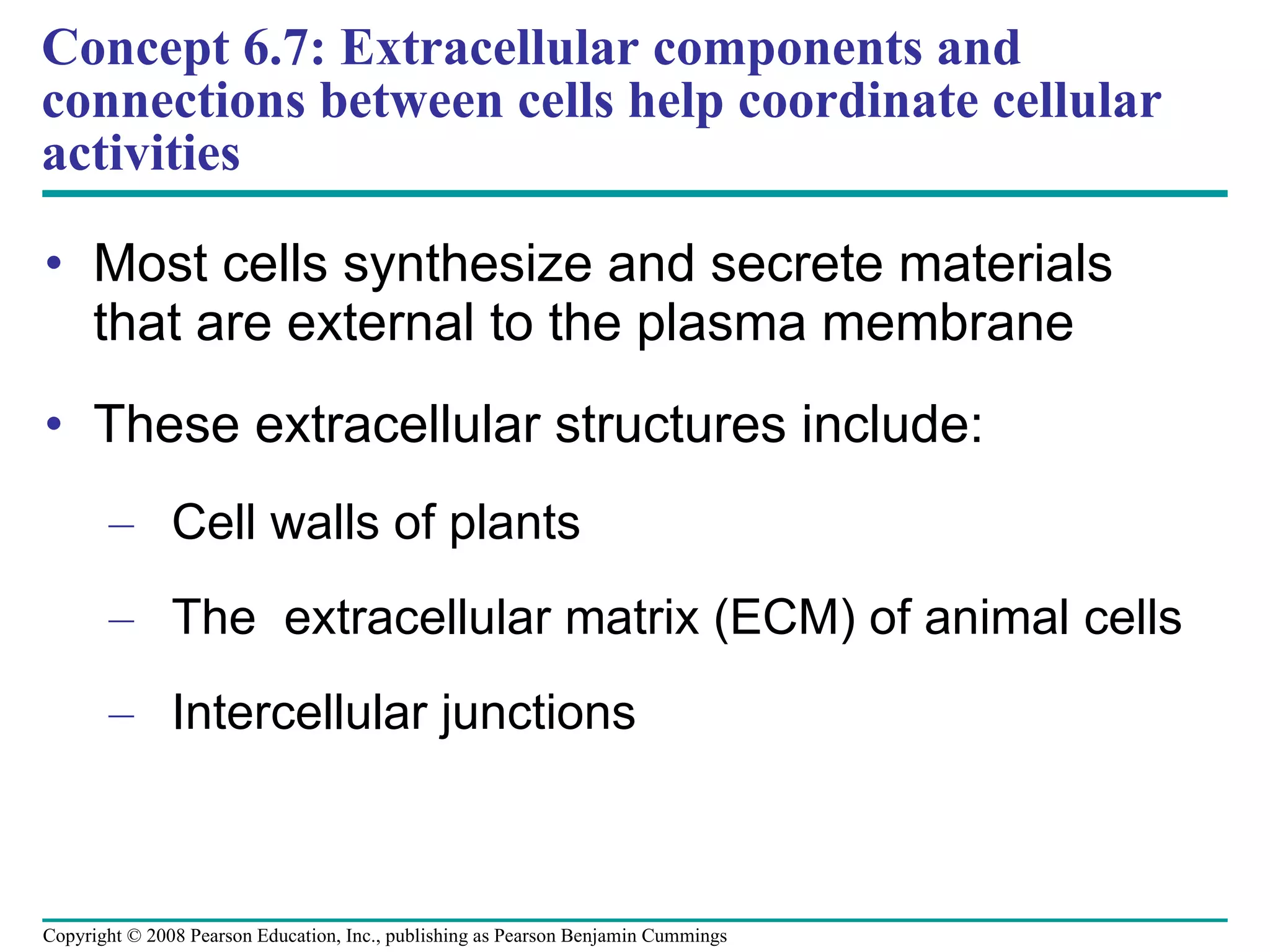
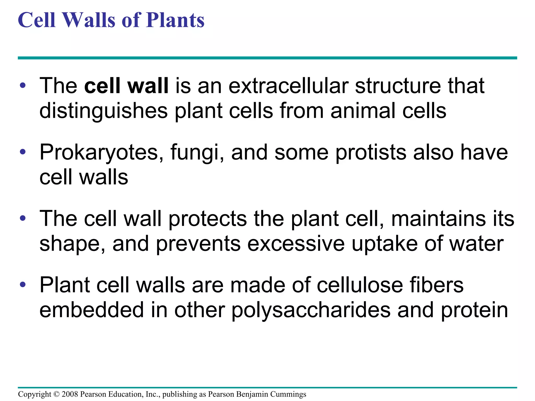
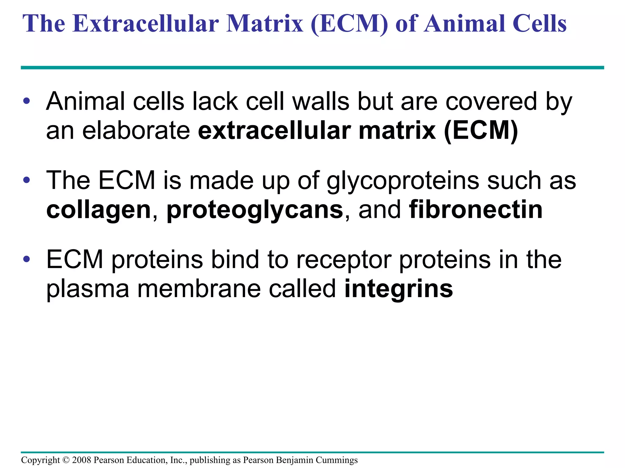
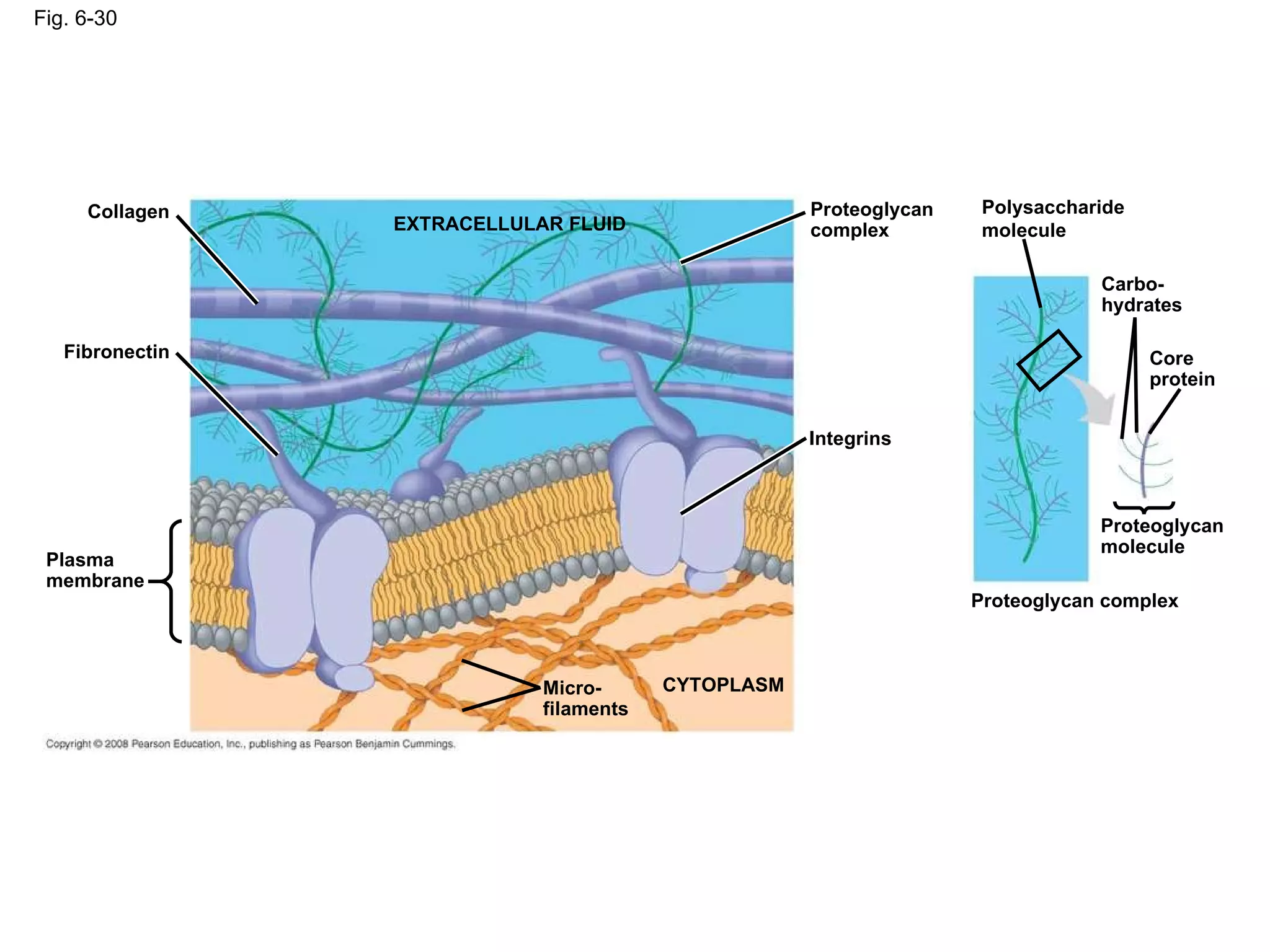
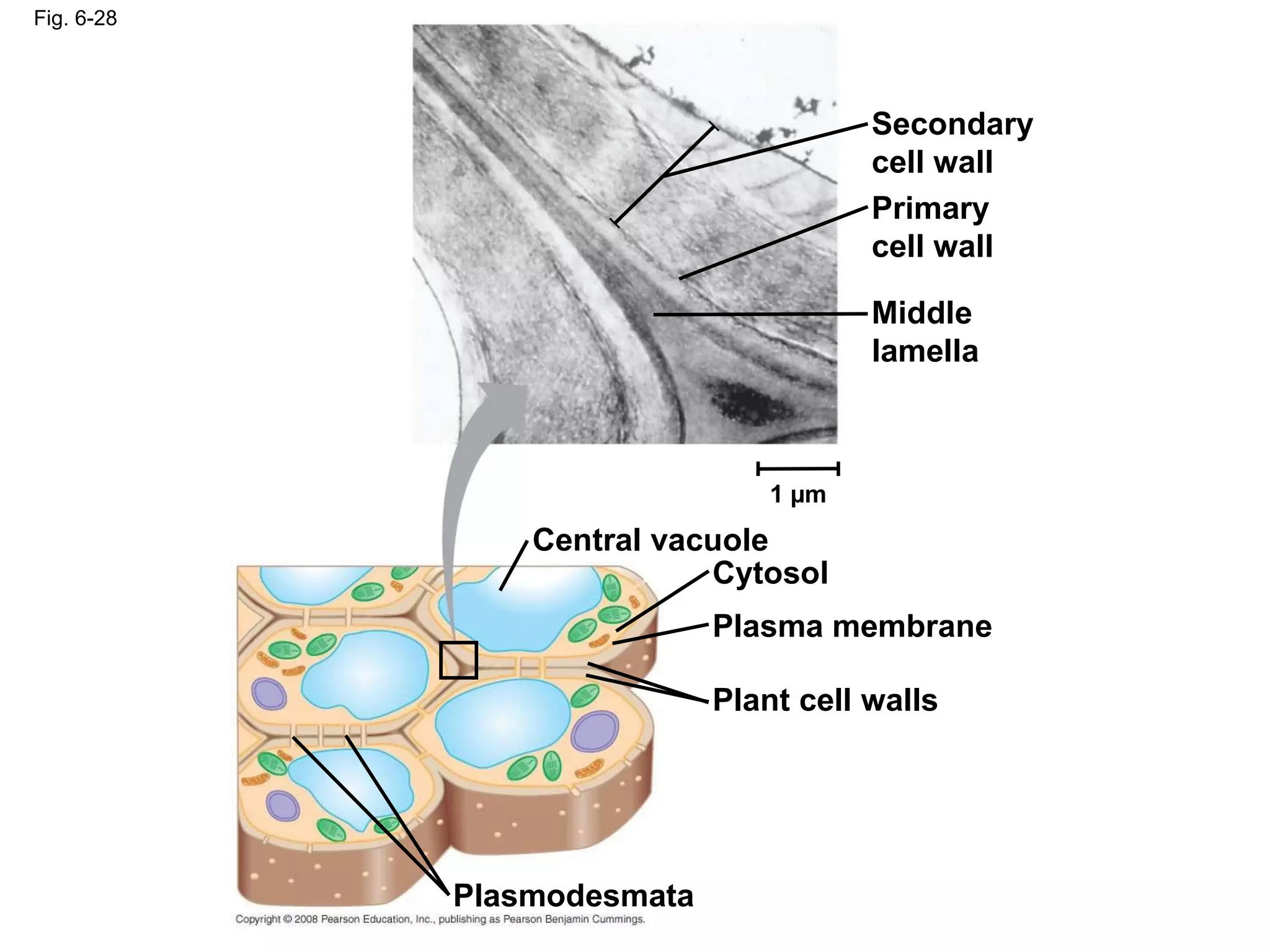
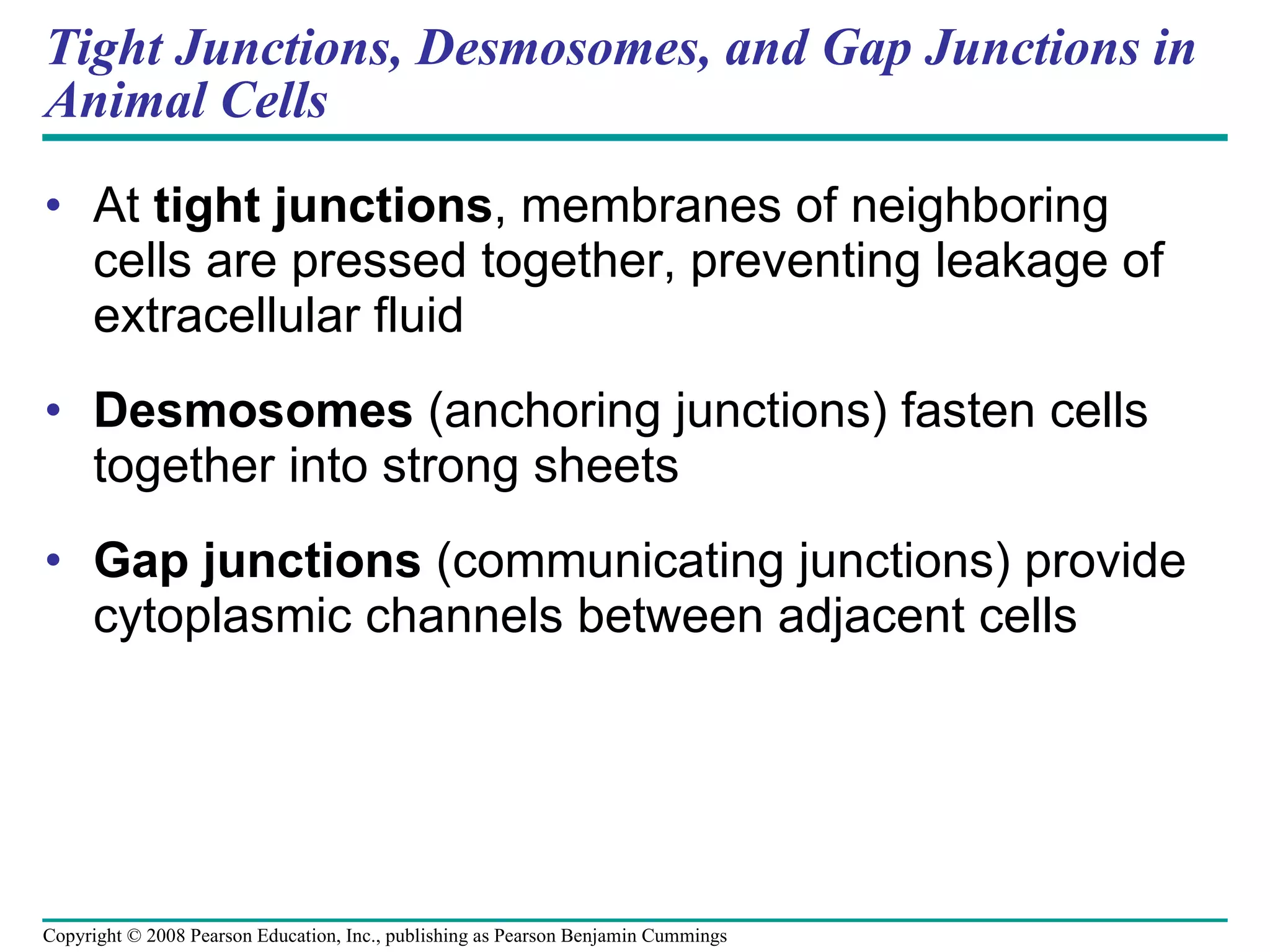
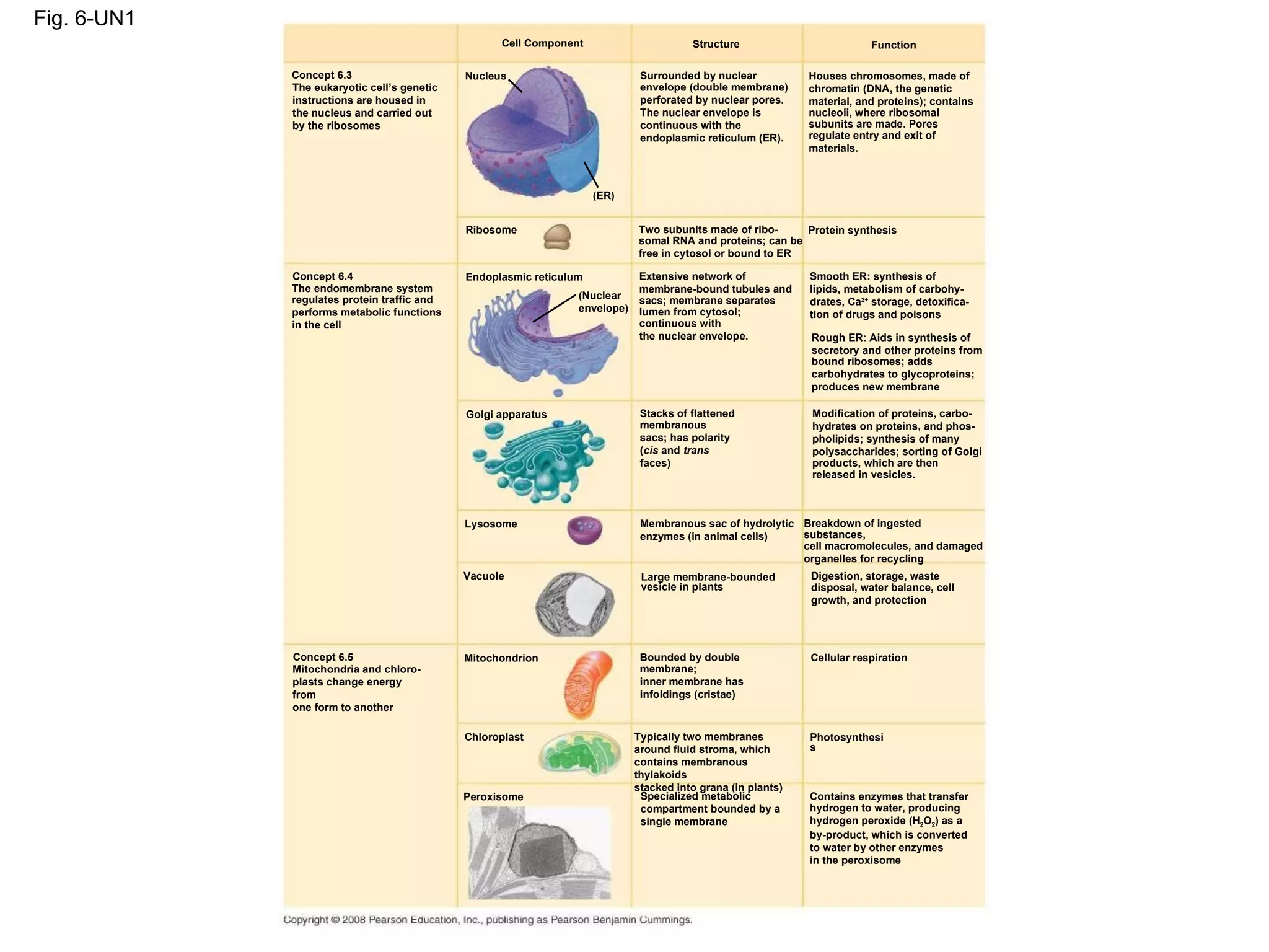
Chapter 6 A Tour of the Cell
Overview: The Fundamental Units of Life All organisms are made of cells The cell is the simplest collection of matter  that can live Cell structure is correlated to cellular function All cells are related by their descent from earlier cells Copyright © 2008 Pearson Education, Inc., publishing as Pearson Benjamin Cummings
Concept 6.2: Eukaryotic cells have internal membranes that compartmentalize their functions The basic structural and functional unit of every organism is one of two types of cells: prokaryotic or eukaryotic Only organisms of the domains Bacteria and Archaea consist of prokaryotic cells Protists, fungi, animals, and plants all consist of eukaryotic cells Copyright © 2008 Pearson Education, Inc., publishing as Pearson Benjamin Cummings
Comparing Prokaryotic and Eukaryotic Cells Basic features of all cells:  Plasma membrane Semifluid substance called  cytosol Chromosomes (carry genes) Ribosomes (make proteins) Copyright © 2008 Pearson Education, Inc., publishing as Pearson Benjamin Cummings
Prokaryotic cells  are characterized by having No nucleus DNA in an unbound region called the  nucleoid No membrane-bound organelles Cytoplasm  bound by the plasma membrane Copyright © 2008 Pearson Education, Inc., publishing as Pearson Benjamin Cummings
Eukaryotic cells  are characterized by having DNA in a nucleus that is bounded by a membranous nuclear envelope Membrane-bound organelles Cytoplasm in the region between the plasma membrane and nucleus Eukaryotic cells are generally much larger than prokaryotic cells Copyright © 2008 Pearson Education, Inc., publishing as Pearson Benjamin Cummings
The  plasma membrane  is a selective barrier that allows sufficient passage of oxygen, nutrients, and waste to service the volume of every cell The general structure of a biological membrane is a double layer of phospholipids Copyright © 2008 Pearson Education, Inc., publishing as Pearson Benjamin Cummings
Fig. 6-7 TEM of a plasma membrane (a) (b) Structure of the plasma membrane Outside of cell Inside of cell 0.1 µm Hydrophilic region Hydrophobic region Hydrophilic region Phospholipid Proteins Carbohydrate side chain
The logistics of carrying out cellular metabolism sets limits on the size of cells  The surface area to volume ratio of a cell is critical Small cells have a greater surface area relative to volume Copyright © 2008 Pearson Education, Inc., publishing as Pearson Benjamin Cummings
Fig. 6-8 Surface area increases while total volume remains constant 5 1 1 6 150 750 125 125 1 6 6 1.2 Total surface area [Sum of the surface areas (height    width) of all boxes sides    number of boxes] Total volume [height    width    length   number of boxes] Surface-to-volume (S-to-V) ratio [surface area ÷ volume]
A Panoramic View of the Eukaryotic Cell A eukaryotic cell has internal membranes that partition the cell into organelles Plant and animal cells have most of the same organelles Copyright © 2008 Pearson Education, Inc., publishing as Pearson Benjamin Cummings
Fig. 6-9a ENDOPLASMIC RETICULUM (ER) Smooth ER Rough ER Flagellum Centrosome CYTOSKELETON: Microfilaments Intermediate filaments Microtubules Microvilli Peroxisome Mitochondrion Lysosome Golgi apparatus Ribosomes Plasma membrane Nuclear envelope Nucleolus Chromatin NUCLEUS
Fig. 6-9b NUCLEUS Nuclear envelope Nucleolus Chromatin Rough endoplasmic reticulum Smooth endoplasmic reticulum Ribosomes Central vacuole Microfilaments Intermediate filaments Microtubules CYTO- SKELETON Chloroplast Plasmodesmata Wall of adjacent cell Cell wall Plasma membrane Peroxisome Mitochondrion Golgi apparatus
Concept 6.3: The eukaryotic cell’s genetic instructions are housed in the nucleus and carried out by the ribosomes The nucleus contains most of the DNA in a eukaryotic cell Ribosomes use the information from the DNA to make proteins Copyright © 2008 Pearson Education, Inc., publishing as Pearson Benjamin Cummings
The Nucleus: Information Central The  nucleus  contains most of the cell’s genes and is usually the most conspicuous organelle The  nuclear envelope  encloses the nucleus, separating it from the cytoplasm The nuclear membrane is a double membrane; each membrane consists of a lipid bilayer Copyright © 2008 Pearson Education, Inc., publishing as Pearson Benjamin Cummings
Fig. 6-10 Nucleolus Nucleus Rough ER Nuclear lamina (TEM) Close-up of nuclear envelope 1 µm 1 µm 0.25 µm Ribosome Pore complex Nuclear pore Outer membrane Inner membrane Nuclear envelope: Chromatin Surface of nuclear envelope Pore complexes (TEM)
In the nucleus, DNA and proteins form genetic material called  chromatin  Chromatin condenses to form discrete  chromosomes The  nucleolus  is located within the nucleus and is the site of ribosomal RNA (rRNA) synthesis Copyright © 2008 Pearson Education, Inc., publishing as Pearson Benjamin Cummings
Ribosomes: Protein Factories Ribosomes  are particles made of ribosomal RNA and protein Ribosomes carry out protein synthesis in two locations: In the cytosol (free ribosomes) On the outside of the endoplasmic reticulum or the nuclear envelope (bound ribosomes) Copyright © 2008 Pearson Education, Inc., publishing as Pearson Benjamin Cummings
Fig. 6-11 Cytosol Endoplasmic reticulum (ER) Free ribosomes Bound ribosomes Large subunit Small subunit Diagram of a ribosome TEM showing ER and ribosomes 0.5 µm
Concept 6.4: The endomembrane system regulates protein traffic and performs metabolic functions in the cell Components of the  endomembrane system : Nuclear envelope Endoplasmic reticulum Golgi apparatus Lysosomes Vacuoles Plasma membrane These components are either continuous or connected via transfer by  vesicles Copyright © 2008 Pearson Education, Inc., publishing as Pearson Benjamin Cummings
The Endoplasmic Reticulum: Biosynthetic Factory The  endoplasmic reticulum (ER)  accounts for more than half of the total membrane in many eukaryotic cells The ER membrane is continuous with the nuclear envelope There are two distinct regions of ER: Smooth ER , which lacks ribosomes Rough ER , with ribosomes studding its surface Copyright © 2008 Pearson Education, Inc., publishing as Pearson Benjamin Cummings
Fig. 6-12 Smooth ER Rough ER Nuclear envelope Transitional ER Rough ER Smooth ER Transport vesicle Ribosomes Cisternae ER lumen 200 nm
Functions of Smooth ER The smooth ER Synthesizes lipids Metabolizes carbohydrates Detoxifies poison Stores calcium Copyright © 2008 Pearson Education, Inc., publishing as Pearson Benjamin Cummings
Functions of Rough ER The rough ER Has bound ribosomes, which secrete  glycoproteins  (proteins covalently bonded to carbohydrates) Distributes  transport vesicles,  proteins surrounded by membranes Is a membrane factory for the cell Copyright © 2008 Pearson Education, Inc., publishing as Pearson Benjamin Cummings
The  Golgi apparatus  consists of flattened membranous sacs called cisternae Functions of the Golgi apparatus: Modifies products of the ER Manufactures certain macromolecules Sorts and packages materials into transport vesicles The Golgi Apparatus: Shipping and  Receiving Center Copyright © 2008 Pearson Education, Inc., publishing as Pearson Benjamin Cummings
Fig. 6-13 cis  face (“receiving” side of Golgi apparatus) Cisternae trans  face (“shipping” side of Golgi apparatus) TEM of Golgi apparatus 0.1 µm
Lysosomes: Digestive Compartments A  lysosome  is a membranous sac of hydrolytic enzymes that can digest macromolecules Lysosomal enzymes can hydrolyze proteins, fats, polysaccharides, and nucleic acids Animation: Lysosome Formation Copyright © 2008 Pearson Education, Inc., publishing as Pearson Benjamin Cummings
Some types of cell can engulf another cell by  phagocytosis ; this forms a food vacuole A lysosome fuses with the food vacuole and digests the molecules Lysosomes also use enzymes to recycle the cell’s own organelles and macromolecules, a process called autophagy Copyright © 2008 Pearson Education, Inc., publishing as Pearson Benjamin Cummings
Fig. 6-14 Nucleus 1 µm Lysosome Digestive enzymes Lysosome Plasma membrane Food vacuole (a) Phagocytosis Digestion (b) Autophagy Peroxisome Vesicle Lysosome Mitochondrion Peroxisome fragment Mitochondrion fragment Vesicle containing two damaged organelles 1 µm Digestion
Vacuoles: Diverse Maintenance Compartments A plant cell or fungal cell may have one or several vacuoles Copyright © 2008 Pearson Education, Inc., publishing as Pearson Benjamin Cummings
Food vacuoles  are formed by phagocytosis Contractile vacuoles , found in many freshwater protists, pump excess water out of cells Central vacuoles , found in many mature plant cells, hold organic compounds and water Video: Paramecium Vacuole Copyright © 2008 Pearson Education, Inc., publishing as Pearson Benjamin Cummings
Fig. 6-15 Central vacuole Cytosol Central vacuole Nucleus Cell wall Chloroplast 5 µm
The Endomembrane System:  A Review The endomembrane system is a complex and dynamic player in the cell’s compartmental organization Copyright © 2008 Pearson Education, Inc., publishing as Pearson Benjamin Cummings
Concept 6.5: Mitochondria and chloroplasts change energy from one form to another Mitochondria  are the sites of cellular respiration, a metabolic process that generates ATP Chloroplasts , found in plants and algae, are the sites of photosynthesis Peroxisomes  are oxidative organelles Copyright © 2008 Pearson Education, Inc., publishing as Pearson Benjamin Cummings
Mitochondria and chloroplasts  Are not part of the endomembrane system Have a double membrane Have proteins made by free ribosomes Contain their own DNA Copyright © 2008 Pearson Education, Inc., publishing as Pearson Benjamin Cummings
Mitochondria: Chemical Energy Conversion Mitochondria are in nearly all eukaryotic cells They have a smooth outer membrane and an inner membrane folded into  cristae The inner membrane creates two compartments: intermembrane space and  mitochondrial matrix Some metabolic steps of cellular respiration are catalyzed in the mitochondrial matrix Cristae present a large surface area for enzymes that synthesize ATP Copyright © 2008 Pearson Education, Inc., publishing as Pearson Benjamin Cummings
Fig. 6-17 Free ribosomes in the mitochondrial matrix Intermembrane space Outer membrane Inner membrane Cristae Matrix 0.1 µm
Chloroplasts: Capture of Light Energy The chloroplast is a member of a family of organelles called  plastids Chloroplasts contain the green pigment chlorophyll, as well as enzymes and other molecules that function in photosynthesis Chloroplasts are found in leaves and other green organs of plants and in algae Copyright © 2008 Pearson Education, Inc., publishing as Pearson Benjamin Cummings
Chloroplast structure includes: Thylakoids , membranous sacs, stacked to form a  granum Stroma , the internal fluid Copyright © 2008 Pearson Education, Inc., publishing as Pearson Benjamin Cummings
Fig. 6-18 Ribosomes Thylakoid Stroma Granum Inner and outer membranes 1 µm
Peroxisomes: Oxidation Peroxisomes are specialized metabolic compartments bounded by a single membrane Peroxisomes produce hydrogen peroxide and convert it to water Oxygen is used to break down different types of molecules Copyright © 2008 Pearson Education, Inc., publishing as Pearson Benjamin Cummings
Concept 6.6: The cytoskeleton is a network of fibers that organizes structures and activities in the cell The  cytoskeleton  is a network of fibers extending throughout the cytoplasm It organizes the cell’s structures and activities, anchoring many organelles It is composed of three types of molecular structures: Microtubules Microfilaments Intermediate filaments Copyright © 2008 Pearson Education, Inc., publishing as Pearson Benjamin Cummings
Roles of the Cytoskeleton: Support, Motility, and Regulation The cytoskeleton helps to support the cell and maintain its shape It interacts with  motor proteins  to produce motility Inside the cell, vesicles can travel along “monorails” provided by the cytoskeleton Recent evidence suggests that the cytoskeleton may help regulate biochemical activities Copyright © 2008 Pearson Education, Inc., publishing as Pearson Benjamin Cummings
Fig. 6-21 Vesicle ATP Receptor for motor protein Microtubule of cytoskeleton Motor protein (ATP powered) (a) Microtubule Vesicles (b) 0.25 µm
Microtubules Microtubules  are hollow rods about 25 nm in diameter and about 200 nm to 25 microns long Functions of microtubules: Shaping the cell Guiding movement of organelles Separating chromosomes during cell division Copyright © 2008 Pearson Education, Inc., publishing as Pearson Benjamin Cummings
Centrosomes and Centrioles In many cells, microtubules grow out from a  centrosome  near the nucleus The centrosome is a “microtubule-organizing center” In animal cells, the centrosome has a pair of  centrioles , each with nine triplets of microtubules arranged in a ring Copyright © 2008 Pearson Education, Inc., publishing as Pearson Benjamin Cummings
Cilia and Flagella Microtubules control the beating of  cilia  and  flagella , locomotor appendages of some cells Cilia and flagella differ in their beating patterns Copyright © 2008 Pearson Education, Inc., publishing as Pearson Benjamin Cummings
Fig. 6-23 5 µm Direction of swimming (a) Motion of flagella Direction of organism’s movement Power stroke Recovery stroke (b) Motion of cilia 15 µm
Intermediate Filaments Intermediate filaments  range in diameter from 8–12 nanometers, larger than microfilaments but smaller than microtubules They support cell shape and fix organelles in place Intermediate filaments are more permanent cytoskeleton fixtures than the other two classes Copyright © 2008 Pearson Education, Inc., publishing as Pearson Benjamin Cummings
Concept 6.7: Extracellular components and connections between cells help coordinate cellular activities Most cells synthesize and secrete materials that are external to the plasma membrane These extracellular structures include: Cell walls of plants The  extracellular matrix (ECM) of animal cells Intercellular junctions Copyright © 2008 Pearson Education, Inc., publishing as Pearson Benjamin Cummings
Cell Walls of Plants The  cell wall  is an extracellular structure that distinguishes plant cells from animal cells Prokaryotes, fungi, and some protists also have cell walls The cell wall protects the plant cell, maintains its shape, and prevents excessive uptake of water Plant cell walls are made of cellulose fibers embedded in other polysaccharides and protein Copyright © 2008 Pearson Education, Inc., publishing as Pearson Benjamin Cummings
The Extracellular Matrix (ECM) of Animal Cells Animal cells lack cell walls but are covered by an elaborate  extracellular matrix (ECM) The ECM is made up of glycoproteins such as  collagen ,  proteoglycans , and  fibronectin ECM proteins bind to receptor proteins in the plasma membrane called  integrins Copyright © 2008 Pearson Education, Inc., publishing as Pearson Benjamin Cummings
Fig. 6-30 EXTRACELLULAR FLUID Collagen Fibronectin Plasma membrane Micro- filaments CYTOPLASM Integrins Proteoglycan complex Polysaccharide molecule Carbo- hydrates Core protein Proteoglycan molecule Proteoglycan complex
Functions of the ECM: Support Adhesion Movement Regulation Copyright © 2008 Pearson Education, Inc., publishing as Pearson Benjamin Cummings
Intercellular Junctions Neighboring cells in tissues, organs, or organ systems often adhere, interact, and communicate through direct physical contact Intercellular junctions facilitate this contact There are several types of intercellular junctions Plasmodesmata Tight junctions Desmosomes Gap junctions Copyright © 2008 Pearson Education, Inc., publishing as Pearson Benjamin Cummings
Plasmodesmata in Plant Cells Plasmodesmata  are channels that perforate plant cell walls Through plasmodesmata, water and small solutes (and sometimes proteins and RNA) can pass from cell to cell Copyright © 2008 Pearson Education, Inc., publishing as Pearson Benjamin Cummings
Fig. 6-28 Secondary cell wall Primary cell wall Middle lamella Central vacuole Cytosol Plasma membrane Plant cell walls Plasmodesmata 1 µm
Tight Junctions, Desmosomes, and Gap Junctions in Animal Cells At  tight junctions , membranes of neighboring cells are pressed together, preventing leakage of extracellular fluid Desmosomes  (anchoring junctions) fasten cells together into strong sheets Gap junctions  (communicating junctions) provide cytoplasmic channels between adjacent cells Copyright © 2008 Pearson Education, Inc., publishing as Pearson Benjamin Cummings
Fig. 6-32 Tight junction 0.5 µm 1 µm Desmosome Gap junction Extracellular matrix 0.1 µm Plasma membranes of adjacent cells Space between cells Gap junctions Desmosome Intermediate filaments Tight junction Tight junctions prevent fluid from moving across a layer of cells
The Cell: A Living Unit Greater Than the Sum of Its Parts Cells rely on the integration of structures and organelles in order to function For example, a macrophage’s ability to destroy bacteria involves the whole cell, coordinating components such as the cytoskeleton, lysosomes, and plasma membrane Copyright © 2008 Pearson Education, Inc., publishing as Pearson Benjamin Cummings
Fig. 6-UN1 Cell Component  Structure  Function  Houses chromosomes, made of chromatin (DNA, the genetic material, and proteins); contains nucleoli, where ribosomal subunits are made. Pores regulate entry and exit of materials.  Nucleus  (ER)  Concept 6.3  The eukaryotic cell’s genetic instructions are housed in the nucleus and carried out by the ribosomes  Ribosome  Concept 6.4  Endoplasmic reticulum  The endomembrane system regulates protein traffic and performs metabolic functions in the cell  (Nuclear envelope)  Concept 6.5  Mitochondria and chloro- plasts change energy from one form to another  Golgi apparatus  Lysosome  Vacuole  Mitochondrion  Chloroplast  Peroxisome Two subunits made of ribo- somal RNA and proteins; can be free in cytosol or bound to ER Extensive network of membrane-bound tubules and sacs; membrane separates lumen from cytosol; continuous with the nuclear envelope. Membranous sac of hydrolytic enzymes (in animal cells)  Large membrane-bounded vesicle in plants Bounded by double membrane; inner membrane has infoldings (cristae) Typically two membranes around fluid stroma, which contains membranous thylakoids stacked into grana (in plants) Specialized metabolic compartment bounded by a single membrane Protein synthesis  Smooth ER: synthesis of lipids, metabolism of carbohy- drates, Ca 2+  storage, detoxifica-tion of drugs and poisons  Rough ER: Aids in synthesis of secretory and other proteins from bound ribosomes; adds carbohydrates to glycoproteins; produces new membrane  Modification of proteins, carbo- hydrates on proteins, and phos- pholipids; synthesis of many polysaccharides; sorting of Golgi products, which are then  released in vesicles.  Breakdown of ingested substances, cell macromolecules, and damaged organelles for recycling  Digestion, storage, waste disposal, water balance, cell growth, and protection Cellular respiration  Photosynthesis  Contains enzymes that transfer hydrogen to water, producing hydrogen peroxide (H 2 O 2 ) as a by-product, which is converted to water by other enzymes in the peroxisome  Stacks of flattened membranous sacs; has polarity ( cis  and  trans faces)  Surrounded by nuclear envelope (double membrane) perforated by nuclear pores. The nuclear envelope is continuous with the endoplasmic reticulum (ER).
You should now be able to: Distinguish between the following pairs of terms: magnification and resolution; prokaryotic and eukaryotic cell; free and bound ribosomes; smooth and rough ER Describe the structure and function of the components of the endomembrane system Briefly explain the role of mitochondria, chloroplasts, and peroxisomes Describe the functions of the cytoskeleton Copyright © 2008 Pearson Education, Inc., publishing as Pearson Benjamin Cummings
Describe the structure of a plant cell wall Describe the structure and roles of the extracellular matrix in animal cells Describe four different intercellular junctions Copyright © 2008 Pearson Education, Inc., publishing as Pearson Benjamin Cummings

Bio Ch 6 Pwpt

  • 1.
    Chapter 6 ATour of the Cell
  • 2.
    Overview: The FundamentalUnits of Life All organisms are made of cells The cell is the simplest collection of matter that can live Cell structure is correlated to cellular function All cells are related by their descent from earlier cells Copyright © 2008 Pearson Education, Inc., publishing as Pearson Benjamin Cummings
  • 3.
    Concept 6.2: Eukaryoticcells have internal membranes that compartmentalize their functions The basic structural and functional unit of every organism is one of two types of cells: prokaryotic or eukaryotic Only organisms of the domains Bacteria and Archaea consist of prokaryotic cells Protists, fungi, animals, and plants all consist of eukaryotic cells Copyright © 2008 Pearson Education, Inc., publishing as Pearson Benjamin Cummings
  • 4.
    Comparing Prokaryotic andEukaryotic Cells Basic features of all cells: Plasma membrane Semifluid substance called cytosol Chromosomes (carry genes) Ribosomes (make proteins) Copyright © 2008 Pearson Education, Inc., publishing as Pearson Benjamin Cummings
  • 5.
    Prokaryotic cells are characterized by having No nucleus DNA in an unbound region called the nucleoid No membrane-bound organelles Cytoplasm bound by the plasma membrane Copyright © 2008 Pearson Education, Inc., publishing as Pearson Benjamin Cummings
  • 6.
    Eukaryotic cells are characterized by having DNA in a nucleus that is bounded by a membranous nuclear envelope Membrane-bound organelles Cytoplasm in the region between the plasma membrane and nucleus Eukaryotic cells are generally much larger than prokaryotic cells Copyright © 2008 Pearson Education, Inc., publishing as Pearson Benjamin Cummings
  • 7.
    The plasmamembrane is a selective barrier that allows sufficient passage of oxygen, nutrients, and waste to service the volume of every cell The general structure of a biological membrane is a double layer of phospholipids Copyright © 2008 Pearson Education, Inc., publishing as Pearson Benjamin Cummings
  • 8.
    Fig. 6-7 TEMof a plasma membrane (a) (b) Structure of the plasma membrane Outside of cell Inside of cell 0.1 µm Hydrophilic region Hydrophobic region Hydrophilic region Phospholipid Proteins Carbohydrate side chain
  • 9.
    The logistics ofcarrying out cellular metabolism sets limits on the size of cells The surface area to volume ratio of a cell is critical Small cells have a greater surface area relative to volume Copyright © 2008 Pearson Education, Inc., publishing as Pearson Benjamin Cummings
  • 10.
    Fig. 6-8 Surfacearea increases while total volume remains constant 5 1 1 6 150 750 125 125 1 6 6 1.2 Total surface area [Sum of the surface areas (height  width) of all boxes sides  number of boxes] Total volume [height  width  length  number of boxes] Surface-to-volume (S-to-V) ratio [surface area ÷ volume]
  • 11.
    A Panoramic Viewof the Eukaryotic Cell A eukaryotic cell has internal membranes that partition the cell into organelles Plant and animal cells have most of the same organelles Copyright © 2008 Pearson Education, Inc., publishing as Pearson Benjamin Cummings
  • 12.
    Fig. 6-9a ENDOPLASMICRETICULUM (ER) Smooth ER Rough ER Flagellum Centrosome CYTOSKELETON: Microfilaments Intermediate filaments Microtubules Microvilli Peroxisome Mitochondrion Lysosome Golgi apparatus Ribosomes Plasma membrane Nuclear envelope Nucleolus Chromatin NUCLEUS
  • 13.
    Fig. 6-9b NUCLEUSNuclear envelope Nucleolus Chromatin Rough endoplasmic reticulum Smooth endoplasmic reticulum Ribosomes Central vacuole Microfilaments Intermediate filaments Microtubules CYTO- SKELETON Chloroplast Plasmodesmata Wall of adjacent cell Cell wall Plasma membrane Peroxisome Mitochondrion Golgi apparatus
  • 14.
    Concept 6.3: Theeukaryotic cell’s genetic instructions are housed in the nucleus and carried out by the ribosomes The nucleus contains most of the DNA in a eukaryotic cell Ribosomes use the information from the DNA to make proteins Copyright © 2008 Pearson Education, Inc., publishing as Pearson Benjamin Cummings
  • 15.
    The Nucleus: InformationCentral The nucleus contains most of the cell’s genes and is usually the most conspicuous organelle The nuclear envelope encloses the nucleus, separating it from the cytoplasm The nuclear membrane is a double membrane; each membrane consists of a lipid bilayer Copyright © 2008 Pearson Education, Inc., publishing as Pearson Benjamin Cummings
  • 16.
    Fig. 6-10 NucleolusNucleus Rough ER Nuclear lamina (TEM) Close-up of nuclear envelope 1 µm 1 µm 0.25 µm Ribosome Pore complex Nuclear pore Outer membrane Inner membrane Nuclear envelope: Chromatin Surface of nuclear envelope Pore complexes (TEM)
  • 17.
    In the nucleus,DNA and proteins form genetic material called chromatin Chromatin condenses to form discrete chromosomes The nucleolus is located within the nucleus and is the site of ribosomal RNA (rRNA) synthesis Copyright © 2008 Pearson Education, Inc., publishing as Pearson Benjamin Cummings
  • 18.
    Ribosomes: Protein FactoriesRibosomes are particles made of ribosomal RNA and protein Ribosomes carry out protein synthesis in two locations: In the cytosol (free ribosomes) On the outside of the endoplasmic reticulum or the nuclear envelope (bound ribosomes) Copyright © 2008 Pearson Education, Inc., publishing as Pearson Benjamin Cummings
  • 19.
    Fig. 6-11 CytosolEndoplasmic reticulum (ER) Free ribosomes Bound ribosomes Large subunit Small subunit Diagram of a ribosome TEM showing ER and ribosomes 0.5 µm
  • 20.
    Concept 6.4: Theendomembrane system regulates protein traffic and performs metabolic functions in the cell Components of the endomembrane system : Nuclear envelope Endoplasmic reticulum Golgi apparatus Lysosomes Vacuoles Plasma membrane These components are either continuous or connected via transfer by vesicles Copyright © 2008 Pearson Education, Inc., publishing as Pearson Benjamin Cummings
  • 21.
    The Endoplasmic Reticulum:Biosynthetic Factory The endoplasmic reticulum (ER) accounts for more than half of the total membrane in many eukaryotic cells The ER membrane is continuous with the nuclear envelope There are two distinct regions of ER: Smooth ER , which lacks ribosomes Rough ER , with ribosomes studding its surface Copyright © 2008 Pearson Education, Inc., publishing as Pearson Benjamin Cummings
  • 22.
    Fig. 6-12 SmoothER Rough ER Nuclear envelope Transitional ER Rough ER Smooth ER Transport vesicle Ribosomes Cisternae ER lumen 200 nm
  • 23.
    Functions of SmoothER The smooth ER Synthesizes lipids Metabolizes carbohydrates Detoxifies poison Stores calcium Copyright © 2008 Pearson Education, Inc., publishing as Pearson Benjamin Cummings
  • 24.
    Functions of RoughER The rough ER Has bound ribosomes, which secrete glycoproteins (proteins covalently bonded to carbohydrates) Distributes transport vesicles, proteins surrounded by membranes Is a membrane factory for the cell Copyright © 2008 Pearson Education, Inc., publishing as Pearson Benjamin Cummings
  • 25.
    The Golgiapparatus consists of flattened membranous sacs called cisternae Functions of the Golgi apparatus: Modifies products of the ER Manufactures certain macromolecules Sorts and packages materials into transport vesicles The Golgi Apparatus: Shipping and Receiving Center Copyright © 2008 Pearson Education, Inc., publishing as Pearson Benjamin Cummings
  • 26.
    Fig. 6-13 cis face (“receiving” side of Golgi apparatus) Cisternae trans face (“shipping” side of Golgi apparatus) TEM of Golgi apparatus 0.1 µm
  • 27.
    Lysosomes: Digestive CompartmentsA lysosome is a membranous sac of hydrolytic enzymes that can digest macromolecules Lysosomal enzymes can hydrolyze proteins, fats, polysaccharides, and nucleic acids Animation: Lysosome Formation Copyright © 2008 Pearson Education, Inc., publishing as Pearson Benjamin Cummings
  • 28.
    Some types ofcell can engulf another cell by phagocytosis ; this forms a food vacuole A lysosome fuses with the food vacuole and digests the molecules Lysosomes also use enzymes to recycle the cell’s own organelles and macromolecules, a process called autophagy Copyright © 2008 Pearson Education, Inc., publishing as Pearson Benjamin Cummings
  • 29.
    Fig. 6-14 Nucleus1 µm Lysosome Digestive enzymes Lysosome Plasma membrane Food vacuole (a) Phagocytosis Digestion (b) Autophagy Peroxisome Vesicle Lysosome Mitochondrion Peroxisome fragment Mitochondrion fragment Vesicle containing two damaged organelles 1 µm Digestion
  • 30.
    Vacuoles: Diverse MaintenanceCompartments A plant cell or fungal cell may have one or several vacuoles Copyright © 2008 Pearson Education, Inc., publishing as Pearson Benjamin Cummings
  • 31.
    Food vacuoles are formed by phagocytosis Contractile vacuoles , found in many freshwater protists, pump excess water out of cells Central vacuoles , found in many mature plant cells, hold organic compounds and water Video: Paramecium Vacuole Copyright © 2008 Pearson Education, Inc., publishing as Pearson Benjamin Cummings
  • 32.
    Fig. 6-15 Centralvacuole Cytosol Central vacuole Nucleus Cell wall Chloroplast 5 µm
  • 33.
    The Endomembrane System: A Review The endomembrane system is a complex and dynamic player in the cell’s compartmental organization Copyright © 2008 Pearson Education, Inc., publishing as Pearson Benjamin Cummings
  • 34.
    Concept 6.5: Mitochondriaand chloroplasts change energy from one form to another Mitochondria are the sites of cellular respiration, a metabolic process that generates ATP Chloroplasts , found in plants and algae, are the sites of photosynthesis Peroxisomes are oxidative organelles Copyright © 2008 Pearson Education, Inc., publishing as Pearson Benjamin Cummings
  • 35.
    Mitochondria and chloroplasts Are not part of the endomembrane system Have a double membrane Have proteins made by free ribosomes Contain their own DNA Copyright © 2008 Pearson Education, Inc., publishing as Pearson Benjamin Cummings
  • 36.
    Mitochondria: Chemical EnergyConversion Mitochondria are in nearly all eukaryotic cells They have a smooth outer membrane and an inner membrane folded into cristae The inner membrane creates two compartments: intermembrane space and mitochondrial matrix Some metabolic steps of cellular respiration are catalyzed in the mitochondrial matrix Cristae present a large surface area for enzymes that synthesize ATP Copyright © 2008 Pearson Education, Inc., publishing as Pearson Benjamin Cummings
  • 37.
    Fig. 6-17 Freeribosomes in the mitochondrial matrix Intermembrane space Outer membrane Inner membrane Cristae Matrix 0.1 µm
  • 38.
    Chloroplasts: Capture ofLight Energy The chloroplast is a member of a family of organelles called plastids Chloroplasts contain the green pigment chlorophyll, as well as enzymes and other molecules that function in photosynthesis Chloroplasts are found in leaves and other green organs of plants and in algae Copyright © 2008 Pearson Education, Inc., publishing as Pearson Benjamin Cummings
  • 39.
    Chloroplast structure includes:Thylakoids , membranous sacs, stacked to form a granum Stroma , the internal fluid Copyright © 2008 Pearson Education, Inc., publishing as Pearson Benjamin Cummings
  • 40.
    Fig. 6-18 RibosomesThylakoid Stroma Granum Inner and outer membranes 1 µm
  • 41.
    Peroxisomes: Oxidation Peroxisomesare specialized metabolic compartments bounded by a single membrane Peroxisomes produce hydrogen peroxide and convert it to water Oxygen is used to break down different types of molecules Copyright © 2008 Pearson Education, Inc., publishing as Pearson Benjamin Cummings
  • 42.
    Concept 6.6: Thecytoskeleton is a network of fibers that organizes structures and activities in the cell The cytoskeleton is a network of fibers extending throughout the cytoplasm It organizes the cell’s structures and activities, anchoring many organelles It is composed of three types of molecular structures: Microtubules Microfilaments Intermediate filaments Copyright © 2008 Pearson Education, Inc., publishing as Pearson Benjamin Cummings
  • 43.
    Roles of theCytoskeleton: Support, Motility, and Regulation The cytoskeleton helps to support the cell and maintain its shape It interacts with motor proteins to produce motility Inside the cell, vesicles can travel along “monorails” provided by the cytoskeleton Recent evidence suggests that the cytoskeleton may help regulate biochemical activities Copyright © 2008 Pearson Education, Inc., publishing as Pearson Benjamin Cummings
  • 44.
    Fig. 6-21 VesicleATP Receptor for motor protein Microtubule of cytoskeleton Motor protein (ATP powered) (a) Microtubule Vesicles (b) 0.25 µm
  • 45.
    Microtubules Microtubules are hollow rods about 25 nm in diameter and about 200 nm to 25 microns long Functions of microtubules: Shaping the cell Guiding movement of organelles Separating chromosomes during cell division Copyright © 2008 Pearson Education, Inc., publishing as Pearson Benjamin Cummings
  • 46.
    Centrosomes and CentriolesIn many cells, microtubules grow out from a centrosome near the nucleus The centrosome is a “microtubule-organizing center” In animal cells, the centrosome has a pair of centrioles , each with nine triplets of microtubules arranged in a ring Copyright © 2008 Pearson Education, Inc., publishing as Pearson Benjamin Cummings
  • 47.
    Cilia and FlagellaMicrotubules control the beating of cilia and flagella , locomotor appendages of some cells Cilia and flagella differ in their beating patterns Copyright © 2008 Pearson Education, Inc., publishing as Pearson Benjamin Cummings
  • 48.
    Fig. 6-23 5µm Direction of swimming (a) Motion of flagella Direction of organism’s movement Power stroke Recovery stroke (b) Motion of cilia 15 µm
  • 49.
    Intermediate Filaments Intermediatefilaments range in diameter from 8–12 nanometers, larger than microfilaments but smaller than microtubules They support cell shape and fix organelles in place Intermediate filaments are more permanent cytoskeleton fixtures than the other two classes Copyright © 2008 Pearson Education, Inc., publishing as Pearson Benjamin Cummings
  • 50.
    Concept 6.7: Extracellularcomponents and connections between cells help coordinate cellular activities Most cells synthesize and secrete materials that are external to the plasma membrane These extracellular structures include: Cell walls of plants The extracellular matrix (ECM) of animal cells Intercellular junctions Copyright © 2008 Pearson Education, Inc., publishing as Pearson Benjamin Cummings
  • 51.
    Cell Walls ofPlants The cell wall is an extracellular structure that distinguishes plant cells from animal cells Prokaryotes, fungi, and some protists also have cell walls The cell wall protects the plant cell, maintains its shape, and prevents excessive uptake of water Plant cell walls are made of cellulose fibers embedded in other polysaccharides and protein Copyright © 2008 Pearson Education, Inc., publishing as Pearson Benjamin Cummings
  • 52.
    The Extracellular Matrix(ECM) of Animal Cells Animal cells lack cell walls but are covered by an elaborate extracellular matrix (ECM) The ECM is made up of glycoproteins such as collagen , proteoglycans , and fibronectin ECM proteins bind to receptor proteins in the plasma membrane called integrins Copyright © 2008 Pearson Education, Inc., publishing as Pearson Benjamin Cummings
  • 53.
    Fig. 6-30 EXTRACELLULARFLUID Collagen Fibronectin Plasma membrane Micro- filaments CYTOPLASM Integrins Proteoglycan complex Polysaccharide molecule Carbo- hydrates Core protein Proteoglycan molecule Proteoglycan complex
  • 54.
    Functions of theECM: Support Adhesion Movement Regulation Copyright © 2008 Pearson Education, Inc., publishing as Pearson Benjamin Cummings
  • 55.
    Intercellular Junctions Neighboringcells in tissues, organs, or organ systems often adhere, interact, and communicate through direct physical contact Intercellular junctions facilitate this contact There are several types of intercellular junctions Plasmodesmata Tight junctions Desmosomes Gap junctions Copyright © 2008 Pearson Education, Inc., publishing as Pearson Benjamin Cummings
  • 56.
    Plasmodesmata in PlantCells Plasmodesmata are channels that perforate plant cell walls Through plasmodesmata, water and small solutes (and sometimes proteins and RNA) can pass from cell to cell Copyright © 2008 Pearson Education, Inc., publishing as Pearson Benjamin Cummings
  • 57.
    Fig. 6-28 Secondarycell wall Primary cell wall Middle lamella Central vacuole Cytosol Plasma membrane Plant cell walls Plasmodesmata 1 µm
  • 58.
    Tight Junctions, Desmosomes,and Gap Junctions in Animal Cells At tight junctions , membranes of neighboring cells are pressed together, preventing leakage of extracellular fluid Desmosomes (anchoring junctions) fasten cells together into strong sheets Gap junctions (communicating junctions) provide cytoplasmic channels between adjacent cells Copyright © 2008 Pearson Education, Inc., publishing as Pearson Benjamin Cummings
  • 59.
    Fig. 6-32 Tightjunction 0.5 µm 1 µm Desmosome Gap junction Extracellular matrix 0.1 µm Plasma membranes of adjacent cells Space between cells Gap junctions Desmosome Intermediate filaments Tight junction Tight junctions prevent fluid from moving across a layer of cells
  • 60.
    The Cell: ALiving Unit Greater Than the Sum of Its Parts Cells rely on the integration of structures and organelles in order to function For example, a macrophage’s ability to destroy bacteria involves the whole cell, coordinating components such as the cytoskeleton, lysosomes, and plasma membrane Copyright © 2008 Pearson Education, Inc., publishing as Pearson Benjamin Cummings
  • 61.
    Fig. 6-UN1 CellComponent Structure Function Houses chromosomes, made of chromatin (DNA, the genetic material, and proteins); contains nucleoli, where ribosomal subunits are made. Pores regulate entry and exit of materials. Nucleus (ER) Concept 6.3 The eukaryotic cell’s genetic instructions are housed in the nucleus and carried out by the ribosomes Ribosome Concept 6.4 Endoplasmic reticulum The endomembrane system regulates protein traffic and performs metabolic functions in the cell (Nuclear envelope) Concept 6.5 Mitochondria and chloro- plasts change energy from one form to another Golgi apparatus Lysosome Vacuole Mitochondrion Chloroplast Peroxisome Two subunits made of ribo- somal RNA and proteins; can be free in cytosol or bound to ER Extensive network of membrane-bound tubules and sacs; membrane separates lumen from cytosol; continuous with the nuclear envelope. Membranous sac of hydrolytic enzymes (in animal cells) Large membrane-bounded vesicle in plants Bounded by double membrane; inner membrane has infoldings (cristae) Typically two membranes around fluid stroma, which contains membranous thylakoids stacked into grana (in plants) Specialized metabolic compartment bounded by a single membrane Protein synthesis Smooth ER: synthesis of lipids, metabolism of carbohy- drates, Ca 2+ storage, detoxifica-tion of drugs and poisons Rough ER: Aids in synthesis of secretory and other proteins from bound ribosomes; adds carbohydrates to glycoproteins; produces new membrane Modification of proteins, carbo- hydrates on proteins, and phos- pholipids; synthesis of many polysaccharides; sorting of Golgi products, which are then released in vesicles. Breakdown of ingested substances, cell macromolecules, and damaged organelles for recycling Digestion, storage, waste disposal, water balance, cell growth, and protection Cellular respiration Photosynthesis Contains enzymes that transfer hydrogen to water, producing hydrogen peroxide (H 2 O 2 ) as a by-product, which is converted to water by other enzymes in the peroxisome Stacks of flattened membranous sacs; has polarity ( cis and trans faces) Surrounded by nuclear envelope (double membrane) perforated by nuclear pores. The nuclear envelope is continuous with the endoplasmic reticulum (ER).
  • 62.
    You should nowbe able to: Distinguish between the following pairs of terms: magnification and resolution; prokaryotic and eukaryotic cell; free and bound ribosomes; smooth and rough ER Describe the structure and function of the components of the endomembrane system Briefly explain the role of mitochondria, chloroplasts, and peroxisomes Describe the functions of the cytoskeleton Copyright © 2008 Pearson Education, Inc., publishing as Pearson Benjamin Cummings
  • 63.
    Describe the structureof a plant cell wall Describe the structure and roles of the extracellular matrix in animal cells Describe four different intercellular junctions Copyright © 2008 Pearson Education, Inc., publishing as Pearson Benjamin Cummings

Editor's Notes

  • #3 For the Discovery Video Cells, go to Animation and Video Files.
  • #9 Figure 6.7 The plasma membrane
  • #11 Figure 6.8 Geometric relationships between surface area and volume
  • #13 Figure 6.9 Animal and plant cells—animal cell
  • #14 Figure 6.9 Animal and plant cells—plant cell
  • #17 Figure 6.10 The nucleus and its envelope
  • #19 For the Cell Biology Video Staining of Endoplasmic Reticulum, go to Animation and Video Files.
  • #20 Figure 6.11 Ribosomes
  • #22 For the Cell Biology Video ER and Mitochondria in Leaf Cells, go to Animation and Video Files.
  • #23 Figure 6.12 Endoplasmic reticulum (ER)
  • #26 For the Cell Biology Video ER to Golgi Traffic, go to Animation and Video Files. For the Cell Biology Video Golgi Complex in 3D, go to Animation and Video Files. For the Cell Biology Video Secretion From the Golgi, go to Animation and Video Files.
  • #27 Figure 6.13 The Golgi apparatus
  • #29 For the Cell Biology Video Phagocytosis in Action, go to Animation and Video Files.
  • #30 Figure 6.14a Lysosomes
  • #33 Figure 6.15 The plant cell vacuole
  • #35 For the Cell Biology Video ER and Mitochondria in Leaf Cells, go to Animation and Video Files. For the Cell Biology Video Mitochondria in 3D, go to Animation and Video Files. For the Cell Biology Video Chloroplast Movement, go to Animation and Video Files.
  • #38 Figure 6.17 The mitochondrion, site of cellular respiration
  • #41 Figure 6.18 The chloroplast, site of photosynthesis
  • #43 For the Cell Biology Video The Cytoskeleton in a Neuron Growth Cone, go to Animation and Video Files For the Cell Biology Video Cytoskeletal Protein Dynamics, go to Animation and Video Files.
  • #45 Figure 6.21 Motor proteins and the cytoskeleton
  • #46 For the Cell Biology Video Transport Along Microtubules, go to Animation and Video Files. For the Cell Biology Video Movement of Organelles in Vivo, go to Animation and Video Files. For the Cell Biology Video Movement of Organelles in Vitro, go to Animation and Video Files.
  • #49 Figure 6.23a A comparison of the beating of flagella and cilia — motion of flagella
  • #50 For the Cell Biology Video Interphase Microtubule Dynamics, go to Animation and Video Files. For the Cell Biology Video Microtubule Sliding in Flagellum Movement, go to Animation and Video Files. For the Cell Biology Video Microtubule Dynamics, go to Animation and Video Files.
  • #51 For the Cell Biology Video Ciliary Motion, go to Animation and Video Files.
  • #53 For the Cell Biology Video Cartoon Model of a Collagen Triple Helix, go to Animation and Video Files. For the Cell Biology Video Staining of the Extracellular Matrix, go to Animation and Video Files. For the Cell Biology Video Fibronectin Fibrils, go to Animation and Video Files.
  • #54 Figure 6.30 Extracellular matrix (ECM) of an animal cell, part 1
  • #58 Figure 6.28 Plant cell walls
  • #60 Figure 6.32 Intercellular junctions in animal tissues