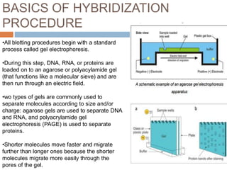
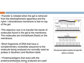
Downloaded 195 times
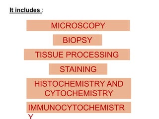
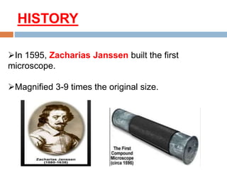
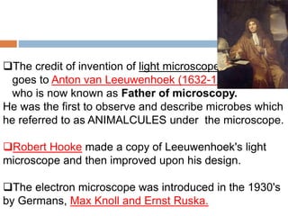
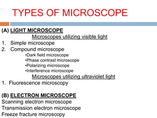
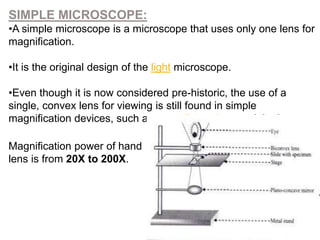
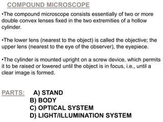
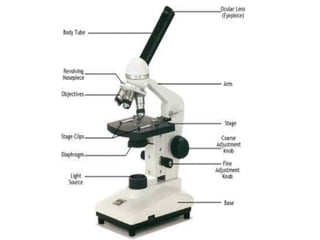
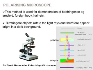


This document discusses various histological tools used to study tissues at the microscopic level. It describes light microscopes, which use visible light and magnification to examine thin tissue slices stained with histological dyes. Electron microscopes are also covered, using electron beams instead of light for higher resolution imaging of cell structures. Specific techniques covered include fluorescence microscopy using fluorescent dyes, polarizing microscopy examining birefringence, and transmission electron microscopy producing 2D images of cell organelles. The history and development of microscopy from early simple microscopes to modern compound and electron microscopes is summarized.