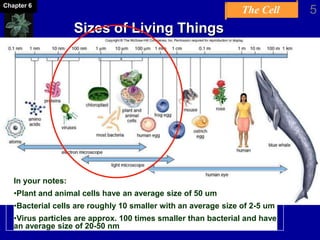
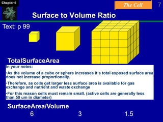
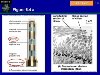
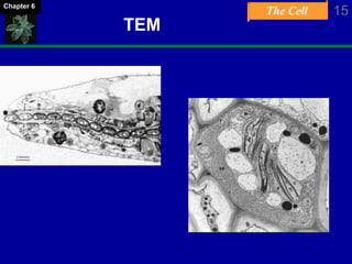
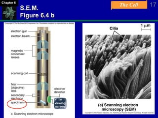
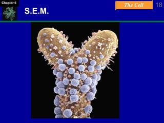
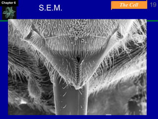
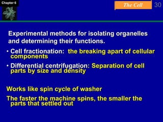
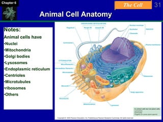
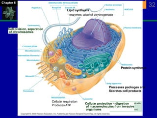
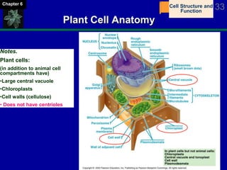
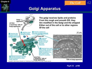
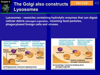
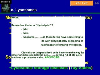
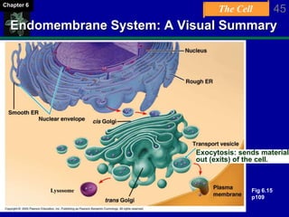
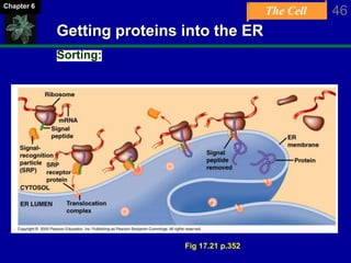
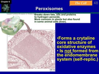
A Tour of the
Cell
Chapter 6
The Cell
Learning Objectives
• Describe principles that limit cell size
• Describe fundamental differences between
prokaryotic and eukaryotic cell types
• Ability to recognize models of eukaryotic
cellular compartments and ascribe functions
to each
• Describe essential similarities and difference
between animal and plant cells
• Outline intracellular and extracellular
protein structures and describe their
functions.
2Chapter 6
The Cell 3Chapter 6
Cell Theory
A unifying concept in biology
Originated from the work of biologists
Schleiden and Schwann in 1838-9
States that:
All organisms are composed of cells
-German botanist Matthais Schleiden in 1838
-German zoologist Theodor Schwann in 1839
All cells come only from preexisting cells
-German physician Rudolph Virchow in 1850’s
Smallest unit of life
The Cell 4Chapter 6
Organisms and Cells
Pallisade cells
Squamous epithelial and striated muscle
400X 400X
The Cell 5Chapter 6
Sizes of Living Things
In your notes:
•Plant and animal cells have an average size of 50 um
•Bacterial cells are roughly 10 smaller with an average size of 2-5 um
•Virus particles are approx. 100 times smaller than bacterial and have
an average size of 20-50 nm
The Cell 6Chapter 6
Cell Size
Size restricted by Surface/Volume (S/V) ratio
Surface is membrane, across which cell
acquires nutrients and expels wastes
Volume is living cytoplasm, which demands
nutrients and produces wastes
As cell grows, volume increases faster than
surface
Cells specialized in absorption modified to
greatly increase surface area per unit volume
In your notes:
•The cell membrane is like the lungs and intestines of a cell.
Therefore the total surface area of the cell membrane must remain
large with respect to the cell contents / volume.
•This will only happen if the cell remains small.
The Cell 7Chapter 6
Surface to Volume Ratio
TotalSurfaceArea
6 cm2 (X 4) 24 cm2 (x 4) 96 cm2
TotalVolume
1cm3 ( x 8) 8 cm3 ( x 8) 64 cm3
SurfaceArea/Volume
6 3 1.5
In your notes:
•As the volume of a cube or sphere increases it s total exposed surface area
does not increase proportionally.
•Therefore, as cells get larger less surface area is available for gas
exchange and nutrient and waste exchange
•For this reason cells must remain small. (active cells are generally less
than 50 um in diameter)
Text: p 99
The Cell 8Chapter 6
Know your metric measures!
1 m = 100 cm
10-2 m = 1 cm = 10 mm (millimeters)
10-3 m = 1 mm = 1000 um (micrometers)
10-6 m = 1 um = 1000 nm (nanometers)
10-9 m
The Cell 9Chapter 6
Microscopes:
Summary:
“There are many different technologies
applied to microscopy”
You will need to differentiate between:
• Light microscopes
• Two types of electron microscopes
• A couple of others, such as Phase contrast,
Imunoflourescence, video enhanced.
The Cell 10Chapter 6
Different techniques for viewing cells:
1. Compound Light Microscope
Light passed through specimen
Focused by glass lenses
Image formed on human retina
Max magnification about 1000X
Resolves objects separated by 0.2 mm,
1000X better than human eye
The Cell 11Chapter 6
Figure 4Aa
The Cell 12Chapter 6
Light Microscopy (1)
The Cell 13Chapter 6
Electron Microscopy
A. Transmission Electron Microscope
Abbreviated T.E.M.
Electrons passed through specimen
Focused by magnetic lenses
Image formed on fluorescent screen
Similar to TV screen
Image is then photographed
Max magnification 1,000,000s X
Resolves objects separated by 0.00002 mm,
100,000X better than human eye
The Cell 14Chapter 6
Figure 6.4 a
The Cell 15Chapter 6
TEM
The Cell 16Chapter 6
b. Scanning Electron Microscope
Abbreviated S.E.M.
Specimen sprayed with thin coat of metal
Electron beam scanned across surface of
specimen
Metal emits secondary electrons
Emitted electrons focused by magnetic lenses
Image formed on fluorescent screen
Similar to TV screen
Image is then photographed
The Cell 17Chapter 6
S.E.M.
Figure 6.4 b
The Cell 18Chapter 6
S.E.M.
The Cell 19Chapter 6
S.E.M.
The Cell 20Chapter 6
Two classes of cells.
1. Prokaryotic cells
Lack a membrane-bound nucleus
Structurally simple
Two of lifes Domains are prokaryotes.
A. Bacteria
Three Shapes
Bacillus (rod)
Coccus (spherical)
Spirilla (spiral)
B. Archaea (“Ancient Bacteria)
Live in extreme habitats (High temp., High salt, toxic gas)
The Cell 21Chapter 6
Shapes of Bacterial Cells
In your notes:
Bacteria have three general shapes
1. Cocci = round. 2. Bacilli = rod shaped. 3. Spirochete = spiral shaped.
The Cell 22Chapter 6
Prokaryotic Cells: Visual Summary
Particles, not organelles,
Very similar to eukaryotic
But smaller
Fig. 6.5 p97
The Cell 23Chapter 6
Prokaryotic Cells:
The Envelope
Cell Envelopes (some have a cell envelope!)
Glycocalyx
- Layer of polysaccharides outside cell wall
- May be slimy and easily removed, or
- Well organized and resistant to removal (capsule)
Cell wall – peptidoyglycans (recall = struct. Carb.)
Plasma membrane (all have plasma membrane)
- Like in eukaryotes
- Form internal pouches (mesosomes)
The Cell 24Chapter 6
Cytoplasm
Semifluid solution within the cell
 No organelles – only small granules of
stored nutrients called inclusion bodies
Appendages
Flagella – Provide motility
Fimbriae – small, bristle-like fibers that
sprout from the cell surface
Sex pili – rigid tubular structures used to
pass DNA from cell to cell
Prokaryotic Cells:
Cytoplasm & Appendages
The Cell 25Chapter 6
2. Eukaryotic Cells
Domain Eukarya
Protists
Fungi
Plants
Animals
Cells are subdivided into specialized
compartments
- All other life forms on earth are eukaryotes
The Cell 26Chapter 6
Eukaryotic Cells :
Organelles
Compartmentalization:
 Isolates reactions from eachother…..therefore….
 Increased efficiency and specialization of reactions
 Allows eukaryotic cells to be larger than prokaryotic cells
 Two classes of eukaryotic compartments:
1.Endomembrane system:
Organelles that communicate with one another
via membrane channels
Via small vesicles: includes Golgi, Endoplasmic reticulum,
Nucleus, lysosomes, transport vessicles
2. Energy related organelles
Mitochondria & chloroplasts
Have their own DNA and ribosomes
The important advancement of
eukaryotes:
** copy to your notes!
The Cell 27Chapter 6
The membrane system of cells
consists of a phospholipid bilayer
Fig. 6.6 p98
The Cell 28Chapter 6
Label Diagram!
Integral transmembrane
protein
Peripheral (surface) protein
Phospholipid (f)
(e)
(g)
(i)
The Cell 29Chapter 6
Origin of
Eukaryotic Cells
Mesosomes
Surround genetic
material
In your notes:
The endosymbiont hypothesis
for the origin of eukaryotes.
A prokaryote predecessor is
thought to have given rise to
eukaryotes via incorporation
of other advantageous
prokaryotes.
Defn:
Symbiosis = two or more
organisms providing each
other with some advantages.
(p. 517 Chap. 25)
The Cell 30Chapter 6
Experimental methods for isolating organelles
and determining their functions.
• Cell fractionation: the breaking apart of cellular
components
• Differential centrifugation: Separation of cell
parts by size and density
Works like spin cycle of washer
The faster the machine spins, the smaller the
parts that settled out
The Cell 31Chapter 6
Animal Cell Anatomy
Notes:
Animal cells have
•Nuclei
•Mitochondria
•Golgi bodies
•Lysosomes
•Endoplasmic reticulum
•Centrioles
•Microtubules
•ribosomes
•Others
The Cell 32Chapter 6
Cell division, separation
of chromosomes
Cellular protection – digestion
of macromolecules from invasive
organisms
Lipid synthesis
Processes packages and
Secretes cell products
Protein synthesis
Cellular respiration
Produces ATP
- enzymes: alcohol deydrogenase
The Cell 33Chapter 6
Plant Cell Anatomy
Notes.
Plant cells:
(in addition to animal cell
compartments have)
•Large central vacuole
•Chloroplasts
•Cell walls (cellulose)
• Does not have centrioles
Cell Structure and
Function
The Cell 34Chapter 6
Functions
A. Nucleus
The genetic command center of cell, usually
near center
Separated from cytoplasm by nuclear
envelope
Consists of double layer of membrane
Nuclear pores permit exchange between
nucleoplasm & cytoplasm
Contains:
Chromatin = semi fluid form of DNA
Chromosomes. Before cells divide, DNA must condense in to
chromosomes
Nucleolus: production center for rRNA
-Produces subunits of ribosomes.
The Cell 35Chapter 6
Anatomy of the Nucleus
Fig. 6.9
p103
The Cell 36Chapter 6
B. Ribosomes
Serve in protein synthesis
Composed of rRNA
Consists of a large subunit and a small
subunit
Subunits made in nucleolus
May be located:
On the endoplasmic reticulum (thereby
making it “rough”), or
Free in the cytoplasm, either singly or in
groups called polyribosomes
The Cell 37Chapter 6
Anatomy of a Ribosome
The Cell 38Chapter 6
C. Endomembrane System
(Improves efficiency of eukaryotic cells by isolating
mechanisms and reactions to specialized
compartments)
Consists of:
a. Endoplasmic reticulum (both smooth and rough)
b. Golgi apparatus
c. Vesicles
-Several types
-Transport materials between organelles of
system
The Cell 39Chapter 6
a. The Endoplasmic Reticulum
i. Rough ER
Studded with ribosomes on cytoplasmic side
Protein anabolism (building proteins)
-Synthesizes proteins
-Modifies proteins
 Adds sugar to protein
 Results in glycoproteins
ii. Smooth ER
No ribosomes
Synthesis of lipids (triglycerides, steroids, phospholipids)
-modifications occur
In golgi apparatus
The Cell 40Chapter 6
Endoplasmic Reticulum
Lipids
In smooth ER
Proteins
In rough ER
To golgi apparatus
Fig. 6.11 p105
The Cell 41Chapter 6
The Golgi Apparatus
b. Golgi Apparatus
Consists of 3-20 flattened, curved saccules
Resembles stack of hollow pancakes
Modifies proteins and lipids
-Packages them in vesicles
-Receives vesicles from ER on cis face
-Prepares for “shipment” in vesicles from
trans face
 Within cell
 Export from cell (secretion, exocytosis)
Note:
Learn to recognize in a cellular cartoon.
Remember its function:
Packages and modifies proteins and fats for
shipment elsewhere inside and outside of the cell.
The Cell 42Chapter 6
Golgi Apparatus
The golgi receives lipids and proteins
From the rough and smooth ER; they
Are modified in the Golgi and the shipped
Either out of the cell or to other regions
Of the cell
Fig 6.12 p106
The Cell 43Chapter 6
The Golgi also constructs
Lysosomes
Lysosomes : vessicles containing hydrolytic enzymes that can digest
cellular debris (damaged organelles), incoming food particles,
phagocytosed foreign cells and viruses.
The Cell 44Chapter 6
c. Lysosomes
Membrane-bound vesicles (not in plants)
Produced by the Golgi apparatus
Low pH
Contain lytic enzymes
-Digestion of large molecules
-Recycling of cellular resources
-Apoptosis (programmed cell death, like
tadpole losing tail)
Some genetic diseases
Caused by defect in lysosomal enzyme
Lysosomal storage diseases (Tay-Sachs)
Notes:
Remember the term “Hydrolysis” ?
- lytic
- lysis
- lysosome……….all these terms have something to
do with enzymatically degrading or
taking apart of organic molecules.
Old cells or unspecialized cells have to make way for
newer or more specialized cell………getting rid of old cells
involves a procecess called APOPTOSIS.
The Cell 45Chapter 6
Endomembrane System: A Visual Summary
Exocytosis: sends material
out (exits) of the cell.
Lysosome Fig 6.15
p109
The Cell 46Chapter 6
Getting proteins into the ER
Sorting:
Fig 17.21 p.352
The Cell 47Chapter 6
Plant Cells:
A. Peroxisomes
Similar to lysosomes
Membrane-bounded vesicles
Enclose enzymes
However
Enzymes synthesized by free ribosomes in
cytoplasm (instead of ER)
Active in lipid metabolism (break down fats)
Catalyze reactions that produce hydrogen
peroxide H2O2
-Toxic
-Broken down to water & O2 by catalase
The Cell 48Chapter 6
Peroxisomes
Breaks down fats, oils and proteins
to hydrogen peroxide.
More common in plants but also found
in some animal cells (liver).
•Forms a crytaline
core structure of
oxidative enzymes
• Is not formed from
the endomembrane
system (self-replic.)
The Cell 49Chapter 6
Plants:
B. Vacuoles
Membranous sacs that are larger than
vesicles
Store materials that occur in excess
Others very specialized (contractile vacuole)
Plants cells typically have a central vacuole
Up to 90% volume of some cells
Functions in:
-Storage of water, nutrients, pigments, and
waste products
-Development of turgor pressure (ie: plants can be turgid or
flacid)
-Some functions performed by lysosomes in
other eukaryotes
The Cell 50Chapter 6
Vacuoles
Fig. 6.14, p. 108
The Cell 51Chapter 6
Energy-Related Organelles:
1. Chloroplasts
Captures light energy to drive cellular
machinery
Photosynthesis
Synthesizes carbohydrates from CO2 & H2O
Makes own food using CO2 as only carbon
source Autotrophic
The Cell 52Chapter 6
Chloroplast Structure
Bounded by double membrane
Inner membrane infolded
Forms disc-like thylakoids, which are
stacked to form grana
Suspended in semi-fluid stroma
Green due to chlorophyll
Green photosynthetic pigment
Found ONLY in inner membranes of
chloroplast
The Cell 53Chapter 6
Energy-Related Organelles:
Chloroplast Structure
?
Fig. 6.18 p111
The Cell 54Chapter 6
Energy-Related Organelles:
2. Mitochondria
Bounded by double membrane
Cristae – Infoldings of inner membrane that
encloses matrix
Matrix – Inner semifluid containing respiratory
enzymes
Involved in cellular respiration
Produce most of ATP utilized by the cell
The Cell 55Chapter 6
Energy-Related Organelles:
Mitochondrial Structure
Fig. 6.17
p111
The Cell 56Chapter 6
The Cytoskeleton
Maintains cell shape
Assists in movement of cell and
organelles
Three types of macromolecular fibers
1. Actin Filaments (small)
2. Intermediate Filaments (medium)
3. Microtubules (bigger)
Assemble and disassemble as needed
The Cell 57Chapter 6
The Cytoskeleton:
Actin Filament Operation
Function in cellular movement through
flow and movement of the plasma
membrane.
The Cell 58Chapter 6
Intermediate filaments
Bigger – hold shape of organelles – used in cell attachment to other cells.
• Myosin
• Dynein
• Kinesin
The Cell 59Chapter 6
The Cytoskeleton:
Microtubule Operation
Alpha and Beta tubulin pairs form hollow cylinders
Can form, unform and reform again. In this way they move
chromosomes into the correct positions for cell division.
Also form major structural component of flagella
The Cell 60Chapter 6
Moving chromosomes: Mitotic Spindle
metaphase
anaphase
telophase
The Cell 61Chapter 6
Microtubular arrays:
Cilia and Flagella
Microtubules can form into specialized
structures called:
- Cilia
- Flagella
- Both have a unique 9 + 2 pattern.
The Cell 62Chapter 6
Structure of a Flagellum
9 & 2
The Cell 63Chapter 6
Review
Cell Theory
Cell Size What restricts cell size?
Prokaryotic Cells
Eukaryotic Cells
Organelles Know the structure and functions of the following:
-Nucleus
-Endomembrane System
-Cytoskeleton
-Centrioles, Cilia, and Flagella
How are these cells different?
A Tour of the
Cell
Chapter 6
Review Chapter  Practice Quiz  WebCT Quiz

Chapter 06 A Tour of the Cell

  • 1.
    A Tour ofthe Cell Chapter 6
  • 2.
    The Cell Learning Objectives •Describe principles that limit cell size • Describe fundamental differences between prokaryotic and eukaryotic cell types • Ability to recognize models of eukaryotic cellular compartments and ascribe functions to each • Describe essential similarities and difference between animal and plant cells • Outline intracellular and extracellular protein structures and describe their functions. 2Chapter 6
  • 3.
    The Cell 3Chapter6 Cell Theory A unifying concept in biology Originated from the work of biologists Schleiden and Schwann in 1838-9 States that: All organisms are composed of cells -German botanist Matthais Schleiden in 1838 -German zoologist Theodor Schwann in 1839 All cells come only from preexisting cells -German physician Rudolph Virchow in 1850’s Smallest unit of life
  • 4.
    The Cell 4Chapter6 Organisms and Cells Pallisade cells Squamous epithelial and striated muscle 400X 400X
  • 5.
    The Cell 5Chapter6 Sizes of Living Things In your notes: •Plant and animal cells have an average size of 50 um •Bacterial cells are roughly 10 smaller with an average size of 2-5 um •Virus particles are approx. 100 times smaller than bacterial and have an average size of 20-50 nm
  • 6.
    The Cell 6Chapter6 Cell Size Size restricted by Surface/Volume (S/V) ratio Surface is membrane, across which cell acquires nutrients and expels wastes Volume is living cytoplasm, which demands nutrients and produces wastes As cell grows, volume increases faster than surface Cells specialized in absorption modified to greatly increase surface area per unit volume In your notes: •The cell membrane is like the lungs and intestines of a cell. Therefore the total surface area of the cell membrane must remain large with respect to the cell contents / volume. •This will only happen if the cell remains small.
  • 7.
    The Cell 7Chapter6 Surface to Volume Ratio TotalSurfaceArea 6 cm2 (X 4) 24 cm2 (x 4) 96 cm2 TotalVolume 1cm3 ( x 8) 8 cm3 ( x 8) 64 cm3 SurfaceArea/Volume 6 3 1.5 In your notes: •As the volume of a cube or sphere increases it s total exposed surface area does not increase proportionally. •Therefore, as cells get larger less surface area is available for gas exchange and nutrient and waste exchange •For this reason cells must remain small. (active cells are generally less than 50 um in diameter) Text: p 99
  • 8.
    The Cell 8Chapter6 Know your metric measures! 1 m = 100 cm 10-2 m = 1 cm = 10 mm (millimeters) 10-3 m = 1 mm = 1000 um (micrometers) 10-6 m = 1 um = 1000 nm (nanometers) 10-9 m
  • 9.
    The Cell 9Chapter6 Microscopes: Summary: “There are many different technologies applied to microscopy” You will need to differentiate between: • Light microscopes • Two types of electron microscopes • A couple of others, such as Phase contrast, Imunoflourescence, video enhanced.
  • 10.
    The Cell 10Chapter6 Different techniques for viewing cells: 1. Compound Light Microscope Light passed through specimen Focused by glass lenses Image formed on human retina Max magnification about 1000X Resolves objects separated by 0.2 mm, 1000X better than human eye
  • 11.
    The Cell 11Chapter6 Figure 4Aa
  • 12.
    The Cell 12Chapter6 Light Microscopy (1)
  • 13.
    The Cell 13Chapter6 Electron Microscopy A. Transmission Electron Microscope Abbreviated T.E.M. Electrons passed through specimen Focused by magnetic lenses Image formed on fluorescent screen Similar to TV screen Image is then photographed Max magnification 1,000,000s X Resolves objects separated by 0.00002 mm, 100,000X better than human eye
  • 14.
    The Cell 14Chapter6 Figure 6.4 a
  • 15.
  • 16.
    The Cell 16Chapter6 b. Scanning Electron Microscope Abbreviated S.E.M. Specimen sprayed with thin coat of metal Electron beam scanned across surface of specimen Metal emits secondary electrons Emitted electrons focused by magnetic lenses Image formed on fluorescent screen Similar to TV screen Image is then photographed
  • 17.
    The Cell 17Chapter6 S.E.M. Figure 6.4 b
  • 18.
  • 19.
  • 20.
    The Cell 20Chapter6 Two classes of cells. 1. Prokaryotic cells Lack a membrane-bound nucleus Structurally simple Two of lifes Domains are prokaryotes. A. Bacteria Three Shapes Bacillus (rod) Coccus (spherical) Spirilla (spiral) B. Archaea (“Ancient Bacteria) Live in extreme habitats (High temp., High salt, toxic gas)
  • 21.
    The Cell 21Chapter6 Shapes of Bacterial Cells In your notes: Bacteria have three general shapes 1. Cocci = round. 2. Bacilli = rod shaped. 3. Spirochete = spiral shaped.
  • 22.
    The Cell 22Chapter6 Prokaryotic Cells: Visual Summary Particles, not organelles, Very similar to eukaryotic But smaller Fig. 6.5 p97
  • 23.
    The Cell 23Chapter6 Prokaryotic Cells: The Envelope Cell Envelopes (some have a cell envelope!) Glycocalyx - Layer of polysaccharides outside cell wall - May be slimy and easily removed, or - Well organized and resistant to removal (capsule) Cell wall – peptidoyglycans (recall = struct. Carb.) Plasma membrane (all have plasma membrane) - Like in eukaryotes - Form internal pouches (mesosomes)
  • 24.
    The Cell 24Chapter6 Cytoplasm Semifluid solution within the cell  No organelles – only small granules of stored nutrients called inclusion bodies Appendages Flagella – Provide motility Fimbriae – small, bristle-like fibers that sprout from the cell surface Sex pili – rigid tubular structures used to pass DNA from cell to cell Prokaryotic Cells: Cytoplasm & Appendages
  • 25.
    The Cell 25Chapter6 2. Eukaryotic Cells Domain Eukarya Protists Fungi Plants Animals Cells are subdivided into specialized compartments - All other life forms on earth are eukaryotes
  • 26.
    The Cell 26Chapter6 Eukaryotic Cells : Organelles Compartmentalization:  Isolates reactions from eachother…..therefore….  Increased efficiency and specialization of reactions  Allows eukaryotic cells to be larger than prokaryotic cells  Two classes of eukaryotic compartments: 1.Endomembrane system: Organelles that communicate with one another via membrane channels Via small vesicles: includes Golgi, Endoplasmic reticulum, Nucleus, lysosomes, transport vessicles 2. Energy related organelles Mitochondria & chloroplasts Have their own DNA and ribosomes The important advancement of eukaryotes: ** copy to your notes!
  • 27.
    The Cell 27Chapter6 The membrane system of cells consists of a phospholipid bilayer Fig. 6.6 p98
  • 28.
    The Cell 28Chapter6 Label Diagram! Integral transmembrane protein Peripheral (surface) protein Phospholipid (f) (e) (g) (i)
  • 29.
    The Cell 29Chapter6 Origin of Eukaryotic Cells Mesosomes Surround genetic material In your notes: The endosymbiont hypothesis for the origin of eukaryotes. A prokaryote predecessor is thought to have given rise to eukaryotes via incorporation of other advantageous prokaryotes. Defn: Symbiosis = two or more organisms providing each other with some advantages. (p. 517 Chap. 25)
  • 30.
    The Cell 30Chapter6 Experimental methods for isolating organelles and determining their functions. • Cell fractionation: the breaking apart of cellular components • Differential centrifugation: Separation of cell parts by size and density Works like spin cycle of washer The faster the machine spins, the smaller the parts that settled out
  • 31.
    The Cell 31Chapter6 Animal Cell Anatomy Notes: Animal cells have •Nuclei •Mitochondria •Golgi bodies •Lysosomes •Endoplasmic reticulum •Centrioles •Microtubules •ribosomes •Others
  • 32.
    The Cell 32Chapter6 Cell division, separation of chromosomes Cellular protection – digestion of macromolecules from invasive organisms Lipid synthesis Processes packages and Secretes cell products Protein synthesis Cellular respiration Produces ATP - enzymes: alcohol deydrogenase
  • 33.
    The Cell 33Chapter6 Plant Cell Anatomy Notes. Plant cells: (in addition to animal cell compartments have) •Large central vacuole •Chloroplasts •Cell walls (cellulose) • Does not have centrioles Cell Structure and Function
  • 34.
    The Cell 34Chapter6 Functions A. Nucleus The genetic command center of cell, usually near center Separated from cytoplasm by nuclear envelope Consists of double layer of membrane Nuclear pores permit exchange between nucleoplasm & cytoplasm Contains: Chromatin = semi fluid form of DNA Chromosomes. Before cells divide, DNA must condense in to chromosomes Nucleolus: production center for rRNA -Produces subunits of ribosomes.
  • 35.
    The Cell 35Chapter6 Anatomy of the Nucleus Fig. 6.9 p103
  • 36.
    The Cell 36Chapter6 B. Ribosomes Serve in protein synthesis Composed of rRNA Consists of a large subunit and a small subunit Subunits made in nucleolus May be located: On the endoplasmic reticulum (thereby making it “rough”), or Free in the cytoplasm, either singly or in groups called polyribosomes
  • 37.
    The Cell 37Chapter6 Anatomy of a Ribosome
  • 38.
    The Cell 38Chapter6 C. Endomembrane System (Improves efficiency of eukaryotic cells by isolating mechanisms and reactions to specialized compartments) Consists of: a. Endoplasmic reticulum (both smooth and rough) b. Golgi apparatus c. Vesicles -Several types -Transport materials between organelles of system
  • 39.
    The Cell 39Chapter6 a. The Endoplasmic Reticulum i. Rough ER Studded with ribosomes on cytoplasmic side Protein anabolism (building proteins) -Synthesizes proteins -Modifies proteins  Adds sugar to protein  Results in glycoproteins ii. Smooth ER No ribosomes Synthesis of lipids (triglycerides, steroids, phospholipids) -modifications occur In golgi apparatus
  • 40.
    The Cell 40Chapter6 Endoplasmic Reticulum Lipids In smooth ER Proteins In rough ER To golgi apparatus Fig. 6.11 p105
  • 41.
    The Cell 41Chapter6 The Golgi Apparatus b. Golgi Apparatus Consists of 3-20 flattened, curved saccules Resembles stack of hollow pancakes Modifies proteins and lipids -Packages them in vesicles -Receives vesicles from ER on cis face -Prepares for “shipment” in vesicles from trans face  Within cell  Export from cell (secretion, exocytosis) Note: Learn to recognize in a cellular cartoon. Remember its function: Packages and modifies proteins and fats for shipment elsewhere inside and outside of the cell.
  • 42.
    The Cell 42Chapter6 Golgi Apparatus The golgi receives lipids and proteins From the rough and smooth ER; they Are modified in the Golgi and the shipped Either out of the cell or to other regions Of the cell Fig 6.12 p106
  • 43.
    The Cell 43Chapter6 The Golgi also constructs Lysosomes Lysosomes : vessicles containing hydrolytic enzymes that can digest cellular debris (damaged organelles), incoming food particles, phagocytosed foreign cells and viruses.
  • 44.
    The Cell 44Chapter6 c. Lysosomes Membrane-bound vesicles (not in plants) Produced by the Golgi apparatus Low pH Contain lytic enzymes -Digestion of large molecules -Recycling of cellular resources -Apoptosis (programmed cell death, like tadpole losing tail) Some genetic diseases Caused by defect in lysosomal enzyme Lysosomal storage diseases (Tay-Sachs) Notes: Remember the term “Hydrolysis” ? - lytic - lysis - lysosome……….all these terms have something to do with enzymatically degrading or taking apart of organic molecules. Old cells or unspecialized cells have to make way for newer or more specialized cell………getting rid of old cells involves a procecess called APOPTOSIS.
  • 45.
    The Cell 45Chapter6 Endomembrane System: A Visual Summary Exocytosis: sends material out (exits) of the cell. Lysosome Fig 6.15 p109
  • 46.
    The Cell 46Chapter6 Getting proteins into the ER Sorting: Fig 17.21 p.352
  • 47.
    The Cell 47Chapter6 Plant Cells: A. Peroxisomes Similar to lysosomes Membrane-bounded vesicles Enclose enzymes However Enzymes synthesized by free ribosomes in cytoplasm (instead of ER) Active in lipid metabolism (break down fats) Catalyze reactions that produce hydrogen peroxide H2O2 -Toxic -Broken down to water & O2 by catalase
  • 48.
    The Cell 48Chapter6 Peroxisomes Breaks down fats, oils and proteins to hydrogen peroxide. More common in plants but also found in some animal cells (liver). •Forms a crytaline core structure of oxidative enzymes • Is not formed from the endomembrane system (self-replic.)
  • 49.
    The Cell 49Chapter6 Plants: B. Vacuoles Membranous sacs that are larger than vesicles Store materials that occur in excess Others very specialized (contractile vacuole) Plants cells typically have a central vacuole Up to 90% volume of some cells Functions in: -Storage of water, nutrients, pigments, and waste products -Development of turgor pressure (ie: plants can be turgid or flacid) -Some functions performed by lysosomes in other eukaryotes
  • 50.
    The Cell 50Chapter6 Vacuoles Fig. 6.14, p. 108
  • 51.
    The Cell 51Chapter6 Energy-Related Organelles: 1. Chloroplasts Captures light energy to drive cellular machinery Photosynthesis Synthesizes carbohydrates from CO2 & H2O Makes own food using CO2 as only carbon source Autotrophic
  • 52.
    The Cell 52Chapter6 Chloroplast Structure Bounded by double membrane Inner membrane infolded Forms disc-like thylakoids, which are stacked to form grana Suspended in semi-fluid stroma Green due to chlorophyll Green photosynthetic pigment Found ONLY in inner membranes of chloroplast
  • 53.
    The Cell 53Chapter6 Energy-Related Organelles: Chloroplast Structure ? Fig. 6.18 p111
  • 54.
    The Cell 54Chapter6 Energy-Related Organelles: 2. Mitochondria Bounded by double membrane Cristae – Infoldings of inner membrane that encloses matrix Matrix – Inner semifluid containing respiratory enzymes Involved in cellular respiration Produce most of ATP utilized by the cell
  • 55.
    The Cell 55Chapter6 Energy-Related Organelles: Mitochondrial Structure Fig. 6.17 p111
  • 56.
    The Cell 56Chapter6 The Cytoskeleton Maintains cell shape Assists in movement of cell and organelles Three types of macromolecular fibers 1. Actin Filaments (small) 2. Intermediate Filaments (medium) 3. Microtubules (bigger) Assemble and disassemble as needed
  • 57.
    The Cell 57Chapter6 The Cytoskeleton: Actin Filament Operation Function in cellular movement through flow and movement of the plasma membrane.
  • 58.
    The Cell 58Chapter6 Intermediate filaments Bigger – hold shape of organelles – used in cell attachment to other cells. • Myosin • Dynein • Kinesin
  • 59.
    The Cell 59Chapter6 The Cytoskeleton: Microtubule Operation Alpha and Beta tubulin pairs form hollow cylinders Can form, unform and reform again. In this way they move chromosomes into the correct positions for cell division. Also form major structural component of flagella
  • 60.
    The Cell 60Chapter6 Moving chromosomes: Mitotic Spindle metaphase anaphase telophase
  • 61.
    The Cell 61Chapter6 Microtubular arrays: Cilia and Flagella Microtubules can form into specialized structures called: - Cilia - Flagella - Both have a unique 9 + 2 pattern.
  • 62.
    The Cell 62Chapter6 Structure of a Flagellum 9 & 2
  • 63.
    The Cell 63Chapter6 Review Cell Theory Cell Size What restricts cell size? Prokaryotic Cells Eukaryotic Cells Organelles Know the structure and functions of the following: -Nucleus -Endomembrane System -Cytoskeleton -Centrioles, Cilia, and Flagella How are these cells different?
  • 64.
    A Tour ofthe Cell Chapter 6 Review Chapter  Practice Quiz  WebCT Quiz