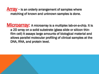
Array - isan orderly arrangement of samples where
matching of known and unknown samples is done.
Microarray: A microarray is a multiplex lab-on-a-chip. It is
a 2D array on a solid substrate (glass slide or silicon thin-
film cell) It assays large amounts of biological material and
allows parallel molecular profiling of clinical samples at the
DNA, RNA, and protein level.
4.
History
The conceptand methodology of microarrays was first
illustrated in antibody microarrays by Tse Wen Chang
in 1983
Multi-tumor tissue block" in 1986 by H.Battifora
Originally used in the Brown Laboratory at Stanford
University in late 1990s
Steadily developed since Patrick Brown and his
colleagues first published their work in 1995
5.
Need For Microarrays
Conventional investigation of tissues was labour intensive,
too expensive and time consuming to be applied to the
characterisation of hundreds or thousands of genes or
gene clusters associated with distinct tumour entities or
other diseases.
Thus, there was a need for techniques that could facilitate
research on
• large series of tissues
• in parallel
• in a single experiment
6.
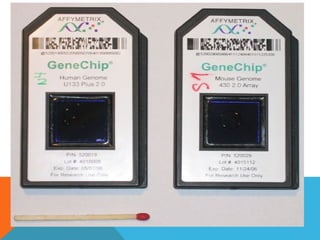
DNA Microarray
DNAchip or Biochip or Gene chip.
Analyze thousands of genes in one experiment
commercially prepared small plates of glass (or silicon or
nylon) about 2 or 3 cm square, coated with probes
is composed of pieces of DNA ranging from 20 to 5000
base pairs concentrated on specific areas.
7.
Used to measurethe expression levels of large numbers
of genes simultaneously or to genotype multiple regions of
a genome.
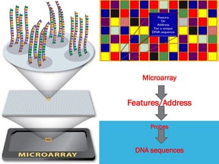
Each DNA spot contains picomoles of a specific DNA
sequence/ gene, known as probes. Each spot may
contain a few million copies of identical probe molecule.
8.
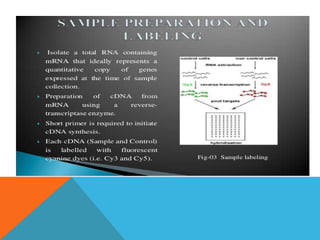
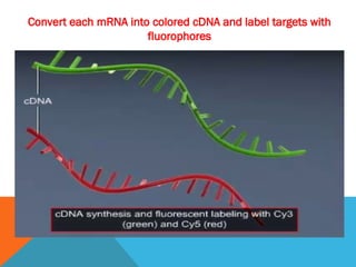
Definitions:
Target -the nucleicacid (cDNA) sample who’s identity
and quantity are being measured.
Fluorophore –usually green and red labels attached to
the target to enable visualizing expression.
Microarray -works as reverse hybridization method
converting from mRNA to cDNA 3’-5’ with TTTT…end.
Probe–an attached nucleic acid with a known sequence
(the DNA chip).
9.
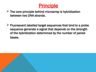
Principle
The coreprinciple behind microarray is hybridization
between two DNA strands.
Fluorescent labelled target sequences that bind to a probe
sequence generate a signal that depends on the strength
of the hybridization determined by the number of paired
bases.
10.
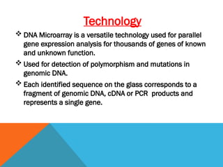
Technology
DNA Microarrayis a versatile technology used for parallel
gene expression analysis for thousands of genes of known
and unknown function.
Used for detection of polymorphism and mutations in
genomic DNA.
Each identified sequence on the glass corresponds to a
fragment of genomic DNA, cDNA or PCR products and
represents a single gene.
11.
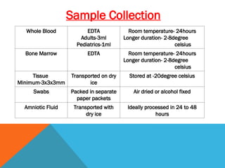
Sample Collection
Whole BloodEDTA
Adults-3ml
Pediatrics-1ml
Room temperature- 24hours
Longer duration- 2-8degree
celsius
Bone Marrow EDTA Room temperature- 24hours
Longer duration- 2-8degree
celsius
Tissue
Minimum-3x3x3mm
Transported on dry
ice
Stored at -20degree celsius
Swabs Packed in separate
paper packets
Air dried or alcohol fixed
Amniotic Fluid Transported with
dry ice
Ideally processed in 24 to 48
hours
12.
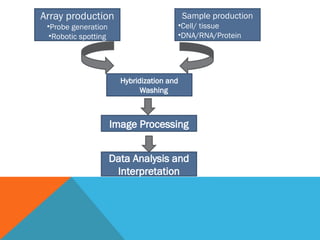
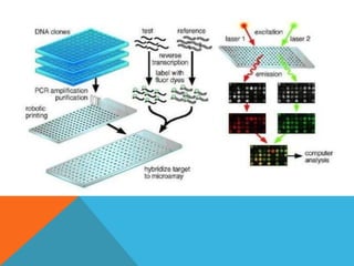
Array production
•Probe generation
•Roboticspotting
Sample production
•Cell/ tissue
•DNA/RNA/Protein
Hybridization and
Washing
Image Processing
Data Analysis and
Interpretation
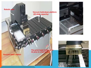
ARRAYING ROBOT
Vaccum washstation
The print head holds up to
32 pins in a 8x4 format
Vaccum hold-down platform
(50 slide capacity)
Robotic arm
17.
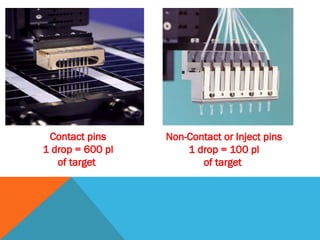
Contact pins
1 drop= 600 pl
of target
Non-Contact or Inject pins
1 drop = 100 pl
of target
18.
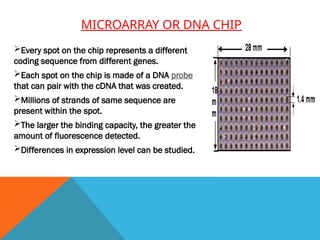
MICROARRAY OR DNACHIP
Every spot on the chip represents a different
coding sequence from different genes.
Each spot on the chip is made of a DNA probe
that can pair with the cDNA that was created.
Millions of strands of same sequence are
present within the spot.
The larger the binding capacity, the greater the
amount of fluorescence detected.
Differences in expression level can be studied.
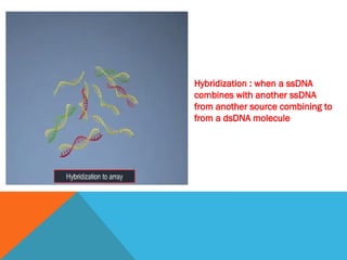
Hybridization : whena ssDNA
combines with another ssDNA
from another source combining to
from a dsDNA molecule
27.
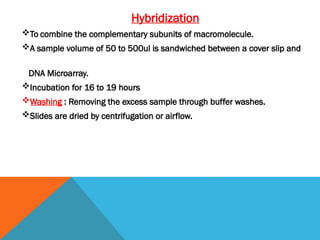
Hybridization
To combine thecomplementary subunits of macromolecule.
A sample volume of 50 to 500ul is sandwiched between a cover slip and
DNA Microarray.
Incubation for 16 to 19 hours
Washing : Removing the excess sample through buffer washes.
Slides are dried by centrifugation or airflow.
29.
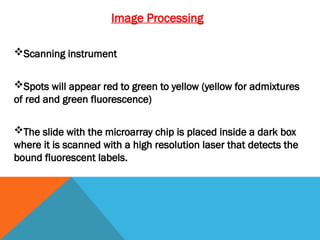
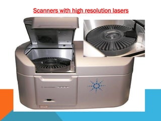
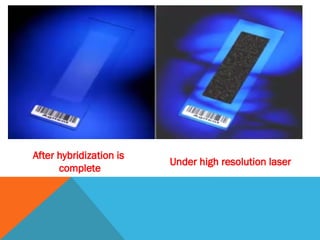
Image Processing
Scanning instrument
Spotswill appear red to green to yellow (yellow for admixtures
of red and green fluorescence)
The slide with the microarray chip is placed inside a dark box
where it is scanned with a high resolution laser that detects the
bound fluorescent labels.
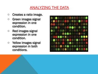
ANALYZING THE DATA
oCreates a ratio image.
o Green images signal
expression in one
condition.
o Red images signal
expression in one
condition.
o Yellow images signal
expression in both
conditions.
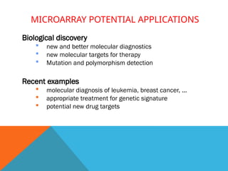
MICROARRAY POTENTIAL APPLICATIONS
Biologicaldiscovery
new and better molecular diagnostics
new molecular targets for therapy
Mutation and polymorphism detection
Recent examples
molecular diagnosis of leukemia, breast cancer, ...
appropriate treatment for genetic signature
potential new drug targets
36.
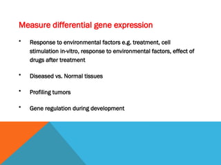
Measure differential geneexpression
• Response to environmental factors e.g. treatment, cell
stimulation in-vitro, response to environmental factors, effect of
drugs after treatment
• Diseased vs. Normal tissues
• Profiling tumors
• Gene regulation during development
37.
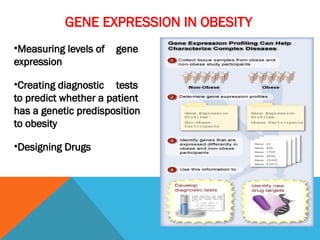
GENE EXPRESSION INOBESITY
•Measuring levels of gene
expression
•Creating diagnostic tests
to predict whether a patient
has a genetic predisposition
to obesity
•Designing Drugs
38.
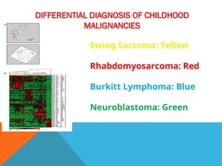
DIFFERENTIAL DIAGNOSIS OFCHILDHOOD
MALIGNANCIES
Ewing Sarcoma: Yellow
Rhabdomyosarcoma: Red
Burkitt Lymphoma: Blue
Neuroblastoma: Green
RESOURCES:
1. Schena, M.,Shalon, D., Heller, R., Chai, A., Brown, P.O. & Davis, R.O. (1996) Parallel
human genome analysis: Microarray-based expression monitoring of 1000 genes. Proc.
Natl. Acad. Sci. U.S.A. 93, 10614-10619. MEDLINE
2. Lipshutz, R.J., Fodor, S.P.A., Gingeras, T.R. & Lockhart, D.J. (1999) High density
synthetic oligonucleotide arrays. Nat. Genet. 21, 20-24. MEDLINE
3. Lowe David, Underwood James; Recent Advances in Histopathology:The Royal society
of medicine press;2004
4. Nazar M.T. Jawhar; Tissue Microarray: A rapidly evolving diagnostic and research
tool;Ann Saudi Med. 2009 Mar-Apr; 29(2): 123–127
5. Simon R1, Mirlacher M, Sauter G.;Immunohistochemical analysis of tissue microarrays;
Methods Mol Biol.2010;664:113-26
6. Rashmil Saxena, BFA Sunil Badve; Tissue Microarray – Construction and Quality
Assurance, Part II: The Potentials and Pitfalls


![ONFH[AVN HIP] -TRIPLE REGIME -A NOVAL SURGICAL CONCEPT .pptx](https://cdn.slidesharecdn.com/ss_thumbnails/onfhavnhip2026koaconcalicutdrgokuldevdrmashraf-260210064517-213ec005-thumbnail.jpg?width=640&height=640&fit=bounds)


