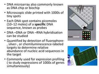
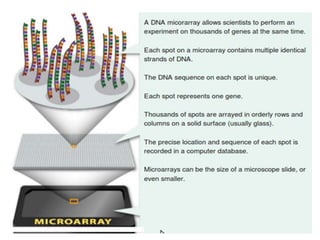
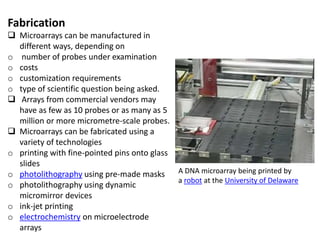
A microarray is a laboratory tool used to detect the expression of thousands of genes at the same time. DNA microarrays are microscope slides that are printed with thousands of tiny spots in defined positions, with each spot containing a known DNA sequence or gene.
This presentation was given to me during my higher education in the Lebanese University, Faculty of Education. It includes detailed explanation about DNA microarray.
A micro-array is a tool for analyzing gene expression that consists of a small membrane or glass slide containing samples of many genes arranged in a regular pattern.
This was made by me while I was in Masters. I have made few animations. I hope it makes understanding better.
The content is made by searching through internet and referencing books. I do not claim any content in whole presentation except the animations made on the subject.
If a microbiologist is studying bacteria that premeditate, or break down, toxic wastes and wants to know which specific genes are active when that bacterium is degrading, say, PCBs, he would likely use a tool called the DNA microarray.
Microarrays enable scientists to monitor the activities of hundreds or thousands of genes at once. All microarrays (also called DNA chips or gene chips) work on the basic principle that complementary nucleotide sequences in DNA (and RNA) match up like the two halves of a piece of Velcro coming together.
Pattern of gene activity on a microarray chip.
A microarray consists of an orderly arrangement of bits of genetic material in super-tiny spots laid down in a grid on a suitable surface, often a glass slide with a specially chemically treated surface.
Molecular Biology research evolves through the development of the technologies used for carrying them out. It is not possible to research on a large number of genes using traditional methods
A DNA microarray (also commonly known as DNA chip or biochip) is a collection of microscopic DNA spots attached to a solid surface.
The core principle behind microarrays is hybridization between two DNA strands, the property of complementary nucleic acid sequences to specifically pair with each other by forming hydrogen bonds between complementary nucleotide base pairs.
Seminar of U.V. Spectroscopy by SAMIR PANDASAMIR PANDA
Spectroscopy is a branch of science dealing the study of interaction of electromagnetic radiation with matter.
Ultraviolet-visible spectroscopy refers to absorption spectroscopy or reflect spectroscopy in the UV-VIS spectral region.
Ultraviolet-visible spectroscopy is an analytical method that can measure the amount of light received by the analyte.
Introduction:
RNA interference (RNAi) or Post-Transcriptional Gene Silencing (PTGS) is an important biological process for modulating eukaryotic gene expression.
It is highly conserved process of posttranscriptional gene silencing by which double stranded RNA (dsRNA) causes sequence-specific degradation of mRNA sequences.
dsRNA-induced gene silencing (RNAi) is reported in a wide range of eukaryotes ranging from worms, insects, mammals and plants.
This process mediates resistance to both endogenous parasitic and exogenous pathogenic nucleic acids, and regulates the expression of protein-coding genes.
What are small ncRNAs?
micro RNA (miRNA)
short interfering RNA (siRNA)
Properties of small non-coding RNA:
Involved in silencing mRNA transcripts.
Called “small” because they are usually only about 21-24 nucleotides long.
Synthesized by first cutting up longer precursor sequences (like the 61nt one that Lee discovered).
Silence an mRNA by base pairing with some sequence on the mRNA.
Discovery of siRNA?
The first small RNA:
In 1993 Rosalind Lee (Victor Ambros lab) was studying a non- coding gene in C. elegans, lin-4, that was involved in silencing of another gene, lin-14, at the appropriate time in the
development of the worm C. elegans.
Two small transcripts of lin-4 (22nt and 61nt) were found to be complementary to a sequence in the 3' UTR of lin-14.
Because lin-4 encoded no protein, she deduced that it must be these transcripts that are causing the silencing by RNA-RNA interactions.
Types of RNAi ( non coding RNA)
MiRNA
Length (23-25 nt)
Trans acting
Binds with target MRNA in mismatch
Translation inhibition
Si RNA
Length 21 nt.
Cis acting
Bind with target Mrna in perfect complementary sequence
Piwi-RNA
Length ; 25 to 36 nt.
Expressed in Germ Cells
Regulates trnasposomes activity
MECHANISM OF RNAI:
First the double-stranded RNA teams up with a protein complex named Dicer, which cuts the long RNA into short pieces.
Then another protein complex called RISC (RNA-induced silencing complex) discards one of the two RNA strands.
The RISC-docked, single-stranded RNA then pairs with the homologous mRNA and destroys it.
THE RISC COMPLEX:
RISC is large(>500kD) RNA multi- protein Binding complex which triggers MRNA degradation in response to MRNA
Unwinding of double stranded Si RNA by ATP independent Helicase
Active component of RISC is Ago proteins( ENDONUCLEASE) which cleave target MRNA.
DICER: endonuclease (RNase Family III)
Argonaute: Central Component of the RNA-Induced Silencing Complex (RISC)
One strand of the dsRNA produced by Dicer is retained in the RISC complex in association with Argonaute
ARGONAUTE PROTEIN :
1.PAZ(PIWI/Argonaute/ Zwille)- Recognition of target MRNA
2.PIWI (p-element induced wimpy Testis)- breaks Phosphodiester bond of mRNA.)RNAse H activity.
MiRNA:
The Double-stranded RNAs are naturally produced in eukaryotic cells during development, and they have a key role in regulating gene expression .
Observation of Io’s Resurfacing via Plume Deposition Using Ground-based Adapt...Sérgio Sacani
Since volcanic activity was first discovered on Io from Voyager images in 1979, changes
on Io’s surface have been monitored from both spacecraft and ground-based telescopes.
Here, we present the highest spatial resolution images of Io ever obtained from a groundbased telescope. These images, acquired by the SHARK-VIS instrument on the Large
Binocular Telescope, show evidence of a major resurfacing event on Io’s trailing hemisphere. When compared to the most recent spacecraft images, the SHARK-VIS images
show that a plume deposit from a powerful eruption at Pillan Patera has covered part
of the long-lived Pele plume deposit. Although this type of resurfacing event may be common on Io, few have been detected due to the rarity of spacecraft visits and the previously low spatial resolution available from Earth-based telescopes. The SHARK-VIS instrument ushers in a new era of high resolution imaging of Io’s surface using adaptive
optics at visible wavelengths.
Multi-source connectivity as the driver of solar wind variability in the heli...Sérgio Sacani
The ambient solar wind that flls the heliosphere originates from multiple
sources in the solar corona and is highly structured. It is often described
as high-speed, relatively homogeneous, plasma streams from coronal
holes and slow-speed, highly variable, streams whose source regions are
under debate. A key goal of ESA/NASA’s Solar Orbiter mission is to identify
solar wind sources and understand what drives the complexity seen in the
heliosphere. By combining magnetic feld modelling and spectroscopic
techniques with high-resolution observations and measurements, we show
that the solar wind variability detected in situ by Solar Orbiter in March
2022 is driven by spatio-temporal changes in the magnetic connectivity to
multiple sources in the solar atmosphere. The magnetic feld footpoints
connected to the spacecraft moved from the boundaries of a coronal hole
to one active region (12961) and then across to another region (12957). This
is refected in the in situ measurements, which show the transition from fast
to highly Alfvénic then to slow solar wind that is disrupted by the arrival of
a coronal mass ejection. Our results describe solar wind variability at 0.5 au
but are applicable to near-Earth observatories.
Cancer cell metabolism: special Reference to Lactate PathwayAADYARAJPANDEY1
Normal Cell Metabolism:
Cellular respiration describes the series of steps that cells use to break down sugar and other chemicals to get the energy we need to function.
Energy is stored in the bonds of glucose and when glucose is broken down, much of that energy is released.
Cell utilize energy in the form of ATP.
The first step of respiration is called glycolysis. In a series of steps, glycolysis breaks glucose into two smaller molecules - a chemical called pyruvate. A small amount of ATP is formed during this process.
Most healthy cells continue the breakdown in a second process, called the Kreb's cycle. The Kreb's cycle allows cells to “burn” the pyruvates made in glycolysis to get more ATP.
The last step in the breakdown of glucose is called oxidative phosphorylation (Ox-Phos).
It takes place in specialized cell structures called mitochondria. This process produces a large amount of ATP. Importantly, cells need oxygen to complete oxidative phosphorylation.
If a cell completes only glycolysis, only 2 molecules of ATP are made per glucose. However, if the cell completes the entire respiration process (glycolysis - Kreb's - oxidative phosphorylation), about 36 molecules of ATP are created, giving it much more energy to use.
IN CANCER CELL:
Unlike healthy cells that "burn" the entire molecule of sugar to capture a large amount of energy as ATP, cancer cells are wasteful.
Cancer cells only partially break down sugar molecules. They overuse the first step of respiration, glycolysis. They frequently do not complete the second step, oxidative phosphorylation.
This results in only 2 molecules of ATP per each glucose molecule instead of the 36 or so ATPs healthy cells gain. As a result, cancer cells need to use a lot more sugar molecules to get enough energy to survive.
introduction to WARBERG PHENOMENA:
WARBURG EFFECT Usually, cancer cells are highly glycolytic (glucose addiction) and take up more glucose than do normal cells from outside.
Otto Heinrich Warburg (; 8 October 1883 – 1 August 1970) In 1931 was awarded the Nobel Prize in Physiology for his "discovery of the nature and mode of action of the respiratory enzyme.
WARNBURG EFFECT : cancer cells under aerobic (well-oxygenated) conditions to metabolize glucose to lactate (aerobic glycolysis) is known as the Warburg effect. Warburg made the observation that tumor slices consume glucose and secrete lactate at a higher rate than normal tissues.
This presentation explores a brief idea about the structural and functional attributes of nucleotides, the structure and function of genetic materials along with the impact of UV rays and pH upon them.
Richard's aventures in two entangled wonderlandsRichard Gill
Since the loophole-free Bell experiments of 2020 and the Nobel prizes in physics of 2022, critics of Bell's work have retreated to the fortress of super-determinism. Now, super-determinism is a derogatory word - it just means "determinism". Palmer, Hance and Hossenfelder argue that quantum mechanics and determinism are not incompatible, using a sophisticated mathematical construction based on a subtle thinning of allowed states and measurements in quantum mechanics, such that what is left appears to make Bell's argument fail, without altering the empirical predictions of quantum mechanics. I think however that it is a smoke screen, and the slogan "lost in math" comes to my mind. I will discuss some other recent disproofs of Bell's theorem using the language of causality based on causal graphs. Causal thinking is also central to law and justice. I will mention surprising connections to my work on serial killer nurse cases, in particular the Dutch case of Lucia de Berk and the current UK case of Lucy Letby.
2. • DNA microarray also commonly known
as DNA chip or biochip
• Microscopic slide printed with 1000s of
tiny spots
• Each DNA spot contains picomoles
(10−12 moles) of a specific DNA
sequence, known as probes
• DNA –DNA or DNA –RNA hybridisation
can be studied
• Quantified by detection of fluorophore-
, silver-, or chemiluminescence-labeled
targets to determine relative
abundance of nucleic acid sequences in
the target
• Commonly used for expression profiling
( to study expressions of 1000s of genes
simultaneously)
3.
4. Fabrication
Microarrays can be manufactured in
different ways, depending on
o number of probes under examination
o costs
o customization requirements
o type of scientific question being asked.
Arrays from commercial vendors may
have as few as 10 probes or as many as 5
million or more micrometre-scale probes.
Microarrays can be fabricated using a
variety of technologies
o printing with fine-pointed pins onto glass
slides
o photolithography using pre-made masks
o photolithography using dynamic
micromirror devices
o ink-jet printing
o electrochemistry on microelectrode
arrays
A DNA microarray being printed by
a robot at the University of Delaware
5. PRINCIPLE
• Hybridization between two DNA strands, the property
of complementary nucleic acid sequences to specifically pair
with each other by forming hydrogen bonds between
complementary nucleotide base pairs
• A high number of complementary base pairs in a nucleotide
sequence means tighter non-covalent bonding between the
two strands
• Total strength of the signal, from a spot (feature), depends
upon the amount of target sample binding to the probes
present on that spot
• Relative quantitation of intensity of a feature is compared to
the intensity of the same feature under a different
condition(temp), and the identity of the feature is known by its
position.
6. • DNA microarray Experiment
process
1. Collect tissue
2. Isolate RNA
3. Isolate mRNA
4. Make labeled DNA copy
5. Apply DNA
6. Scan microarray
7. Analyze data
8. m RNA levels compared in many
different contexts
Different tissue, same organism (brain v liver)
Same tissue, same organism (tumour v non tumour)
Same tissue, different organism (wt v mutant)
Time course experiments (development)
Two Affymetrix chips. A match is shown
at bottom left for size comparison.
9. Applications
Gene expression profiling
• in different cells /tissues
• During course of development
• Under different environmental/chemical stimuli
• In disease versus healthy
Drug development
• Identification of new targets
Pharmacogenomics
• Individualized medicine
10. Comparative genomic hybridisation
• Assessing genomic content in closely related
organisms
SNP detection
• Identifying SNPs among populations
ChIP studies(Chromatin immunoprecipitation)
• Determining protein binding site occupancy
throughout genome by ChIP on chip technology
GeneID
• Small microarrays to check IDs of organisms in food
and feed or pathogens for disease detection
12. DNA microarrays: a powerful genomic
tool for biomedical and clinical research.
Trevino V1, Falciani F, Barrera-Saldaña HA.
• Microarrays initially designed to measure gene transcriptional level and for
comparing other genome features among individuals and their tissues and
cells.
• Results provide valuable information on disease subcategories, disease prognosis,
and treatment outcome.
• Microarray reveal differences in genetic makeup, regulatory mechanisms
o Relating Gene Expression to Physiology:
o Biomarker Detection: Supervised Classification
o Describing the Relationship between the Molecular State of Biological Samples
13. o Identification of Prognostic Genes Associated to Risk and Survival
o Association of Genes to disease surrogate markers
o Genetic Disorders: Gene Copy Number and Comparative Genomic Hybridization
o Genetic Disorders: Epigenetics and Methylation
o Genetic Disorders and Variability: Gene Polymorphism and Single Nucleotide
Polymorphism
o Chromatin Immunoprecipitation: Genetic Control and Transcriptional Regulation
o Pathogen detection
DNA microarrays are a powerful, mature versatile and easy-to-use genomic tool
that can be applied for biomedical and clinical research
The main advantage is the genomic-wide information provided at reasonable
costs
14. Monitoring the Expression Pattern of 1300 Arabidopsis
Genes under Drought and Cold Stresses by Using a Full-
Length cDNA Microarray
Motoaki Seki,a,b Mari Narusaka,a Hiroshi Abe,c Mie Kasuga,c Kazuko Yamaguchi-
Shinozaki,c Piero Carninci,dYoshihide Hayashizaki,d and Kazuo Shinozaki1,a,b
15.
16. Microarray analysis of diurnal and circadian-regulated genes
in Arabidopsis.
Schaffer R1, Landgraf J, Accerbi M, Simon V, Larson M, Wisman E.
17. The next generation of microarray research:
applications in evolutionary and ecological
genomics
Analysis of microarray experiments of gene
expression profiling