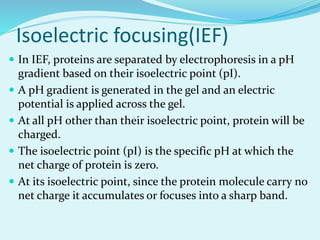
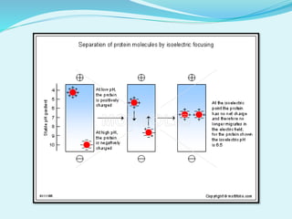
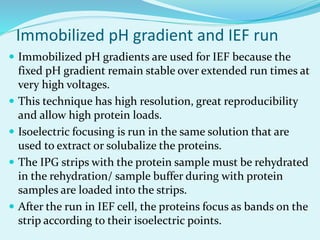
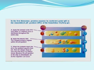
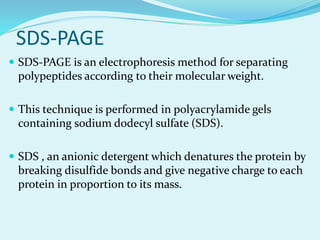
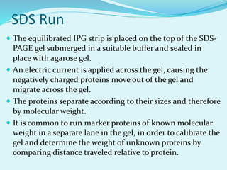
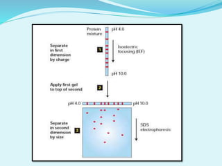
Submitted by-
Vivek kumar
M.Sc Microbiology
Bangalore University
Introduction
 2D-PAGE is a widely used method for the analysis of
complex protein mixture extracted from cells, tissues,
or other biological samples.
 This techniques was first developed by O’Farrell and
Klose in 1975.
 2D-PAGE is used in 2 sequential steps-
1. Isoelectric focusing
2. SDS-PAGE
Sample preparation
 Must select appropriate method to get selected proteins
from cellular compartment of interest.
 Must break all non-covalent protein-protein, protein-DNA,
protein-lipid interaction, disrupt S-S bonds.
 Must prevent proteolysis, accidental phosphorylation,
oxidation, cleavage.
 Must remove substance that might interfere with
separation process such as salts, polar detergents (SDS),
lipids, polysaccharides, nucleic acid.
 Must try to keep proteins soluble during both phases of
electrophoresis process.
Isoelectric focusing(IEF)
 In IEF, proteins are separated by electrophoresis in a pH
gradient based on their isoelectric point (pI).
 A pH gradient is generated in the gel and an electric
potential is applied across the gel.
 At all pH other than their isoelectric point, protein will be
charged.
 The isoelectric point (pI) is the specific pH at which the
net charge of protein is zero.
 At its isoelectric point, since the protein molecule carry no
net charge it accumulates or focuses into a sharp band.
Immobilized pH gradient and IEF run
 Immobilized pH gradients are used for IEF because the
fixed pH gradient remain stable over extended run times at
very high voltages.
 This technique has high resolution, great reproducibility
and allow high protein loads.
 Isoelectric focusing is run in the same solution that are
used to extract or solubalize the proteins.
 The IPG strips with the protein sample must be rehydrated
in the rehydration/ sample buffer during with protein
samples are loaded into the strips.
 After the run in IEF cell, the proteins focus as bands on the
strip according to their isoelectric points.
SDS-PAGE
 SDS-PAGE is an electrophoresis method for separating
polypeptides according to their molecular weight.
 This technique is performed in polyacrylamide gels
containing sodium dodecyl sulfate (SDS).
 SDS , an anionic detergent which denatures the protein by
breaking disulfide bonds and give negative charge to each
protein in proportion to its mass.
 SDS linearizes the protein so that they may be separated
strictly by molecular weight.
 Protein may be further treated with reducing agent, such
as DTT or TRP to break any reformed disulfide bonds and
then alkalated with iodoacetamide to prevent reformation
of disulfide bonds.
 Tracking dye may be added to the protein solution to track
the progress of the protein solution through the gel during
the electrophoretic run.
SDS Run
 The equilibrated IPG strip is placed on the top of the SDS-
PAGE gel submerged in a suitable buffer and sealed in
place with agarose gel.
 An electric current is applied across the gel, causing the
negatively charged proteins move out of the gel and
migrate across the gel.
 The proteins separate according to their sizes and therefore
by molecular weight.
 It is common to run marker proteins of known molecular
weight in a separate lane in the gel, in order to calibrate the
gel and determine the weight of unknown proteins by
comparing distance traveled relative to protein.
Visualization
 After electrophoresis the gel is stained to visualize the
separated proteins.
 Commonly used stained are Coomassie Brilliant Blue or
SYPRO RUBY or Sliver Stain, different proteins will appear
as distinct spot within gel.
Application
 To protein can be separated in pure form from spots which
can be quantified and also analyzed by MS.
 To study of gene products at molecular level.
 To study proteomics.
 Provides important information correlating the absence or
presence of individual protein characteristic of specific
clinical conditions.
Disadvantages
 This technique include a large amount of sample handling,
less reproducibility.
 It is also not automated for high throughput analysis. 2D-
PAGE has limited dynamic range.
 Difficulty to separate low abundance proteins, acidic and
basic proteins, very large and very small proteins and
hydrophobic proteins.
Reference
 Rabilloud T, Lelong C. Two-dimensional gel
electrophoresis in proteomics; a tutorial. 2011; page
1829-1841.
 J Mammary Gland Biol Neoplasia. The application of
2D gel. 2002. page 385-393.

2d Page

  • 1.
    Submitted by- Vivek kumar M.ScMicrobiology Bangalore University
  • 2.
    Introduction  2D-PAGE isa widely used method for the analysis of complex protein mixture extracted from cells, tissues, or other biological samples.  This techniques was first developed by O’Farrell and Klose in 1975.  2D-PAGE is used in 2 sequential steps- 1. Isoelectric focusing 2. SDS-PAGE
  • 3.
    Sample preparation  Mustselect appropriate method to get selected proteins from cellular compartment of interest.  Must break all non-covalent protein-protein, protein-DNA, protein-lipid interaction, disrupt S-S bonds.  Must prevent proteolysis, accidental phosphorylation, oxidation, cleavage.  Must remove substance that might interfere with separation process such as salts, polar detergents (SDS), lipids, polysaccharides, nucleic acid.  Must try to keep proteins soluble during both phases of electrophoresis process.
  • 4.
    Isoelectric focusing(IEF)  InIEF, proteins are separated by electrophoresis in a pH gradient based on their isoelectric point (pI).  A pH gradient is generated in the gel and an electric potential is applied across the gel.  At all pH other than their isoelectric point, protein will be charged.  The isoelectric point (pI) is the specific pH at which the net charge of protein is zero.  At its isoelectric point, since the protein molecule carry no net charge it accumulates or focuses into a sharp band.
  • 6.
    Immobilized pH gradientand IEF run  Immobilized pH gradients are used for IEF because the fixed pH gradient remain stable over extended run times at very high voltages.  This technique has high resolution, great reproducibility and allow high protein loads.  Isoelectric focusing is run in the same solution that are used to extract or solubalize the proteins.  The IPG strips with the protein sample must be rehydrated in the rehydration/ sample buffer during with protein samples are loaded into the strips.  After the run in IEF cell, the proteins focus as bands on the strip according to their isoelectric points.
  • 8.
    SDS-PAGE  SDS-PAGE isan electrophoresis method for separating polypeptides according to their molecular weight.  This technique is performed in polyacrylamide gels containing sodium dodecyl sulfate (SDS).  SDS , an anionic detergent which denatures the protein by breaking disulfide bonds and give negative charge to each protein in proportion to its mass.
  • 9.
     SDS linearizesthe protein so that they may be separated strictly by molecular weight.  Protein may be further treated with reducing agent, such as DTT or TRP to break any reformed disulfide bonds and then alkalated with iodoacetamide to prevent reformation of disulfide bonds.  Tracking dye may be added to the protein solution to track the progress of the protein solution through the gel during the electrophoretic run.
  • 10.
    SDS Run  Theequilibrated IPG strip is placed on the top of the SDS- PAGE gel submerged in a suitable buffer and sealed in place with agarose gel.  An electric current is applied across the gel, causing the negatively charged proteins move out of the gel and migrate across the gel.  The proteins separate according to their sizes and therefore by molecular weight.  It is common to run marker proteins of known molecular weight in a separate lane in the gel, in order to calibrate the gel and determine the weight of unknown proteins by comparing distance traveled relative to protein.
  • 12.
    Visualization  After electrophoresisthe gel is stained to visualize the separated proteins.  Commonly used stained are Coomassie Brilliant Blue or SYPRO RUBY or Sliver Stain, different proteins will appear as distinct spot within gel.
  • 13.
    Application  To proteincan be separated in pure form from spots which can be quantified and also analyzed by MS.  To study of gene products at molecular level.  To study proteomics.  Provides important information correlating the absence or presence of individual protein characteristic of specific clinical conditions.
  • 14.
    Disadvantages  This techniqueinclude a large amount of sample handling, less reproducibility.  It is also not automated for high throughput analysis. 2D- PAGE has limited dynamic range.  Difficulty to separate low abundance proteins, acidic and basic proteins, very large and very small proteins and hydrophobic proteins.
  • 15.
    Reference  Rabilloud T,Lelong C. Two-dimensional gel electrophoresis in proteomics; a tutorial. 2011; page 1829-1841.  J Mammary Gland Biol Neoplasia. The application of 2D gel. 2002. page 385-393.