Intracranial vascular malformations
•Download as PPTX, PDF•
3 likes•402 views

Intracranial vascular malformations by Dr. Gobardhan Thapa

Recommended
Recommended
More Related Content
What's hot
What's hot (20)
Similar to Intracranial vascular malformations
Similar to Intracranial vascular malformations (20)
More from Milan Silwal
More from Milan Silwal (20)
Recently uploaded
Recently uploaded (20)
Intracranial vascular malformations
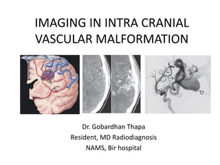
- 1. IMAGING IN INTRA CRANIAL VASCULAR MALFORMATION Dr. Gobardhan Thapa Resident, MD Radiodiagnosis NAMS, Bir hospital
- 2. Etiology • Most intra-cranial vascular malformations (CVMs) are congenital lesions and represent morphogenetic errors affecting arteries, capillaries, veins, or a combination of these elements. • Mutations in various components of the vasculogenesis (angiogenetic systems) have been implicated in the development of various CVMs.
- 3. Classification • CVMs have been traditionally classified by histopathology into four major types: 1. Arteriovenous malformations (AVMs), 2. Venous angiomas (developmental venous anomalies), 3. Cavernous malformations. 4. Capillary telangiectasias (sometimes simply termed “telangiectasia” or “telangiectasis”)
- 4. Functional classification: CVMs that display arteriovenous shunting – Arteriovenous malformation – Dural venous fistula – Vein of Galen malformation CVMs without AV shunting – Developmental venous anomaly (DVA) – Sinus pericranii – Cavernous malformations – Capillary telangiectasia • The former are potentially amenable to endovascular intervention; • the latter are either treated surgically or left alone.
- 5. CVMs with Arteriovenous Shunting: Arteriovenous Malformation (AVM) • tightly packed tangle of thin- walled vessels with direct arterial to venous shunting. • No intervening capillary bed. • Mostly parenchymal lesions and are also called “pial AVMs,” although mixed pial- dural malformations do occur. Fig. Graphic depicts AVM nidus with intranidal aneurysm , feeding artery (“pedicle”) aneurysm , and enlarged draining veins .
- 6. • Mostly solitary (~98%). • Multiple AVMs are almost always syndromic (2%) - Common associations: – hereditary hemorrhagic telangiectasia (HHT, also known as Rendu-Osler-Weber disease) and – segmental neurovascular syndromes called cerebrofacial arteriovenous metameric syndrome (CAMS). • E.g. Wyburn-Mason syndrome (AVMs in retina and brain).
- 7. • Tiny (“micro” AVMs) to giant lesions occupying most of a cerebral hemisphere (usually 2-6 cm). • Grossly, compact ovoid or pyramidal lesions. • Broadest surface at or near the cortex, and the apex toward the ventricles. • The brain surrounding an AVM often appears abnormal e.g. “perinidal” capillary bed in some cases. • Hemorrhagic residue in adjacent brain are common, as are gliosis and secondary ischemic changes. Fig. Autopsy case demonstrates a classic AVM. The nidus contains no normal brain. An intranidal aneurysm is present.
- 8. • Peak age between 20-40 years of age, ~25% patients with an AVM - symptomatic by age 15. • No gender predilection. • Most common presentation: Headache with parenchymal hemorrhage (50%). Seizure and focal neurologic deficits are the initial symptoms in 25% each.
- 9. • Size – Small (< 3 cm) = 1 – Medium (3-6 cm) = 2 – Large (> 6 cm) = 3 • Eloquence of Adjacent Brain – Noneloquent = 0 – Eloquent = 1 • Venous Drainage – Superficial only = 0 – Deep component = 1 Eloquent areas: sensorimotor cortex, visual cortex, deep nuclei, internal capsule, thalamus, hypothalamus, brain stem, cerebellar peduncle AVMs are graded on a scale from 1-5 based on the sum of “scores” Spetzler-Martin (SM) scale
- 11. TREATMENT OPTIONS. • Embolization, • surgery, • stereotactic radiosurgery, • or a combination of treatments are all.
- 12. Imaging Three distinct components: • Feeding arteries, • A central nidus, and • Draining veins
- 13. CT FINDINGS • “bag of worms” formed by a tightly packed tangle of vessels with little or no mass effect on adjacent brain. • NECT - numerous well- delineated, slightly hyperdense serpentine vessels. • Calcification common. • Enhancement of all three AVM components (feeding arteries, nidus, draining veins) -typically intense and uniform on CECT scans Fig. (Left) NECT shows serpentine hyperdensities . (Right) CECT shows strong uniform enhancement. Wedge-shaped configuration is typical for AVM.
- 14. MR FINDINGS • High-flow lesions - tightly packed mass or a “honeycomb” of “flow voids” on both T1- and T2 scans. • Brain parenchyma within an AVM is typically gliotic and hyperintense on T2WI and FLAIR. • Contrast enhancement of AVMs is variable, depending on flow rate and direction. Draining veins typically enhance strongly and uniformly . • Hemorrhagic residue are common - Foci of “blooming” both within and around AVMs. Fig. A. Axial T1WI in a 32-year old man with headache shows a classic wedge-shaped left parietal AVM with multiple serpentine “flow voids” . A few linear foci of T1 shortening represent thrombosed vessels within the nidus. B. T2WI in the same patient nicely demonstrates the wedge of “flow voids” Fig. C. FLAIR scan demonstrates minimal hyperintensity within and around the AVM , suggesting small foci of gliotic brain; D. T1 C+ scan shows some linear and serpentine areas of enhancement that are mostly in draining veins.
- 15. ANGIOGRAPHY (CTA, MRA, DSA) • The feeding arteries - often enlarged and tortuous ; a flow-induced ‘’pedicle” aneurysm’’ (~10-15% of cases). • Nidus - a tightly packed tangle of abnormal arteries and veins without an intervening capillary bed. • ~ 50% - at least one aneurysmally dilated vessel (“intranidal aneurysm”). • Draining veins typically opacify in the mid- to late-arterial phase (“early draining” veins). Veins draining AVMs are typically enlarged, tortuous, and may become so prominent that they form varices and exert local mass effect on the adjacent cortex. • Stenosis of one or more “outlet” draining veins may elevate intranidal pressure and contribute to AVM hemorrhage.
- 16. Fig. E. Lateral DSA shows enlarged MCA, ACA feeding vessels with a tangle of smaller vessels in the wedge shaped nidus . Faint opacification of the superior sagittal sinus represents arteriovenous shunting of contrast. F. Late arterial phase of the DSA shows the nidus and “early draining” veins emptying into the SSS . No deep venous drainage was identified.
- 17. Fig. Three dimensional angiogram showing the brain AVM (arrow). The angiogram permits a more detailed view of the AVM and the blood vessels that surround it.
- 18. Cerebral Proliferative Angiopathy (diffuse nidus type AVM) • rare entity characterized by diffuse angiogenesis and progressive hyper vascular shunting. • Large lesions that can occupy most of a lobe or even an entire cerebral hemisphere. • Unlike classic BAVMs; CPAs - normal brain parenchyma interspersed between the proliferative vascular channels and absence of early venous drainage.
- 19. • presentation with seizure (45%), severe headache (40%), or progressive neurologic deficits. Only 12% present with a hemorrhagic event. • Mean age at symptom onset is 22 years. There is a 2:1 female predominance.
- 20. Imaging • CT/MR - large (usually more than six centimeters), diffusely dispersed network of innumerable dilated vascular spaces intermingled with normal brain parenchyma. Dense enhancement following contrast. Fig. Proliferative type brain AVM in a 27-year-old woman who presented with a 6-year history of headaches and seizures. Axial CECT - enhancing vascular lesion in the left parasagittal frontal lobe, with internal focal isoattenuating areas representing normal brain parenchyma interspersed within the nidus.
- 21. Fig. Proliferative angiopathy in a 26-year-old man with a 6-year history of progressive left-sided weakness. Axial proton-density–weighted (a) and gadolinium-enhanced T1-weighted (b) MR images show multiple flow voids and contrast-enhanced tubular structures representing a large vascular lesion that involves the entire right cerebral hemisphere. The normal brain parenchyma is interspersed between the abnormal vessels.
- 22. DSA • Well-circumscribed nidus - absent. Instead, multiple small caliber non-dominant feeding arteries present. • The draining veins only moderately enlarged relative to the striking extent of the vascular abnormality. • Despite their large size, flow related aneurysms are not a feature of CPA. Fig. DSA of selective internal carotid angiogram in a patient with cerebral proliferative angiopathy shows innumerable dilated vascular spaces with no dominant feeding arteries. Fig. Selective vertebral angiogram in the same patient shows additional small feeding vessels supplying the lesion. Despite its size, there are unopacified spaces within the lesion.
- 23. Dural arteriovenous fistula (dAVF) • second major type of cerebrovascular malformation - Much less common than AVMs • network of tiny, crack- like vessels that shunt blood between meningeal arteries and small venules within the wall of a dural venous sinus. Fig. Graphic depicts dAVF with thrombosed transverse sinus with multiple tiny arteriovenous in the dural wall . Lesion is mostly supplied by transosseous feeders from the external carotid artery.
- 24. Pathology • Most common locations in adults - transverse, sigmoid, and cavernous sinuses. • The superior sagittal sinus more common in children. • Size varies from tiny single vessel shunts to massive complex lesions with multiple feeders and arteriovenous shunts in the sinus wall.
- 25. • 10-15% of all intracranial vascular malformations with arteriovenous shunting. • Peak age is 40-60 years, roughly 20 years older than the peak age for AVMs. • No gender predilection. • Uncomplicated dAVFs in the transverse/sigmoid sinus region - either bruit and/or tinnitus. • dAVFs in the cavernous sinus - pulsatile proptosis, chemosis, retroorbital pain, bruit, and ophthalmoplegia. • “Malignant” dAVFs, lesions with cortical venous drainage - seizures and progressive dementia in addition to focal neurologic deficits.
- 26. Imaging: CT findings • Hemorrhage: uncommon in the absence of cortical venous drainage or dysplastic venous dilatation. • An enlarged dural sinus or draining vein can sometimes seen in NECT. Carotid- cavernous fistulas - an enlarged superior ophthalmic vein. • Dilated transcalvarial channels from enlarged transosseous feeding arteries can occasionally be seen on bone CT images. • CECT - enlarged feeding arteries and draining veins. The involved dural venous sinus is often thrombosed or stenotic. Fig. A. CTA source image in a patient with right- sided tinnitus shows no obvious abnormality, although the right sigmoid sinus looks peculiar. B. Bone CT in the same patient shows multiple enlarged transosseous vascular channels in the squama of the right occipital bone.
- 27. MR FINDINGS • Dilated cortical veins without an identifiable nidus adjacent to normal-appearing brain may suggest the presence of a dAVF. • Mostly, a thrombosed dural venous sinus containing vascular-appearing “flow voids”. • Thrombus is typically isointense with brain on T1- and T2 scans and “blooms” on T2* sequences. Chronically thrombosed sinuses may enhance. Fig. C. CE MRA source image shows dural sinus thrombosis , multiple enhancing vascular channels characteristic of posterior fossa dAVF. D. MRA in the same patient shows innumerable tiny feeding arteries supplying a dAVF at the transverse-sigmoid sinus junction. The sinus has partially recanalized , and the distal sigmoid sinus and jugular bulb are partially opacified.
- 28. ANGIOGRAPHY • CTA/CTV; DSA (best imaging tool with superselective Catheterization) • As most dAVFs arise adjacent to the skull base, multiple enlarged dural and transosseous branches arising from the external carotid artery are usually present. • Dural branches may also arise from the internal carotid and vertebral arteries. Fig. A. DSA of the external carotid artery in a patient with tinnitus, dAVF in the occluded transverse sinus supplied by the middle meningeal artery , transosseous branches from the ECA.
- 29. Cognard Classification • Grade 1: In sinus wall; normal antegrade venous drainage (low risk; benign clinical course) • Grade 2A: In sinus; reflux to sinus, not cortical veins • Grade 2B: Reflux (retrograde drainage) into cortical veins (10-20% hemorrhage) • Grade 3: Direct cortical venous drainage; no venous ectasia (40% hemorrhage) • Grade 4: Direct cortical venous drainage + venous ectasia (65% hemorrhage) • Grade 5: Spinal perimedullary venous drainage Borden Classification • Type I: Dural arterial supply with antegrade drainage into venous sinus • Type Ia: Simple dAVF with single meningeal arterial supply • Type Ib: Complex dAVF with multiple meningeal arteries • Type II: Dural supply + ↑ intrasinus pressure → antegrade sinus, retrograde cortical venous drainage • Type III: Dural arteries drain into cortical veins
- 30. Carotid-Cavernous Fistula • CCFs are divided into two subgroups, direct and indirect fistulas. • “Direct” CCFs are typically high-flow lesions that result from rupture of the cavernous internal carotid artery (ICA) directly into the cavernous sinus (CS), with or without a preexisting ICA aneurysm. • “Indirect” CCFs are usually slow-flow, low-pressure lesions that represent an arteriovenous fistula between dural branches of the cavernous ICA and the cavernous sinus. Fig. Coronal graphic depicts a carotid- cavernous fistula (CCF). The right cavernous sinus is enlarged by numerous dilated arterial and venous channels.
- 31. • CCFs usually acquired lesions; traumatic or non-traumatic in origin. • Most direct CCFs - traumatic, usually secondary to central skull base fractures. Either stretch injury to the ICA or direct puncture from a bony fracture fragment. • A single-hole laceration/transection of the cavernous ICA with direct fistulization into the CS is the typical finding. • Spontaneous (i.e., nontraumatic) rupture of a preexisting cavernous ICA aneurysm is less common. • Indirect CCFs - nontraumatic lesions and thought to be degenerative in origin. In contrast to dAVFs elsewhere, indirect CCFs rarely occur as sequelae of dural sinus thrombosis.
- 32. • Grossly, direct CCF - arterialized flow causes dilatation of the CS with venous hypertension and retrograde flow into the superior and inferior ophthalmic veins. • Indirect CCFs demonstrate enlarged crack-like vessels within the CS that resemble those seen in typical dAVFs elsewhere. Fig. Clinical photograph of a patient with a CCF shows numerous enlarged scleral vessels.
- 33. BARROW CLASSIFICATION OF CAROTID-CAVERNOUS FISTULAS Type A: Direct ICA-cavernous sinus high-flow shunt Type B: Dural ICA branches- cavernous sinus shunt Type C: Dural ECA-cavernous sinus shunt Type D: Both ICA/ECA dural branches shunt to CS
- 34. Clinical Issues • Indirect CCF is the second most common site of intracranial dAVF, following the transverse/sigmoid sinus junction. • Indirect CCFs are most frequent in women 40-60 years of age. • As direct CCFs typically occur with trauma, they are found in both genders and at all ages. • Direct high-flow CCFs are much less common. • Direct CCFs may present within hours to days or even weeks following trauma. Bruit, pulsatile exophthalmos, orbital edema, decreasing vision, glaucoma, and headache are typical. In severe cases, vision loss may be rapid and severe. Cranial neuropathy may occur but is less common.
- 35. Imaging CT FINDINGS • NECT - mild or striking proptosis, a prominent CS with enlarged superior ophthalmic vein (SOV), and enlarged extraocular muscles. • “Dirty” fat secondary to edema and venous engorgement • Occasionally, subarachnoid hemorrhage from trauma or ruptured cortical veins. • CECT - enlarged SOV and CS; Fig. CECT scan shows classic findings of CCF. The right cavernous sinus is enlarged [BULGING CAVERNOUS SINUS], and the ipsilateral superior ophthalmic vein is more than 4 times the size of the left superior ophthalmic vein.
- 36. MR FINDINGS • T1 - prominent “bulging” CS and SOV as well as “dirty” orbital fat. • T2 - multiple “flow voids” in the CS. • Strong, uniform enhancement of the CS and SOV is typical. • Enlarged, tortuous intracranial veins may occur with high-flow, high- pressure shunts. Fig. T2WI shows typical MR findings of CCF with an enlarged right cavernous sinus containing numerous abnormal “flow voids”
- 37. ANGIOGRAPHY • Direct CCFs - rapid flow with very early opacification of the CS. • A single-hole fistula is usually present, typically between the C4 and C5 ICA segments. Fig. Lateral DSA in a case of direct CCF in a 21-year-old woman with multiple skull base fractures shows that the ICA narrows before terminating in a large venous pouch . High-pressure venous reflux into the superior and inferior ophthalmic veins and the sphenoparietal sinus is present.
- 38. Pial arterio-venous fistula (pAVF) • rare vascular malformation; a single dilated pial artery connecting directly to an enlarged cortical draining vein. • No intervening capillary bed or nidus. • Unlike dural AVFs, 80% of pAVFs are supratentorial. • Typically lie on or just within the brain surface or adjacent to the ventricular ependyma. • supplied by branches of the anterior, middle, or posterior cerebral arteries and are usually associated with a venous varix. Fig. Pial AVF with slightly enlarged ACA branches connecting to a venous varix , dilated cortical draining vein
- 39. Fig. Coronal T1 C+ scan shows a pial AVF in the posterior fossa. A small cerebellar artery connects directly to a venous pouch , which in turn drains into a subependymal vein near the fourth ventricle.
- 40. Vein of Galen Aneurysmal Malformation • most common extra-cardiac cause of high output cardiac failure in newborns. • direct arteriovenous fistula between deep choroidal arteries and a persistent embryonic precursor of the vein of Galen. • flow-related aneurysmal dilatation of this primitive vein, forming a large midline venous pouch that lies behind the third ventricle. Fig. Graphic illustrates vein of Galen malformation. Enlarged choroid arteries drain directly into dilated median prosencephalic vein (MPV) , falcine sinus . Torcular herophili (venous sinus confluence) is massively enlarged.
- 41. • Normal fetal development, arterial supply to the choroid plexus drains via a single transient midline vein, the median prosencephalic vein (MPV) of Markowski. • Normally, the developing internal cerebral veins annex drainage of the fetal choroid plexus, and the MPV regresses. • In a VGAM, a high-flow fistula prevents formation of the definitive vein of Galen.
- 42. Pathology • Grossly - enlarged arteries drain directly into a dilated MPV. • “Aneurysmal” dilatation of the persistent MPV forms a large venous pouch behind the third ventricle that often drains into a markedly enlarged superior sagittal sinus via an embryonic falcine sinus. • The ventricles are often markedly dilated. • The brain is frequently atrophic. • Ischemic changes are common.
- 43. • < 1% of all CVM but ~30% of symptomatic vascular malformations in children. • Neonatal VGAMs are more common than those presenting in infancy or childhood. Adult presentation is rare. • (M:F = 2:1). • Neonates, high-output congestive heart failure and a loud cranial bruit are typical. Older infants may present with macrocrania and hydrocephalus, with or without heart failure. • VGAMs in older children are often associated with developmental delay and seizures. • VGAMs in young adults typically present with headache with or without hemorrhage and hydrocephalus.
- 44. Imaging: CT findings • NECT - enlarged, well delineated, mildly hyperdense mass at the tentorial apex, usually compressing the third ventricle and causing severe obstructive hydrocephalus. • Variable encephalomalacia, hemorrhage, and/or dystrophic calcification in the brain parenchyma. • CECT - strong uniform enhancement. Fig. CECT scan in a newborn demonstrates a massive VGAM draining into an enlarged falcine sinus, causing obstructive hydrocephalus.
- 45. MR FINDINGS • Enlarged arterial feeders are usually seen as serpentine “flow voids” adjacent to the lesion. • Thrombus of varying ages may be present lining the VGAM. Fig. Sagittal T2WI shows prominent arteries supplying an enlarged median prosencephalic vein. Note enlarged falcine sinus .
- 46. ANGIOGRAPHY • Two forms of VGAM are recognized based on their specific angioarchitecture. • In more than 50% of all VGAMs, the straight sinus is hypoplastic or absent and venous drainage is into a persistent embryonic “falcine sinus.” • The falcine sinus is easily identified as it angles posterosuperiorly toward the superior sagittal sinus. Fig. DSA in the same patient shows that the VGAM is supplied by multiple direct arterial fistulas
- 47. ULTRASOUND diagnosed antenatally • hypoechoic to mildly echogenic midline mass behind the third ventricle is typical. • Color Doppler shows bidirectional turbulent flow within the VGAM Fig. Neonatal transcranial US shows a large VGAM posterior to the 3rd ventricle. Prominent vessels with arterial flow supply the lesion.
- 48. CVMs without Arteriovenous Shunting Developmental Venous Anomaly • With the advent of contrast-enhanced MR, developmental venous anomalies have become the most frequently diagnosed intracranial vascular malformation (60% of CVM of brain). • Once thought to be rare lesions with substantial risk of hemorrhage, the vast majority of venous “angiomas” are now recognized as asymptomatic and incidental imaging findings. • Neurologic complications are rare.
- 49. • an umbrella-shaped congenital cerebral vascular malformation composed of angiogenically mature venous elements. • Dilated, thin-walled venous channels lie within (and are separated by) normal brain parenchyma. Fig. Autopsy case shows left frontal DVA as dilated medullary veins interspersed with normal brain. Fig. Graphic depicts DVA with enlarged medullary veins draining into a single transmantle collector vein .
- 50. • In the deep white matter (WM), adjacent to the frontal horn of the lateral ventricle. • The second most common location is next to the fourth ventricle. • Size varies from tiny, almost imperceptible lesions to giant DVAs that can involve most of the hemispheric WM. • A cluster of variably sized enlarged medullary (WM) veins embedded within brain parenchyma
- 51. • found in patients of all ages without gender predilection. • Mostly incidentally at autopsy or on imaging studies. • 98% of all DVAs are asymptomatic. • Two percent - hemorrhage or infarct, probably caused by stenosis or spontaneous thrombosis of the outlet collector vein.
- 52. • Most DVAs solitary; unless associated with a vascular neurocutaneous syndrome such as blue rubber bleb nevus syndrome. • DVAs may coexist with a sinus pericranii. Sinus pericranii is typically the cutaneous sign of an underlying venous anomaly. DVAs are also associated with periorbital lymphatic/lymphaticovenous malformations.
- 53. • No treatment is required or recommended for solitary DVAs (they are “leave me alone!” lesions). • If a DVA is histologically mixed, treatment is determined by the coexisting lesion. Preoperative identification of such mixed malformations is important as ligating the collector vein or removing its tributaries may result in venous infarction.
- 54. Imaging • NECT - usually normal unless the DVA is very large and a prominent draining vein is present. • CECT - numerous linear and/or punctate enhancing foci that converge on a well- delineated tubular collector vein. Fig. CECT, CTA depict classic DVA in the left cerebellar hemisphere
- 55. MR FINDINGS • Small DVA - may be undetectable unless contrast-enhanced scans are obtained. • T1 C+ sequences - a stellate collection of linear enhancing structures converging on the transparenchymal or subependymal collector vein. • The collector vein may show variable high-velocity signal loss. Because flow in the venous radicles of a DVA is typically slow, blood deoxygenates and T2* scans (GRE, SWI) show striking linear hypointensities. • If a DVA is mixed with a cavernous malformation, blood products in various stages of degradation may be present and “bloom” on T2* sequences. Fig. T1 C+ scan shows a classic DVA with enlarged WM veins and a collector vein draining into the anterior aspect of the superior sagittal sinus.
- 56. ANGIOGRAPHY • The arterial phase is normal. • The venous phase shows the typical hair-like collection (“Medusa head/inverted umbrella”) of dilated medullary veins within the white matter. Fig. 3D SSD demonstrates a classic DVA with enlarged medullary veins draining into the collector vein. The appearance resembles a “Medusa head,” “upside- down willow tree” or “umbrella.”
- 57. Sinus Pericranii • large transcalvarial communication between the intra- and extracranial venous drainage systems. • A bluish sac beneath or just above the periosteum of the calvaria is typical. The dilated, blood-filled sac connects through an enlarged emissary vein with the intracranial circulation. Fig. Coronal graphic depicts a classic sinus pericranii (SP) with an expanded venous pouch under the scalp connecting to the intracranial venous system through a transcalvarial channel . Some SPs are associated with a developmental venous anomaly .
- 58. • The frontal lobe is the most common site, followed by the parietal and occipital lobes. • SPs in the middle and posterior cranial fossae are rare. • SP may be associated with single or multiple intracranial DVAs.
- 59. • Rare lesions (<10% of patients who present for treatment of craniofacial vascular malformations and 4% of patients with palpable scalp/cranial vault lesions). • Mostly in children or young adults; No gender predilection. • A nontender, non-pulsatile somewhat bluish compressible scalp mass that increases with Valsalva maneuver and reduces in the upright position is typical. • A history of “forgotten trauma” is not uncommon. • Mostly asymptomatic, other than their cosmetic effect. • SP with multiple DVAs is associated with blue rubber bleb nevus syndrome.
- 60. • Most SPs behave benignly and remain stable in size. • Surgical removal of the extracranial component with cranioplasty is occasionally performed for cosmetic purposes. • Surgery without adequate imaging may result in potentially lethal complications including hemorrhage, venous infarction (if the SP is associated with a DVA), and air embolism.
- 61. Imaging • A vascular or subperiosteal scalp mass overlies a well-defined bone defect. The mass communicates directly with the intracranial venous system through the bony defect. • CT - iso- or hyperdense on NECT and shows strong uniform enhancement after contrast administration. • Occasionally an SP may contain calcifications (phleboliths) or thrombi. Fig. Sagittal CTV shows a small sinus pericranii connecting to the superior sagittal sinus through an adjacent skull defect.
- 62. MR FINDINGS • Most SPs are isointense on T1WI and hyperintense to brain on T2WI. • “Puddling” of contrast within the SP on T1 C+ is typical unless the lesion is unusually large and flow is rapid. • MRV is helpful in delineating both the intra- and extracranial components. Fig. Coronal contrast enhanced MR scan shows a classic sinus pericranii that connects to the superior sagittal sinus via a small transcalvarial venous channel.
- 63. ANGIOGRAPHY • The arterial and capillary phases are normal. Mostly seen only on the very late venous phase. • well-defined rounded pools of contrast that slowly accumulate within and adjacent to the skull defect containing the transcalvarial vein. • Flow is variable and often bidirectional. ULTRASOUND • Color Doppler may delineate the extracranial component and define flow direction. • US does not define the intracranial component of an SP. Fig. Late venous phase DSA shows angiographic findings of sinus pericranii with enlarged venous pouches connecting directly to the superior sagittal sinus through a transcalvarial channel.
- 64. Cerebral Cavernous Malformation • Intracranial vascular malformation characterized by repeated “intralesional” hemorrhages into thin-walled, angiogenically immature, blood- filled locules called “caverns.” • Discrete, well-marginated lesions that do not contain normal brain parenchyma. • Most are surrounded by a complete hemosiderin rim. Fig. Subacute , classic “popcorn ball” appearances of CCMs. Microhemorrhages are seen as multifocal “blooming black dots”
- 65. • relatively common cause of spontaneous nontraumatic intracranial hemorrhage in young and middle-aged adults, although they can occur at any age. • Occur throughout CNS • Solitary (2/3), multiple (1/3, familial)
- 66. • Third most common CVM • At any age; peak = 40-60 years -- like DAVF • Course variable, unpredictable • Repeated intralesional hemorrhages typical Hemorrhage risk = 0.25-0.75% per lesion per year • Patients with familial CCM develop de novo lesions.
- 67. ZABRAMSKI CLASSIFICATION OF Cerebral CMs Type 1: Subacute hemorrhage Hyperintense on T1, hyper-/hypointense on T2 Type 2: Different age hemorrhages Classic = “popcorn ball” Mixed signal with hyper/hypo on both T1 and T2 Blood-filled locules with fluid-fluid levels Type 3: Chronic hemorrhage Type 4: Punctate microhemorrhages “Blooming black dots” on T2* (GRE, SWI)
- 68. Imaging • NECT: Hyperdense ± scattered Ca++ • MR: Appearance varies • “Popcorn ball” with fluid-fluid levels, hemosiderin rim • Multifocal “blooming black dots” • DSA usually negative Fig. T2WI shows classic “popcorn ball” appearance with locules of blood in different stages of evolution surrounded by hemosiderin rim
- 69. Capillary Telangiectasia • collection of enlarged, thin-walled vessels resembling capillaries. • The vessels are surrounded and separated by normal brain parenchyma. Fig. Graphic depicts pontine capillary telangiectasia with tiny dilated capillaries interspersed with normal brain.
- 70. • Cluster of thin-walled, dilated capillaries • Normal brain interspersed between vascular channels. • Can be found throughout CNS; Pons, cerebellum, spinal cord most common sites. Fig. Autopsy specimen shows a large pontine capillary telangiectasia . Note the transverse pontine fibers passing through the lesion.
- 71. • 10-20% of all cerebrovascular malformations • All ages; peak = 30-40 years • Rarely symptomatic • Most discovered incidentally at imaging
- 72. Imaging • NECT, CECT usually normal • MR - T1/T2 usually normal • T2* key sequence (dark gray hypointensity) • Brush-like enhancement on T1 C+
- 73. Fig. Series of images demonstrates classic findings of pontine capillary telangiectasia. A. Axial T1WI is normal. B. Axial T2WI in the same patient likewise shows no abnormality. C. FLAIR scan shows faint patchy hyperintensity in the pons. D. T2* GRE scan shows susceptibility effect with grayish hypointensity in the mid pons.
- 74. Fig. E. T1 C+ scan shows the brush-like faint enhancement that is characteristic of capillary telangiectasia. F. DTI fiber tracking is normal. The transverse pontine fibers cross undisturbed through the lesion.
- 75. summary TYPE ETIOLOG Y PATHOLO GY NUMB ER LOCATION PREVALE NCE AGE HHG RISK IMAGIN G CLUES AVM Congenit al Nidus+ar terial feeders, draining veins; no capillary bed Solitar y <2% multipl e) Parenchym a (85% supratento rial, 15% posterior fossa) 0.04- 0.5% populati on; 85- 90% of CVM with AV shunting Peak = 20-40 yrs Very high; 2-4% per yr cumulative Bag of worms, flow voids on MR
- 76. Dural AV Fistula (dAVF) Acquired (trauma; Dural sinus thrombo sis) Network of multiple AV microfist ula Solitar y Skull base; Dural sinus wall 10- 15% of CVMs with AV shunting Peak = 40- 60 years Varies With venous Draining (increased if Cortical veins involved) Enlarged Meninge al Arteries with network of Tiny vessels in wall of thrombo sed Dural venous sinus
- 77. Vein of Galen malformat ion (VGAM) Congenital (fetal arterial Fistula to Primitive precursor of vein of Galen) Large venous pouch Solitary Behind Third ventricle < 1% of CVMs with AV shunting Newborn > >infant, Older Child Low (but hydroceph alus brain damage common) Large midline venous varix in neonate With high output congestive Heart Failure
- 78. Develo pmental Venous anomaly (DVA) Congenit al (arrested Fetal medullar y Vein develop ment) Dilated WM veins; Normal brain in between Solitary (unless BRBNS) Deep WM, Usually near ventricle Most com mon CVM (60% of all), between 2-9% of the populati on Any Age Extremel y Low unless mixed with Caverno us malfora mtion “Medusa Head” of dilated WM veins Convergi ng on enlarged Collector Vein
- 79. Sinus pericranii (SP) Congenit al Bluish blood filled subcutan eous scalp Mass Solitary Scalp Rare Any age (usually child hood) Extremel y Low unless Direct trauma Vascular Scalp mass connecti ng through skull defect to intracran ial Venous circulatio n
- 80. Cavernous Malformat ion (CM) Congenital Collection Of blood filled “cave rns” with no normal Brain; complete Hemosider in Rim 2/3 solit ary (spor adic) ; 1/3 Multiple (familial) Throughou t Brain Any age (peak = 40- 60 years; younger in Familial CCM syndrome) High (0.25- 0.75% per Year; 1% per lesion Per year in familial) Varies; most common is solitary “popcorn Ball” (locules with blood fluid levels, Hemos iderin rim); multifocal “black dots” In familial
- 81. Capilla ry telangi ectasia (CT) Conge nital Dilated capillar ies; normal Brain in betwe en Solitar y > > > multIpl e Anywh ere But pons; medull a Most comm on 15-20% of all CVMs Any age (peak = 30-40 years) Extrem ely low unless mixed with cavern ous malfor mation Faint Brush- like enhanc ement, becom es Hypoin tense on T2*
- 82. References • Osborn's Brain Imaging; Anne G Osborn. • Radiologic Assessment of Brain Arteriovenous Malformations: What Clinicians Need to Know; Geibprasert et al, RadioGraphics 2010; 30:483–501
- 83. Thank you !!
Editor's Notes
- Development of the human fetal vascular system occurs via two related processes: Vasculogenesis and angiogenesis. In vasculogenesis, capillary-like tubes develop first and constitute the primary vascular plexus. This primary capillary network is subsequently remodeled into large caliber vessels (arteries, veins) and small capillaries. Angiogenesis is regulated by a number of inter-cell signaling and growth factors.
- Many interventional neuroradiologists and neurosurgeons group CVMs by function, not histopathology.
- Vessels comprising the AVM nidus are of variable caliber and wall thickness. Some appear dysplastic and thin-walled without normal subendothelial support. Others exhibit intimal hyperplasia and fibrosis/hyalinization. There are no capillaries and no normal brain parenchyma within an AVM nidus. Instead, varying amounts of laminated thrombus, dystrophic calcification, and hemorrhagic residua are often present. Small amounts of brain parenchyma within the nidus are occasionally identified but are typically gliotic and nonfunctional.
- NATURAL HISTORY - The lifelong risk of hemorrhage is estimated at 2-4% per year, cumulative. Annual hemorrhage risk increases with increasing age, deep brain location, and deep venous drainage. Risk ranges from 1% per year (in patients whose initial presentation was non-hemorrhagic) to nearly 35% per year for patients harboring all three risk factors.
- Spontaneous regression of sporadic brain AVMs is rare and unpredictable, occurring in approximately 1% of cases. Most “obliterated” AVMs follow a hemorrhagic episode, often with venous stasis, thrombosis, and elevated intracranial pressure. Rare non-hemorrhagic cases of spontaneous AVM regression have been reported.
- Flow-related angiopathy may be present, ranging from simple dilatation to endothelial thickening, stenosis, and occasionally even thrombosis and occlusion. Displacement of angiographic midline markers (e.g., the anterior cerebral arteries and internal cerebral veins) is therefore usually absent unless an acute hematoma is present.
- It is unclear whether CPA is a completely different disorder or an unusual subtype of AVM.
- Patients may have laboratory evidence of ongoing angiogenesis with elevated CSF levels of VEGF and bFGF. Bevacizumab, an antiangiogenesis monoclonal antibody that binds to VEGF, has been used in a few patients with inconclusive results.
- pCT and pMR - prolonged mean transit time and hypoperfusion abnormalities (“steal” phenomena) that extend far beyond the morphologic abnormalities.
- AVMs are approximately 10 times as common as dAVFs. Patients who present with intracranial hemorrhage or non-hemorrhagic neurologic deficits also have a higher risk of new adverse events than those with an asymptomatic fistula.
- Parenchymal hyperintensity on T2WI and FLAIR indicates venous congestion or ischemia, usually secondary to retrograde cortical venous drainage.
- An enlarged tentorial branch of the meningo-hypophyseal trunk commonly contributes to dAVFs at the transverse/sigmoid sinus junction.
- special type of arteriovenous shunt that develops within the cavernous sinus.
- Most indirect CCFs are found in the dural wall of the cavernous sinus and supplied via intracavernous branches of the ICA and deep (maxillary) branches of the ECA.
- In rare cases, rupture of an intracavernous ICA aneurysm may cause life-threatening epistaxis.
- Inferior drainage into a prominent pterygoid venous plexus and posterior drainage into the clival venous plexus are sometimes present.
- Rare cases of high-flow, aggressive direct CCFs with prominent pontomesencephalic and perimedullary venous drainage causing progressive myelopathy.
- Selective ICA injection with very rapid image acquisition is often necessary to localize the fistula site precisely.
- MICROSCOPIC FEATURES. The wall of the venous pouch may become significantly thickened and dysplastic.
- Rapid but turbulent flow in the VGAM causes inhomogeneous signal loss and phase artifact (signal misregistration in the phase-encoding direction).
- The most common is the “choroidal” form - multiple branches from the pericallosal, choroidal, and thalamoperforating arteries drain directly into an enlarged, aneurysmally dilated midline venous sac. In the rare “mural” form - single or a few enlarged branches from collicular or posterior choroidal arteries drain into the sinus wall.
- Developmental venous anomaly (DVA), also called venous “angioma” or “venous malformation,” Very rarely, a dilated or tortuous venous pouch without discernible arterial or venous tributaries occurs. In these unusual cases, the term venous varix is appropriate.
- Multiple cerebral venous malformations have been reported in blue rubber bleb nevus syndrome(BRBNS). MICROSCOPIC FEATURES. Thin-walled, somewhat dilated venous channels are interspersed in normal-appearing white matter. Occasionally, the vessel walls are thickened and hyalinized. Hemorrhage and calcification are uncommon unless the DVA is associated with a CCM.
- DVAs are occasionally associated with cortical dysplasia. In such cases, the cortical malformation may cause seizures. DVAs may coexist with other vascular lesions that cause symptomatic intracranial hemorrhage. The most common “histologically mixed” cerebrovascular malformation is a cavernous-venous malformation. Occasionally a “triad” malformation that consists of cavernous, venous, and capillary components is identified.
- In atypical DVAs, perfusion CT may show a venous congestion pattern with increased CBV, CBF, and MTT in the adjacent brain parenchyma.
- A faint, prolonged “blush” or capillary “stain” may be present in some cases. A transitional form of venous-arteriovenous malformation with enlarged feeders and AV shunting (“early draining” vein) occurs but is uncommon. Rarely, a true venous varix may occur with a DVA.
- Some investigators consider SP the cutaneous manifestation of an intracranial developmental venous anomaly (DVA) as the two lesions are often—but not invariably—associated.
- There is a very small lifetime risk of air embolism or hemorrhage from direct trauma to the SP.
- The underlying calvarial defect varies in size but is typically well-demarcated.
- In “closed” SPs, blood flows from and back into the adjacent dural venous sinus. “Drainer” SPs have unidirectional drainage into the venous pouch and adjacent pericranial scalp veins.