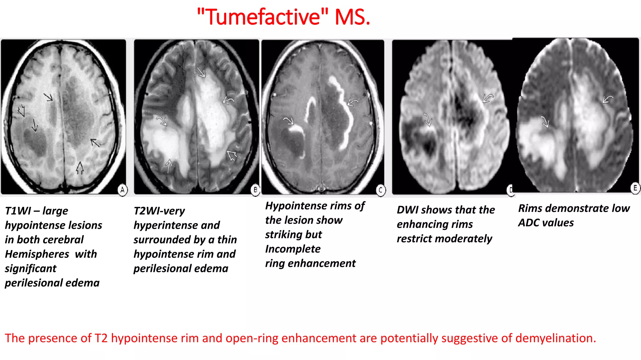
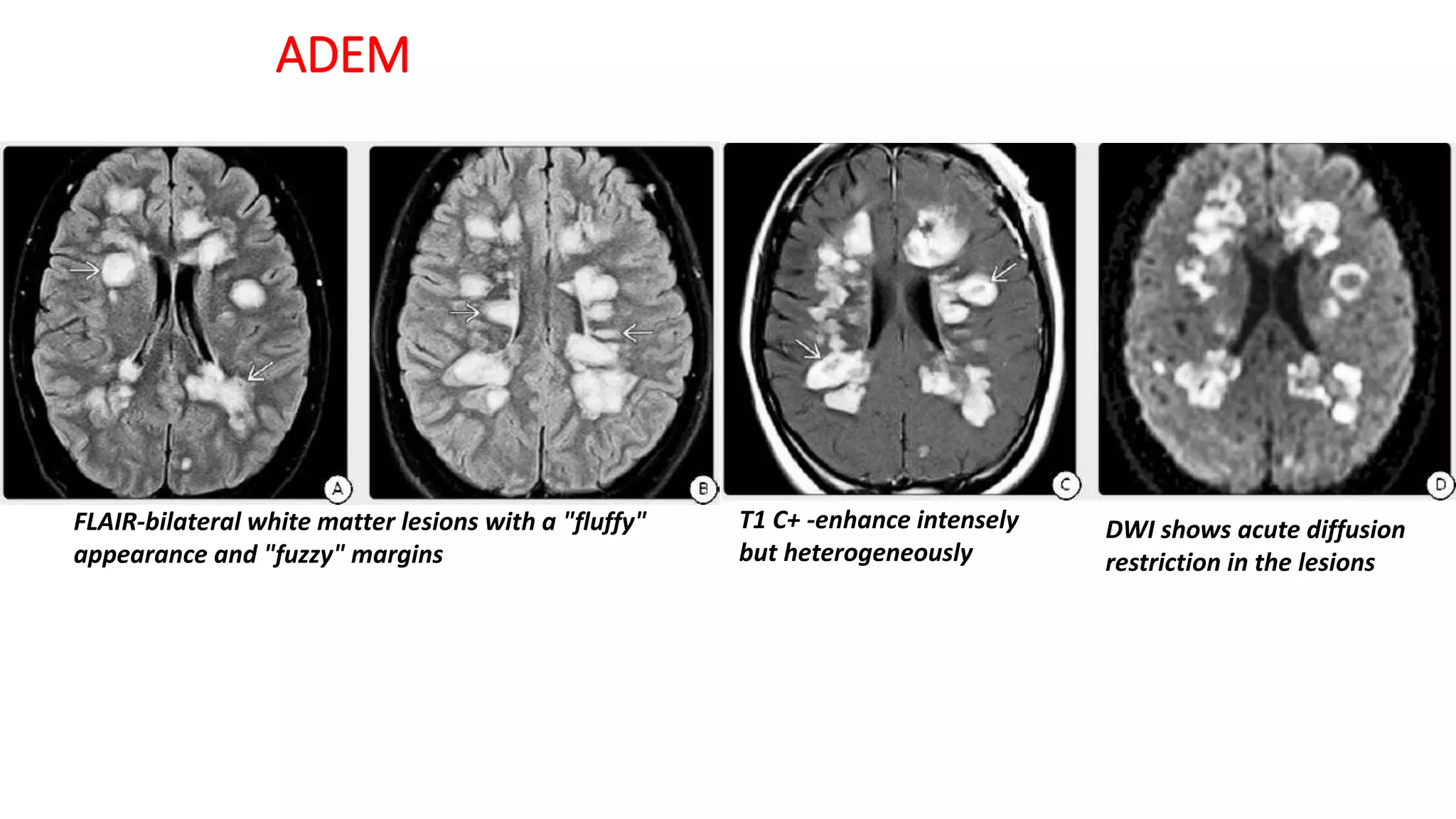
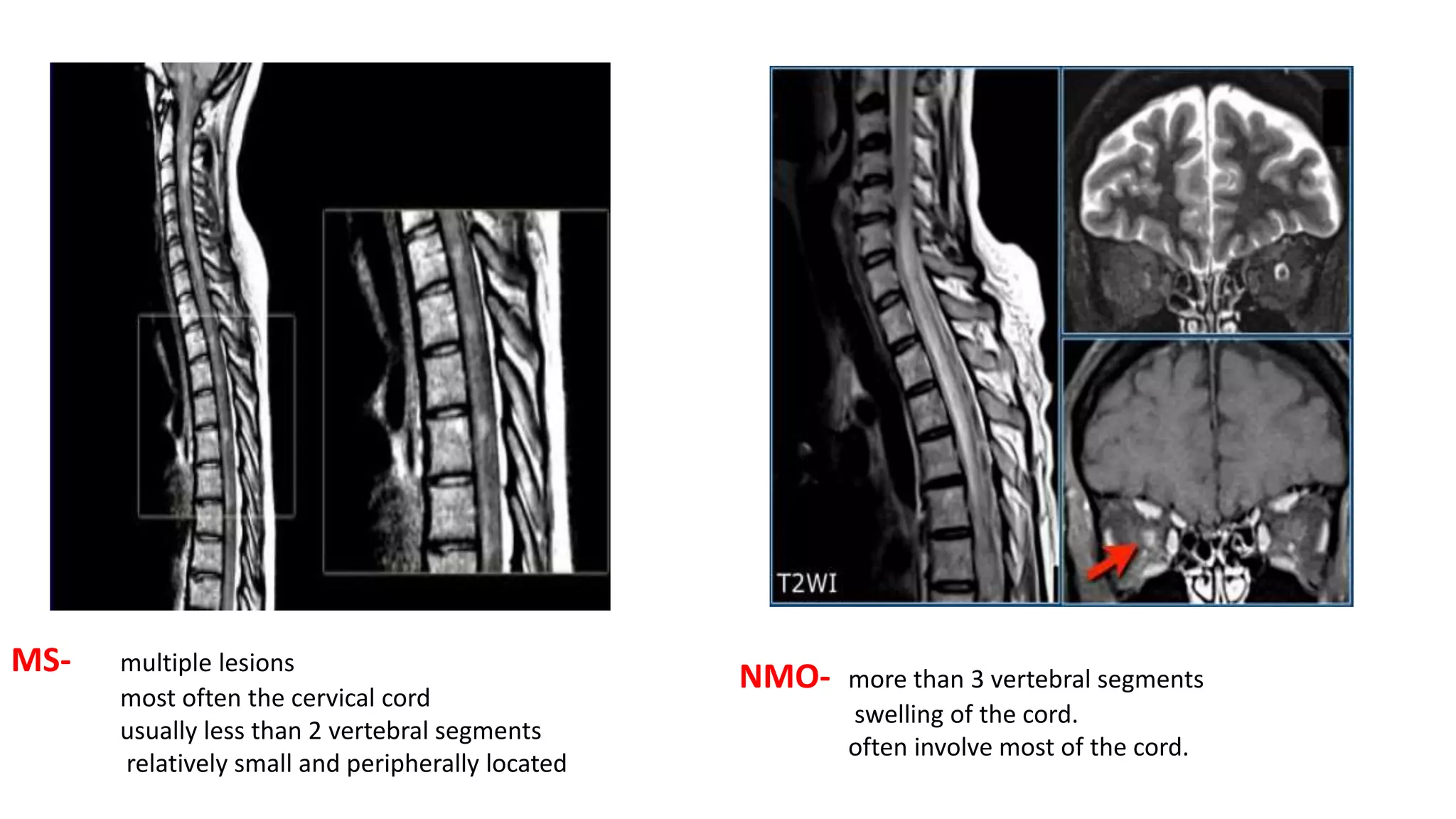
IMAGING IN NEUROLOGY
DEMYELINATING DISEASES
Dr. Rahi Kiran.B
DM Resident
Dept. of Neurology
GMC Kota
Normal myelination
• does not reach maturity until 2 years
• myelination progresses from central to peripheral
• caudal to rostral
• dorsal to ventral
• sensory then motor
• As myelination progresses myelin deposition causes an increase in T1 signal that
completes at 1 year of age - adult T1 myelination pattern
• Between 1-2 years of age the drawing out of water from myelinating areas results
in a decrease in T2 signal - adult myelination pattern achieved by 3 years
A
D
B
E F
C
Multiple Sclerosis
• A- FLAIR best for periventricular and juxtacortical lesions.
• B- T2 images are often best for viewing infratentorial lesions.
• C- Homogenous uptake of contrast.
• D- Black hole sign On MRI T1W
• E- dawsons fingers
• F- Open-ring pattern, specific for demyelinating lesions.
Multiple Sclerosis
Multiple Sclerosis
• Over 95% CDMS have positive findings on MRI
• linear, round, or ovoid lesions surrounding the medullary
veins that radiate centripetally away from the lateral ventricles
• One of the earliest findings is alternating areas of linear
hyperintensity along the ependyma on sagittal FLAIR
- "ependymal 'dot-dash'" sign
• Spinal cord lesions can be found in 50% to 90% of CDMS
• The most common site - cervical cord.
• Typical MS lesions –
• do not extend beyond 2 vertebral segments,
• tend to involve the posterior and lateral regions,
• occupy less than half the area of the cord on axial images.
triangle-shaped hyperintensities . at the
callososeptal interface and a "dot-dash"
appearance . along the ventricle.
multiple foci of punctate and ring enhancement in the cerebral white matter.
Note "target" appearance
• Before steroids after steroids
"Tumefactive" MS.
T1WI – large
hypointense lesions
in both cerebral
Hemispheres with
significant
perilesional edema
T2WI-very
hyperintense and
surrounded by a thin
hypointense rim and
perilesional edema
Hypointense rims of
the lesion show
striking but
Incomplete
ring enhancement
DWI shows that the
enhancing rims
restrict moderately
Rims demonstrate low
ADC values
The presence of T2 hypointense rim and open-ring enhancement are potentially suggestive of demyelination.
MS: DIFFERENTIAL DIAGNOSIS
• MULTIFOCAL T2/FLAIR HYPERINTENSITIES
• ADEM-history of viral prodrome or recent vaccination
• Hypoxic-ischemic lesions- small-vessel disorders- cortical infarcts, border zone, or watershed
lesions, lacunes
• Lyme disease-Cranial nerve enhancement is more common than in MS.
• Vascular-preferentially involves the basal ganglia and spares the callososeptal interface
• Susac syndrome-preferentially involve the middle of the corpus callosum, not the callososeptal
interface.
• MASS-LIKE ("TUMEFACTIVE") LESION(S)
• Neoplasm
• ○ Glioblastoma multiforme
• ○ Metastases
• • PML/PML-IRIS
• ○ HIV/AIDS
• ○ Natalizumab-treated MS
• • Medication-related-Enbrel
MS: DIFFERENTIAL DIAGNOSIS
Brainstem - symmetrical and central peripheral
Multiple Sclerosis Variants-Marburg disease
Axial FLAIR- Large
heterogeneously
hyperintense lesion in the
right parietal WM with
a smaller lesion on the left
Axial T1 C+- multiple
bilateral incomplete ring-
enhancing lesions in the
deep and periventricular
WM.
T1 C+ -through ventricles
shows the necrotic,
cavitating, acutely
enhancing right
parietal "tumefactive"
mass
Coronal T1C+ -shows
extension around the left
ventricle
Multiple Sclerosis Variants-Marburg disease
• acute, severe fulminant MS
• Multifocal > solitary disease
• ○ Characterized by coalescent white matter plaques - Brain , spinal cord
• Lesions characterized by massive inflammation, necrosis
• • diffusely disseminated disease
• ○ Large cavitating lesions
• ○ Incomplete ("open") enhancing rim
• ○ Multiple other patchy enhancing foci
• monophasic with a low rate of recurrence
• Signs of increased ICP, aphasia, and behavioral symptoms are typical.
• CSF is usually normal
• no history to suggest acute disseminated encephalomyelitis (ADEM)
• Approximately 15% of cases progress to MS.
• Solitary unilateral masses are present in two-thirds
• Differential Diagnosis-Tumefactive" MS, Pyogenic abscess, neoplasm, metastasis
and glioblastoma multiforme.
Multiple Sclerosis Variants-Schilder Disease
Multiple Sclerosis Variants-Balo Concentric Sclerosis
Acute lesions-hyperintense on FLAIR, restrict
on DWI, show concentric "onion bulb"
enhancement
Follow-up scans show alternating rings
of iso- and hyperintensity on T1 and T2WI,
no enhancement
• • Concentric rings of demyelination/myelin preservation
• ○ Resemble tree trunk or onion bulb
• ○ Solitary > multifocal
• • "Whirlpool" hyperintense concentric rings on T2WI
• ○ Minimal mass effect, edema
• • Actively demyelinating layers enhance
Multiple Sclerosis Variants-Balo Concentric Sclerosis
Chronic Relapsing Inflammatory Optic Neuropathy
(CRION)
• Young middle age female with recurrent sequential optic neuritis(Steroid responsive)
T2 - high-signal intensity foci -
left optic nerve
T1C+ enhancement DWI - expanded left optic nerve
with bright signal
Chronic Relapsing Inflammatory Optic Neuropathy
(CRION)
• Relapsing ON without known involvement in other areas of the CNS.
• unilateral relapsing optic neuritis, sequential relapses of both optic nerves, and
simultaneous, bilateral optic neuritis.
• Characteristic features –
• Middle age female
• history of optic neuritis with at least one relapse,
• objective loss of visual function
• NMO IgG negative
• contrast enhancement of the affected optic nerve on MRI
• Response to steroid treatment followed by relapse with dose reduction
• Compared to inflammatory ON – Severity less, response more
ADEM
FLAIR-bilateral white matter lesions with a "fluffy"
appearance and "fuzzy" margins
T1 C+ -enhance intensely
but heterogeneously
DWI shows acute diffusion
restriction in the lesions
ADEM numerous, large, same stage , include
white(asymmetric) and gray matter
(symmetric-thalamus and basal
ganglia ),do not always show
gadolinium enhancement, also show
good resolution compared with MS
lesions.
Spinal cord lesions 10-30% >50%
Acute Hemorrhagic Leukoencephalitis
Axial FLAIR-splenium
and genu of the corpus
Callosum, bifrontal focal
hemispheric white matter
lesions , subtle confluent
hyperintensity in the
occipital subcortical white
matter
DWI shows restricted
diffusion in the corpus
callosum splenium
GRE - punctate
hypointensities in the
corpus callosum with
subtle "blooming" in
the subcortical WM
SWI-Innumerable bilaterally
symmetric punctate and
linear "blooming"
hypointensities are seen
throughout the WM with
striking sparing of cortical
gray matter.
Mortality is 60-80%.
Petechial microhemorrhages similar to those seen in
AHLE
• Diffuse vascular injury,
• disseminated intravascular coagulopathy,
• Fat emboli,
• thrombotic thrombocytopenic purpura,
• sepsis,
• vasculitis,
• hemorrhagic viral fevers,
• malaria, and rickettsial diseases.
A-Axial T2 WI - showing swelling and bright T2
B- Axial CE MRI -no evident post-contrast enhancement.
C- Axial - DWI ADC -bilateral symmetrical parenchymal areas
of bright signal in DWI and low values in ADC
acute necrotizing encephalopathy (ANE)
• young children, is often associated with influenza,
• results from a para- or postinfectious cytokine storm.
• Strikingly symmetric thalamic necrosis with bilateral T2/FLAIR hyperintensities
and bithalamic hemorrhages is common
T2 FLAIR- medulla, hypothalamus,optic chiasma,
midbrain, internal capsule, periventricular white matter,
corpus callosum,
T2 hyperintense C1-5 with patchy
enhance ment
Neuromyelitis Optica Spectrum
Disorder
Neuromyelitis Optica Spectrum Disorder
• NMO-IgG, is 90% specific and 70-75% sensitive ..
• 10-25% of NMOSD patients are seronegative -The F:M ratio - 1:1
• one or both optic nerves are involved together with the spinal cord- most
commonly cervical
• Brain - cluster around the third and fourth ventricles and the dorsal
midbrain/aqueduct of Sylvius.
• 15-20% of patients with NMOSD are over age 60 at onset
• (85- 90%) are relapsing,
• 30% of NMOSD are initially misdiagnosed with MS.
• NMO-IgG seropositivity is detected in 3-5% of patients with CIS
• brain is more involved in MS, whereas multisegmental contiguous spinal cord
disease is typical of NMOSD
-U fibres
MS- multiple lesions
most often the cervical cord
usually less than 2 vertebral segments
relatively small and peripherally located
NMO- more than 3 vertebral segments
swelling of the cord.
often involve most of the cord.
Susac Syndrome
T2WI - hyperintense foci,
the middle of the corpus
callosum genu and
thalamus
Axial T1 C+ shows that
the corpus callosum genu
lesion enhances.
Enhancing lesions in the left temporal lobe, left
thalamus,pons
Susac Syndrome SICRET (Small Infarcts of Cochlear, Retinal, and Encephalic Tissue)
CLIPPERS
Sagittal FLAIR - multiple
punctate hyperintensities
"peppering" the pons,
Medulla extending into the
upper cervical spinal cord
T1 C+ - punctate and
curvilinear foci of
enhancement, extension
into the cerebellum and
superior cerebellar
peduncle
DWI - scattered foci of
restricted diffusion
Axial T2 SWI shows multiple
hemorrhagic foci in the
pons.
CLIPPERS
Chronic Lymphocytic Inflammation with Pontine Perivascular Enhancement Responsive to Steroids.
• onset is is 40-50 years, minor male predominance
• subacute brainstem symptoms such as gait ataxia, diplopia, facial paresthesias, and
nystagmus
• differential diagnosis - autoimmune encephalitis , Bickerstaff brainstem encephalitis,
vasculitis, intravascular lymphoma, lymphomatoid granulomatosis, neuro-Behçet,
neurosarcoidosis, CNS histiocytosis, multiple sclerosis, and NMOSD.
• Dramatic response to glucocorticosteroids (GCSs) supports
• relapses are common
• therapy failure is a strong indication for an alternative diagnosis.
Osmotic demyelination syndrome
T1 C+ (Gd): no enhancement trident shaped appearance FLAIR: hyperintense DWI: hyperintense
THANK YOU

imaging in neurology - demyelinating diseases

  • 1.
    IMAGING IN NEUROLOGY DEMYELINATINGDISEASES Dr. Rahi Kiran.B DM Resident Dept. of Neurology GMC Kota
  • 2.
    Normal myelination • doesnot reach maturity until 2 years • myelination progresses from central to peripheral • caudal to rostral • dorsal to ventral • sensory then motor • As myelination progresses myelin deposition causes an increase in T1 signal that completes at 1 year of age - adult T1 myelination pattern • Between 1-2 years of age the drawing out of water from myelinating areas results in a decrease in T2 signal - adult myelination pattern achieved by 3 years
  • 4.
  • 5.
    Multiple Sclerosis • A-FLAIR best for periventricular and juxtacortical lesions. • B- T2 images are often best for viewing infratentorial lesions. • C- Homogenous uptake of contrast. • D- Black hole sign On MRI T1W • E- dawsons fingers • F- Open-ring pattern, specific for demyelinating lesions.
  • 6.
  • 7.
    Multiple Sclerosis • Over95% CDMS have positive findings on MRI • linear, round, or ovoid lesions surrounding the medullary veins that radiate centripetally away from the lateral ventricles • One of the earliest findings is alternating areas of linear hyperintensity along the ependyma on sagittal FLAIR - "ependymal 'dot-dash'" sign • Spinal cord lesions can be found in 50% to 90% of CDMS • The most common site - cervical cord. • Typical MS lesions – • do not extend beyond 2 vertebral segments, • tend to involve the posterior and lateral regions, • occupy less than half the area of the cord on axial images. triangle-shaped hyperintensities . at the callososeptal interface and a "dot-dash" appearance . along the ventricle.
  • 8.
    multiple foci ofpunctate and ring enhancement in the cerebral white matter. Note "target" appearance • Before steroids after steroids
  • 9.
    "Tumefactive" MS. T1WI –large hypointense lesions in both cerebral Hemispheres with significant perilesional edema T2WI-very hyperintense and surrounded by a thin hypointense rim and perilesional edema Hypointense rims of the lesion show striking but Incomplete ring enhancement DWI shows that the enhancing rims restrict moderately Rims demonstrate low ADC values The presence of T2 hypointense rim and open-ring enhancement are potentially suggestive of demyelination.
  • 10.
    MS: DIFFERENTIAL DIAGNOSIS •MULTIFOCAL T2/FLAIR HYPERINTENSITIES • ADEM-history of viral prodrome or recent vaccination • Hypoxic-ischemic lesions- small-vessel disorders- cortical infarcts, border zone, or watershed lesions, lacunes • Lyme disease-Cranial nerve enhancement is more common than in MS. • Vascular-preferentially involves the basal ganglia and spares the callososeptal interface • Susac syndrome-preferentially involve the middle of the corpus callosum, not the callososeptal interface. • MASS-LIKE ("TUMEFACTIVE") LESION(S) • Neoplasm • ○ Glioblastoma multiforme • ○ Metastases • • PML/PML-IRIS • ○ HIV/AIDS • ○ Natalizumab-treated MS • • Medication-related-Enbrel
  • 11.
    MS: DIFFERENTIAL DIAGNOSIS Brainstem- symmetrical and central peripheral
  • 12.
    Multiple Sclerosis Variants-Marburgdisease Axial FLAIR- Large heterogeneously hyperintense lesion in the right parietal WM with a smaller lesion on the left Axial T1 C+- multiple bilateral incomplete ring- enhancing lesions in the deep and periventricular WM. T1 C+ -through ventricles shows the necrotic, cavitating, acutely enhancing right parietal "tumefactive" mass Coronal T1C+ -shows extension around the left ventricle
  • 13.
    Multiple Sclerosis Variants-Marburgdisease • acute, severe fulminant MS • Multifocal > solitary disease • ○ Characterized by coalescent white matter plaques - Brain , spinal cord • Lesions characterized by massive inflammation, necrosis • • diffusely disseminated disease • ○ Large cavitating lesions • ○ Incomplete ("open") enhancing rim • ○ Multiple other patchy enhancing foci
  • 14.
    • monophasic witha low rate of recurrence • Signs of increased ICP, aphasia, and behavioral symptoms are typical. • CSF is usually normal • no history to suggest acute disseminated encephalomyelitis (ADEM) • Approximately 15% of cases progress to MS. • Solitary unilateral masses are present in two-thirds • Differential Diagnosis-Tumefactive" MS, Pyogenic abscess, neoplasm, metastasis and glioblastoma multiforme. Multiple Sclerosis Variants-Schilder Disease
  • 15.
    Multiple Sclerosis Variants-BaloConcentric Sclerosis Acute lesions-hyperintense on FLAIR, restrict on DWI, show concentric "onion bulb" enhancement Follow-up scans show alternating rings of iso- and hyperintensity on T1 and T2WI, no enhancement
  • 16.
    • • Concentricrings of demyelination/myelin preservation • ○ Resemble tree trunk or onion bulb • ○ Solitary > multifocal • • "Whirlpool" hyperintense concentric rings on T2WI • ○ Minimal mass effect, edema • • Actively demyelinating layers enhance Multiple Sclerosis Variants-Balo Concentric Sclerosis
  • 18.
    Chronic Relapsing InflammatoryOptic Neuropathy (CRION) • Young middle age female with recurrent sequential optic neuritis(Steroid responsive) T2 - high-signal intensity foci - left optic nerve T1C+ enhancement DWI - expanded left optic nerve with bright signal
  • 19.
    Chronic Relapsing InflammatoryOptic Neuropathy (CRION) • Relapsing ON without known involvement in other areas of the CNS. • unilateral relapsing optic neuritis, sequential relapses of both optic nerves, and simultaneous, bilateral optic neuritis. • Characteristic features – • Middle age female • history of optic neuritis with at least one relapse, • objective loss of visual function • NMO IgG negative • contrast enhancement of the affected optic nerve on MRI • Response to steroid treatment followed by relapse with dose reduction • Compared to inflammatory ON – Severity less, response more
  • 20.
    ADEM FLAIR-bilateral white matterlesions with a "fluffy" appearance and "fuzzy" margins T1 C+ -enhance intensely but heterogeneously DWI shows acute diffusion restriction in the lesions
  • 21.
    ADEM numerous, large,same stage , include white(asymmetric) and gray matter (symmetric-thalamus and basal ganglia ),do not always show gadolinium enhancement, also show good resolution compared with MS lesions. Spinal cord lesions 10-30% >50%
  • 22.
    Acute Hemorrhagic Leukoencephalitis AxialFLAIR-splenium and genu of the corpus Callosum, bifrontal focal hemispheric white matter lesions , subtle confluent hyperintensity in the occipital subcortical white matter DWI shows restricted diffusion in the corpus callosum splenium GRE - punctate hypointensities in the corpus callosum with subtle "blooming" in the subcortical WM SWI-Innumerable bilaterally symmetric punctate and linear "blooming" hypointensities are seen throughout the WM with striking sparing of cortical gray matter.
  • 23.
  • 24.
    Petechial microhemorrhages similarto those seen in AHLE • Diffuse vascular injury, • disseminated intravascular coagulopathy, • Fat emboli, • thrombotic thrombocytopenic purpura, • sepsis, • vasculitis, • hemorrhagic viral fevers, • malaria, and rickettsial diseases.
  • 25.
    A-Axial T2 WI- showing swelling and bright T2 B- Axial CE MRI -no evident post-contrast enhancement. C- Axial - DWI ADC -bilateral symmetrical parenchymal areas of bright signal in DWI and low values in ADC
  • 26.
    acute necrotizing encephalopathy(ANE) • young children, is often associated with influenza, • results from a para- or postinfectious cytokine storm. • Strikingly symmetric thalamic necrosis with bilateral T2/FLAIR hyperintensities and bithalamic hemorrhages is common
  • 27.
    T2 FLAIR- medulla,hypothalamus,optic chiasma, midbrain, internal capsule, periventricular white matter, corpus callosum, T2 hyperintense C1-5 with patchy enhance ment Neuromyelitis Optica Spectrum Disorder
  • 28.
    Neuromyelitis Optica SpectrumDisorder • NMO-IgG, is 90% specific and 70-75% sensitive .. • 10-25% of NMOSD patients are seronegative -The F:M ratio - 1:1 • one or both optic nerves are involved together with the spinal cord- most commonly cervical • Brain - cluster around the third and fourth ventricles and the dorsal midbrain/aqueduct of Sylvius. • 15-20% of patients with NMOSD are over age 60 at onset • (85- 90%) are relapsing, • 30% of NMOSD are initially misdiagnosed with MS. • NMO-IgG seropositivity is detected in 3-5% of patients with CIS • brain is more involved in MS, whereas multisegmental contiguous spinal cord disease is typical of NMOSD
  • 29.
  • 30.
    MS- multiple lesions mostoften the cervical cord usually less than 2 vertebral segments relatively small and peripherally located NMO- more than 3 vertebral segments swelling of the cord. often involve most of the cord.
  • 31.
    Susac Syndrome T2WI -hyperintense foci, the middle of the corpus callosum genu and thalamus Axial T1 C+ shows that the corpus callosum genu lesion enhances. Enhancing lesions in the left temporal lobe, left thalamus,pons
  • 32.
    Susac Syndrome SICRET(Small Infarcts of Cochlear, Retinal, and Encephalic Tissue)
  • 33.
    CLIPPERS Sagittal FLAIR -multiple punctate hyperintensities "peppering" the pons, Medulla extending into the upper cervical spinal cord T1 C+ - punctate and curvilinear foci of enhancement, extension into the cerebellum and superior cerebellar peduncle DWI - scattered foci of restricted diffusion Axial T2 SWI shows multiple hemorrhagic foci in the pons.
  • 34.
    CLIPPERS Chronic Lymphocytic Inflammationwith Pontine Perivascular Enhancement Responsive to Steroids. • onset is is 40-50 years, minor male predominance • subacute brainstem symptoms such as gait ataxia, diplopia, facial paresthesias, and nystagmus • differential diagnosis - autoimmune encephalitis , Bickerstaff brainstem encephalitis, vasculitis, intravascular lymphoma, lymphomatoid granulomatosis, neuro-Behçet, neurosarcoidosis, CNS histiocytosis, multiple sclerosis, and NMOSD. • Dramatic response to glucocorticosteroids (GCSs) supports • relapses are common • therapy failure is a strong indication for an alternative diagnosis.
  • 35.
    Osmotic demyelination syndrome T1C+ (Gd): no enhancement trident shaped appearance FLAIR: hyperintense DWI: hyperintense
  • 36.