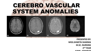
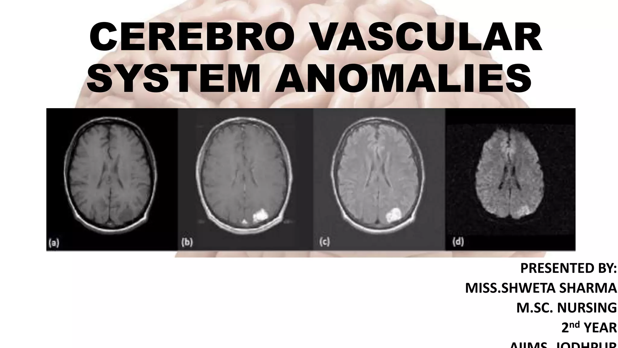
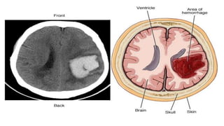
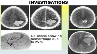
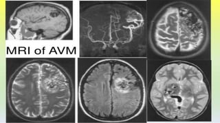
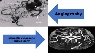
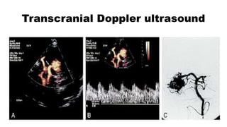
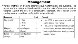
CEREBRO VASCULAR
SYSTEM ANOMALIES
PRESENTED BY:
MISS.SHWETA SHARMA
M.SC. NURSING
2nd YEAR
INTRODUCTION
• Cerebrovascular anomalies or malformations are conditions characterized by
malformed blood vessels that can lead to hemorrhages, stroke, blood clots, and
other complications.
• Some lesions present with serious symptoms such as hemorrhage, seizure, or
headaches, whereas other lesions are quite benign with no symptoms at all.
• The presentation, location, and natural history of these lesions determines
whether management will be surgical, endovascular, radio surgical, or
observation.
CLASSIFICATIONS OF VASCULAR
MALFORMATIONS
Arteriovenous
malformations (AVMs)
Venous angioma
Capillary
telangiectasias
Cavernous
malformation/angioma
Histopathologic
classification
B. Functional classification
CVMs that display shunting:
•AVM
•Fistula
CVMs without AV shunting:
•Venous, capillary, cavernous
malformations.
EPIDEMIOLOGY
• The incidence of vascular malformations varies from 0.1% to 4% in various
autopsy studies. In a large autopsy series, the detection of AVMs was 1.4%
(46 AVMs among 3,200 brain tumour cases).
• Vascular malformations are about one seventh as common as intracranial
saccular aneurysms. AVMs are the most common type of vascular lesion.
• With improvement in cross-sectional imaging in both CT and MRI, higher
detection rates are being reported and in patients with increasing age.
ARTERIOVENOUS MALFORMATIONS
• AVM is an abnormal tangle of blood vessels connecting arteries and veins,
which disrupts normal blood flow and oxygen circulation.
• AVMs may be small and focal, or they may be large, involving an entire
hemisphere. Some are conical, with the apex pointing inward and the base
positioned on the surface of the cerebral cortex. In rare instances, the lesion
is so deep that the ventricles and choroid plexus are involved, thus
predisposing the person to intraventricular hemorrhage.
• They are developmental anomalies of the intracranial
vasculature; they are not neoplastic despite their tendency
to expand with time.
• Dilated arteries feed directly into a tangled mass of blood
vessels of varying calibre; they bypass capillaries and shunt
oxygenated blood directly into the venous system.
• Due to high intraluminal pressure, veins may adopt an
‘aneurysmal’ appearance.
• Arteriovenous malformations occur at any site but are
commonest in the middle cerebral artery.
EPIDEMIOLOGY
• Current incidence rate is about 1.3 per 100, 000 annually. The ratio of AVMs
to intracranial aneurysms is 1:10.
• Approximately 90% of AVMs are located in the supratentorial area and involve
the cerebral hemispheres; only 10% of this group are located in the deep
subcortical areas (e.g., basal ganglia, thalamus, corpus callosum).
• The location of 10% of AVMs is within the cerebellum and brainstem. Of those
patients with AVMs, 80% develop symptoms between the ages of 20 and 40
years; the remaining 20% develop symptoms before the age of 20 years.
RISK FACTORS
•Family history
•Certain hereditary conditions
like hereditary haemorrhagic
telangiectasia (HHT), also
called Osler-Weber-Rendu
syndrome.
CAUSES
• Development of abnormal direct connections between arteries and
veins.
• Genetic changes
• Parenchymal AVMs result from abnormalities in the vasculature during
fetal development from fetal age of 3 to 12 weeks. Failure of normal
involution of embryonic vasculature networks is thought to occur as
AVMs develop. Some AVMs may enlarge by recruitment of new blood
vessels during childhood and early adulthood.
PATHOPHYSIOLOGY
There are two pathophysiologic characteristics related to AVMs.
• The first is the effect of shunting of blood from the arterial to the venous
system without the intervening capillary network. Normally, the capillary
network provides capillary resistance to blood flow, thus decreasing the
intravascular pressure.
• However, when an AVM is present, blood is shunted from the high-resistance
normal vascular bed to the low-resistance vessels within the AVM, thus
exposing the draining venous channels to elevated intravascular pressure.
These dynamics predispose the vessels to rupture and hemorrhage.
• The second characteristic is the effect of impaired perfusion of the
cerebral tissue adjacent to the AVM. Elevated intravascular and
venous pressure impairs cerebral perfusion pressure. When the AVM
is large and has a high flow, the diversion of blood to the AVM may
cause ischemia to the adjacent normal tissue. Clinically, this is
evidenced by slowly progressive neurological deficits. The diversion of
blood to the AVM is called the vascular steal phenomenon.
CLINICAL PRESENTATION
Haemorrhage
• In 40–60% of patients
• AVMs tend to bleed in younger patients, i.e. 20–40 years, and are less likely to
have a fatal outcome.
• Annual risk of haemorrhage: patients with no history of haemorrhage have an
annual risk of bleeding is 2–4%. For those presenting with haemorrhage, the
risk of rebleeding may be higher, particularly in the first year.
• Relatively low mortality rate of approximately 10%.
• Of patients presenting with haemorrhage,
30% have a history of epilepsy.
Neurological deficit
• Large AVMs, especially those involving the
basal ganglia, may present with a slowly
progressive dementia, hemiparesis or visual
field defect.
• The infrequent brain stem AVM may also
produce a motor or sensory deficit, with or
without cranial nerve involvement.
Headache
•Attacks of well localized headache –
unilateral and throbbing – occur in a
proportion of patients subsequently
shown to have a large AVM.
Cranial bruit
•Auscultation, especially over the
eyeball, occasionally reveals a bruit.
INVESTIGATIONS
Angiography
Magnetic resonance
angiography
Transcranial Doppler ultrasound
Management
• Various methods of treating arteriovenous malformations are available. The
urgency of the patient’s clinical condition and the risks of treatment must be
weighed against the risk of a conservative approach. The Spetzler-Martin
grading system provides a useful guide to operative risk:
Parameter Grade Example
Size of AVM < 3 cm
3–6 cm
> 6 cm
1
2
3
 2 cm AVM in non-eloquent area with no
deeply draining veins = grade 1
 4 cm AVM in eloquent area (motor, speech
or visual cortex, thalamus, internal capsule,
basal ganglia, brain stem) with deep venous
drainage = grade 4
Eloquence of
adjacent brain
non-eloquent
eloquent
0
1
Pattern of venous
drainage
superficial only
deep
0
1
INDICATIONS FOR INTERVENTION
• ‘Expanding’ haematoma associated with AVM
• Progressive neurological deficit
• Risk of haemorrhage especially in young patients
with many years at risk and AVMs < 3 cm
METHODS OF TREATMENT
Operation: Excision
• Most effective method of treatment
particularly for small AVMs.
• Larger lesions (> 6 cm) have a greater risk
of postoperative hyper perfusion
syndrome and brain swelling and carry a
40% risk of permanent neurological
deficit.
Stereotactic radiosurgery
• Focused beams from multiple cobalt sources or from a linear
accelerator (25Gy) obliterates about 75% of AVMs < 3 cm in diameter,
but this may take up to 3 years during which time the risk of
haemorrhage persists. In smaller lesions < 1 cm the obliteration rate
with 25 Gy approaches 100%.
• For lesions greater than 3 cm, the lower dose required to minimize the
damaging effect of local tissue destruction, makes obliteration unlikely.
Embolization
•Skilled catheterization permits selective embolization of
feeding vessels with isobutyl-cyanoacrylate, although this
technique is not without risk. Embolization may cure up to 40%
of AVMs when small particularly if supplied by a single feeding
vessel, but filling may persist from collaterals. When used
preoperatively, it may significantly aid operative removal.
CASE STUDY
EL was a 34-year-old woman who experienced a 10/10 headache and right lower
extremity paresthesia. She has a history of migraine headaches and other headaches. A
previous MRI scan revealed a left parietal AVM measuring 1 cm × 1.1 cm × 1.3 cm. After
having a baby recently, she saw a neurosurgeon who recommended an angiogram. She
refused the invasive diagnostic test because of the associated risks. Eight months after
the initial diagnosis, EL presented to the emergency department, with headache and right
lower extremity paraesthesia. She was complaining of mild photophobia and nausea. A
CTA was ordered to evaluate her known AVM. The scan revealed a left parietal
intraparenchymal hematoma measuring 1.7 cm × 1.8 cm × 2 cm. The scan also showed
the presence of subarachnoid hemorrhage.
The bleeding pattern and location was consistent with a haemorrhaged AVM. It
was reported to be a Spetzler–Martin grade I AVM; the neurosurgeon decided to
proceed with open surgery to remove the clot and AVM. The patient was
monitored closely in the neurological intensive care unit (NICU) for several days
after her hemorrhage and surgery to ensure continued stability. A second
diagnostic cerebral angiography was performed immediately after the patient’s
open surgery, confirming the complete removal of the lesion. Two months after
surgery, the patient was seen in clinic and had no neurological deficits and was
doing well. She was prescribed anticonvulsant medication, which she will continue
taking for another 7 to 8 months. She is expected to continue with an excellent
recovery.
VENOUS ANGIOMA
• Developmental venous anomaly (DVA), also known as cerebral venous
angioma, is a congenital malformation of veins which drain normal brain. They
are composed of anomalous veins, separated by normal parenchyma, which
drain into a dilated venous trunk.
• They are the most common cerebral vascular malformation, accounting for
approximately 55% of all such lesions.
• A DVA is characterized by the caput medusae sign of veins draining into a single
larger collecting vein, which in turn drains into either a dural sinus or into a deep
ependymal vein. The appearance has also been likened to a palm tree.
Epidemiology
• DVAs are very common as incidental finding, with an estimated
prevalence of 2.5-9% on contrast-enhanced MRI scans.
Etiology
• Idiopathic
• Arrested development of venous structures
• Histologically they consist of a number of abnormally thickened
veins with normal feeding arteries and capillaries.
Clinical presentation
• Intracranial hemorrhage (1-5%)- Hemorrhage can occur from bleeding
into an infarcted area from venous congestion but most often
associated with CM or even AVM.
• Ischemic stroke
• Epilepsy
Location and classification
• The most common locations are:
frontoparietal region (36-64%), usually draining towards the frontal
horn of the lateral ventricle
cerebellar hemisphere (14-27%) draining towards the fourth ventricle
• However, DVAs can be seen anywhere, draining either superficially or
deep.
Diagnostic evaluation
CT
MRI
Angiography
(DSA)
Management
•Generally, developmental venous anomalies (DVAs) do
not require treatment. These veins do a necessary job of
getting blood in and out of the brain, so they do not
need to be surgically removed or closed. Because they
are normal and not dangerous, long-term imaging is
generally not necessary.
CAPILLARY TELANGIECTASIAS
• Capillary telangiectasias are an
area of dilated capillaries, like a
small petechial patch (0.3 to 1
cm) on the brain surface –
especially in the pons. These
lesions are often only revealed at
autopsy.
Epidemiology
• Most frequently found in middle-aged and elderly adults.
• They account for up to 20% of all intracerebral vascular malformations and are
considered the second most common vascular anomaly after venous angiomas
(developmental venous anomaly).
Clinical presentation
• The vast majority of capillary telangiectasias are completely asymptomatic and
discovered incidentally on MRI when the brain is imaged for other reasons.
• Occasionally there may be associated intracerebral hemorrhage, although a
direct causative relationship has not been established.
Location
•Most capillary telangiectasias occur in the pons,
cerebellum and spinal cord.
Diagnostic evaluation
•They are usually not seen on CT and DSA.
•MRI - They appear as subtle lesions with no mass effect.
Treatment and prognosis
• These lesions are almost always asymptomatic, have interspersed normal brain
tissue and are most frequently located in the pons, making treatment
impractical. Thus, no follow-up is required if the imaging appearances are
characteristic.
Complications
• Stroke
• Cerebral abscesses
• Both cerebral and spinal AVMs
CAVERNOUS MALFORMATIONS
• Also called cavernous hemangiomas, cavernous
angiomas, and cavernomas, are congenital
nodular lesions.
• They resemble a mulberry or “popcorn-like”
appearing lesion in the brain, spinal cord, or
nerve roots and are composed of sinusoidal-
type vessels that are not separated by normal-
appearing parenchyma (neural tissue).
• Microscopic examination often reveals
small haemorrhages with numerous,
hemosiderin-laden macrophages and
gliotic tissue in the adjacent
parenchyma. Elastic fibres are absent in
the walls of these vascular caverns.
Thrombosis may be present in some of
the dilated venules. Calcification within
the lesion is common.
Epidemiology
• Cavernous malformations occur in 0.5% of the population. They are occasionally
multiple and, in a few patients, have a familial basis. CMs are rare in children
and account for about 10% of all symptomatic vascular malformations. The
peak occurrence is in the third and fifth decades of life.
Types
• Sporadic- characterized by one lesion.
• Familial- characterized by multiple lesions with an autosomal dominant mode
of inheritance.
Causes
• Genetic linkage - A locus is present for CM on chromosome 7q. Two additional loci
have also been identified mapping to chromosomes 7p and 3q. The CM gene was
successfully identified as KRIT1.
Clinical manifestations
• Epilepsy
• Haemorrhage
• Focal neurological signs
• Headache
• Paralysis
Diagnostic evaluation
• MRI is the investigation of choice as cavernous
malformations, are often missed on CT scanning
and rarely seen on angiography. Most lesions
show marked signal change around this lesion
due to a rim of hemosiderin deposition.
• Angiography (DSA)- Cavernous malformations
are angiographically occult and do not
demonstrate arteriovenous shunting.
Management
• Seizures may be controlled with anti-epileptic drugs.
• Surgery may be necessary to remove a cerebral cavernous
malformation that is causing symptoms or that suffered multiple
bleedings.
• The hemorrhage can create a clot cavity, thus providing a route for
surgical access and creating a plane for dissection of the lesion.
DURAL ARTERIOVENOUS FISTULA
They are defined by the following criteria:
• the nidus of the arteriovenous (AV) shunting is within the cranial dura mater
(a nidus is defined as the focus of the AVM, i.e., the tangle of abnormal
vessels).
• arterial supply arises exclusively from the extracranial circulation or from the
meningeal branches of the intracranial branches.
• venous drainage is either directly into the dural venous sinus or into nearby
leptomeningeal veins.
DAVMs can occur in either the cranial or spinal areas of the central nervous
system.
Epidemiology
• DAVFs comprise 10% to 15% of all intracranial cavernous malformations. There is an
estimated incidence of occurrence ranging from 0.71 to 12.38 per 100,000 persons
worldwide.
• These lesions most frequently affect adults in their 50 to 60s and rarely effect
children.
Causes
• Trauma
• Surgery
• Infection
Pathophysiology
• The formation of DAVFs is a dynamic
process arising from stenosis or
occlusion of the dural venous sinus.
The stenosis or occlusion creates
increased venous pressure, and
meningeal arteries develop fistula
connections with dural sinus of cortical
veins.
Clinical manifestations
Generalized nervous system signs
• Papilledema
• Headache
• Visual disturbances
• Hydrocephalus due to malabsorption of the cerebrospinal fluid
Focal signs
• Seizure
• Transient ischemic attack
• Intraparenchymal hemorrhage
• Fixed cortical, brainstem, and cerebellar deficits
Diagnostic evaluation
• CT
• MRI
• Visual examination
• Computed tomography angiography
• Four-vessel angiogram with selective external carotid artery (ECA)
injection remains the - gold standard
Management
• Trans arterial embolization, transvenous embolization, and surgical resection of the
lesion may be used alone or in combination depending on the anatomy of the
lesion.
• Prior embolization helps to reduce blood flow and allows for easier identification of
the lesion during surgery. Ideally, treatment should occur at a centre with both
surgical and interventional capabilities for multimodality treatment planning.
• Stereotactic radiosurgery can also be used. This treatment can take 1 to 3 years for
complete lesion obliteration and is therefore used in benign presentations.
VEIN OF GALEN MALFORMATIONS
• A rare high-flow vascular malformation of the brain in which there is marked dilatation of
the venous system. It is a congenital malformation of blood vessels of the brain that
occurs prior to birth, most likely between gestational weeks 6 and 11.
• The vein of Galen is a large deep vein at the base of the brain.
• This malformation shunts blood from the arteries to the brain very
quickly, increasing overall blood flow, thus increasing the work of the
heart. This can result in cardiac failure, which is the most common
presentation of the disease.
• Similar to other AVMs, the capillaries are missing. The high blood
flow can also interfere with the normal blood drainage of the brain,
leading to hydrocephalus.
Classification
• Vein of Galen aneurysmal malformation (VGAM) - lesion of early
embryologic occurrence. The VGAM is a pathologic varix of the
embryologic median vein of the prosencephalon that drains the
flow from the fistulous malformation. There may be single or
multiple arterial feeders.
• Vein of Galen aneurysmal dilatation (VGAD) - lesion seen when
the venous system of the brain is more developed.
Clinical manifestations
• The clinical manifestation of VGAMs and VGADs is determined by the volume of
flow to the fistulous connection, the severity of venous constraints, the stage of
venous development at which the lesion became dominant, and anatomic
configuration.
• The dominant clinical complications in neonates relate to cardiac effects of the
high-flow lesion. Immediate neurological complications in neonates are thought to
be related to brain ischemia due to venous hypertension, tissue edema, and
ischemia compounded by decreased perfusion due to cardiac failure.
Diagnosis
• Prenatal ultrasound
• MRI
• Angiography
Treatment
• Endovascular occlusion of the arterial components of these malformations by either
a transvenous or transarterial approach is the treatment of choice.
• The embolization materials include coils, particulate matter, suture material, and
glue. When surgery follows the embolization, it is targeted at the remaining fistula.
• It may be necessary to partially treat a neonate who presents with cardiac failure
related to a vein of Galen malformation with an endovascular approach in order to
stabilize the cardiac condition.
• Definitive treatment can be performed when the child is older and more stable.
Direct surgical treatment is associated with very high morbidity and mortality, with
rates up to 70%.
Nursing assessment
• Conduct a baseline neurological assessment as well as ongoing
monitoring of neurological signs.
• Monitor vital signs for evidence of hypertension.
• Assess and monitor characteristics of headache, if present.
• Monitor the patient for evidence of seizure activity.
• Administer drug therapy as necessary.
Nursing diagnosis
1. Ineffective tissue perfusion: cerebral related to shunting of
blood from cerebral tissue and/ or intracerebral hemorrhage
(ICH).
2. Impaired physical mobility related to paresthesia.
3. Disturbed Sensory Perception: visual related to papilledema.
4. Risk for injury related to decreased vision.
5. Deficient knowledge related to the post-operative care.
Endovascular Treatment of Cerebellar Arteriovenous Malformations: A Single-
Centre Experience of 75 Consecutive Patients
Yong Sun et al (2020) conducted a study to determine the safety and
effectiveness of endovascular treatment for cerebellar arteriovenous
malformations (AVMs). 75 patients with cerebellar AVMs who underwent
endovascular treatment were included. 61 (81.3%) presented with initial
hemorrhage, and 44 (58.7%) presented with 63 cerebral aneurysms. Immediate
digital subtraction angiography (DSA) after the procedure showed complete
occlusion of the cerebral aneurysms in all the patients, and total occlusion of the
AVM nidus in 32/75 (42.7%) patients, 99–90% occlusion in 31/75 (41.3%)
patients, and <90% occlusion in 12/75 (16.0%) patients. The study concluded that
endovascular treatment of cerebellar AVMs is safe and feasible. The high rate of
associated cerebral aneurysms could explain the tendency of initial hemorrhage
in cerebellar AVMs; targeted embolization of coexisting cerebral aneurysms
should be the first priority. Increasing patient age, eloquent AVM location, and
the size of AVM are independent predictors of poor outcome after endovascular
treatment of cerebellar AVMs.
Cerebro vascular anomalies

Cerebro vascular anomalies

  • 1.
    CEREBRO VASCULAR SYSTEM ANOMALIES PRESENTEDBY: MISS.SHWETA SHARMA M.SC. NURSING 2nd YEAR
  • 2.
    INTRODUCTION • Cerebrovascular anomaliesor malformations are conditions characterized by malformed blood vessels that can lead to hemorrhages, stroke, blood clots, and other complications. • Some lesions present with serious symptoms such as hemorrhage, seizure, or headaches, whereas other lesions are quite benign with no symptoms at all. • The presentation, location, and natural history of these lesions determines whether management will be surgical, endovascular, radio surgical, or observation.
  • 3.
    CLASSIFICATIONS OF VASCULAR MALFORMATIONS Arteriovenous malformations(AVMs) Venous angioma Capillary telangiectasias Cavernous malformation/angioma Histopathologic classification
  • 4.
    B. Functional classification CVMsthat display shunting: •AVM •Fistula CVMs without AV shunting: •Venous, capillary, cavernous malformations.
  • 5.
    EPIDEMIOLOGY • The incidenceof vascular malformations varies from 0.1% to 4% in various autopsy studies. In a large autopsy series, the detection of AVMs was 1.4% (46 AVMs among 3,200 brain tumour cases). • Vascular malformations are about one seventh as common as intracranial saccular aneurysms. AVMs are the most common type of vascular lesion. • With improvement in cross-sectional imaging in both CT and MRI, higher detection rates are being reported and in patients with increasing age.
  • 6.
    ARTERIOVENOUS MALFORMATIONS • AVMis an abnormal tangle of blood vessels connecting arteries and veins, which disrupts normal blood flow and oxygen circulation. • AVMs may be small and focal, or they may be large, involving an entire hemisphere. Some are conical, with the apex pointing inward and the base positioned on the surface of the cerebral cortex. In rare instances, the lesion is so deep that the ventricles and choroid plexus are involved, thus predisposing the person to intraventricular hemorrhage.
  • 8.
    • They aredevelopmental anomalies of the intracranial vasculature; they are not neoplastic despite their tendency to expand with time. • Dilated arteries feed directly into a tangled mass of blood vessels of varying calibre; they bypass capillaries and shunt oxygenated blood directly into the venous system. • Due to high intraluminal pressure, veins may adopt an ‘aneurysmal’ appearance. • Arteriovenous malformations occur at any site but are commonest in the middle cerebral artery.
  • 9.
    EPIDEMIOLOGY • Current incidencerate is about 1.3 per 100, 000 annually. The ratio of AVMs to intracranial aneurysms is 1:10. • Approximately 90% of AVMs are located in the supratentorial area and involve the cerebral hemispheres; only 10% of this group are located in the deep subcortical areas (e.g., basal ganglia, thalamus, corpus callosum). • The location of 10% of AVMs is within the cerebellum and brainstem. Of those patients with AVMs, 80% develop symptoms between the ages of 20 and 40 years; the remaining 20% develop symptoms before the age of 20 years.
  • 10.
    RISK FACTORS •Family history •Certainhereditary conditions like hereditary haemorrhagic telangiectasia (HHT), also called Osler-Weber-Rendu syndrome.
  • 13.
    CAUSES • Development ofabnormal direct connections between arteries and veins. • Genetic changes • Parenchymal AVMs result from abnormalities in the vasculature during fetal development from fetal age of 3 to 12 weeks. Failure of normal involution of embryonic vasculature networks is thought to occur as AVMs develop. Some AVMs may enlarge by recruitment of new blood vessels during childhood and early adulthood.
  • 14.
    PATHOPHYSIOLOGY There are twopathophysiologic characteristics related to AVMs. • The first is the effect of shunting of blood from the arterial to the venous system without the intervening capillary network. Normally, the capillary network provides capillary resistance to blood flow, thus decreasing the intravascular pressure. • However, when an AVM is present, blood is shunted from the high-resistance normal vascular bed to the low-resistance vessels within the AVM, thus exposing the draining venous channels to elevated intravascular pressure. These dynamics predispose the vessels to rupture and hemorrhage.
  • 15.
    • The secondcharacteristic is the effect of impaired perfusion of the cerebral tissue adjacent to the AVM. Elevated intravascular and venous pressure impairs cerebral perfusion pressure. When the AVM is large and has a high flow, the diversion of blood to the AVM may cause ischemia to the adjacent normal tissue. Clinically, this is evidenced by slowly progressive neurological deficits. The diversion of blood to the AVM is called the vascular steal phenomenon.
  • 16.
    CLINICAL PRESENTATION Haemorrhage • In40–60% of patients • AVMs tend to bleed in younger patients, i.e. 20–40 years, and are less likely to have a fatal outcome. • Annual risk of haemorrhage: patients with no history of haemorrhage have an annual risk of bleeding is 2–4%. For those presenting with haemorrhage, the risk of rebleeding may be higher, particularly in the first year. • Relatively low mortality rate of approximately 10%.
  • 18.
    • Of patientspresenting with haemorrhage, 30% have a history of epilepsy. Neurological deficit • Large AVMs, especially those involving the basal ganglia, may present with a slowly progressive dementia, hemiparesis or visual field defect. • The infrequent brain stem AVM may also produce a motor or sensory deficit, with or without cranial nerve involvement.
  • 19.
    Headache •Attacks of welllocalized headache – unilateral and throbbing – occur in a proportion of patients subsequently shown to have a large AVM. Cranial bruit •Auscultation, especially over the eyeball, occasionally reveals a bruit.
  • 20.
  • 22.
  • 23.
  • 24.
    Management • Various methodsof treating arteriovenous malformations are available. The urgency of the patient’s clinical condition and the risks of treatment must be weighed against the risk of a conservative approach. The Spetzler-Martin grading system provides a useful guide to operative risk: Parameter Grade Example Size of AVM < 3 cm 3–6 cm > 6 cm 1 2 3  2 cm AVM in non-eloquent area with no deeply draining veins = grade 1  4 cm AVM in eloquent area (motor, speech or visual cortex, thalamus, internal capsule, basal ganglia, brain stem) with deep venous drainage = grade 4 Eloquence of adjacent brain non-eloquent eloquent 0 1 Pattern of venous drainage superficial only deep 0 1
  • 25.
    INDICATIONS FOR INTERVENTION •‘Expanding’ haematoma associated with AVM • Progressive neurological deficit • Risk of haemorrhage especially in young patients with many years at risk and AVMs < 3 cm
  • 26.
    METHODS OF TREATMENT Operation:Excision • Most effective method of treatment particularly for small AVMs. • Larger lesions (> 6 cm) have a greater risk of postoperative hyper perfusion syndrome and brain swelling and carry a 40% risk of permanent neurological deficit.
  • 27.
    Stereotactic radiosurgery • Focusedbeams from multiple cobalt sources or from a linear accelerator (25Gy) obliterates about 75% of AVMs < 3 cm in diameter, but this may take up to 3 years during which time the risk of haemorrhage persists. In smaller lesions < 1 cm the obliteration rate with 25 Gy approaches 100%. • For lesions greater than 3 cm, the lower dose required to minimize the damaging effect of local tissue destruction, makes obliteration unlikely.
  • 28.
    Embolization •Skilled catheterization permitsselective embolization of feeding vessels with isobutyl-cyanoacrylate, although this technique is not without risk. Embolization may cure up to 40% of AVMs when small particularly if supplied by a single feeding vessel, but filling may persist from collaterals. When used preoperatively, it may significantly aid operative removal.
  • 30.
    CASE STUDY EL wasa 34-year-old woman who experienced a 10/10 headache and right lower extremity paresthesia. She has a history of migraine headaches and other headaches. A previous MRI scan revealed a left parietal AVM measuring 1 cm × 1.1 cm × 1.3 cm. After having a baby recently, she saw a neurosurgeon who recommended an angiogram. She refused the invasive diagnostic test because of the associated risks. Eight months after the initial diagnosis, EL presented to the emergency department, with headache and right lower extremity paraesthesia. She was complaining of mild photophobia and nausea. A CTA was ordered to evaluate her known AVM. The scan revealed a left parietal intraparenchymal hematoma measuring 1.7 cm × 1.8 cm × 2 cm. The scan also showed the presence of subarachnoid hemorrhage.
  • 31.
    The bleeding patternand location was consistent with a haemorrhaged AVM. It was reported to be a Spetzler–Martin grade I AVM; the neurosurgeon decided to proceed with open surgery to remove the clot and AVM. The patient was monitored closely in the neurological intensive care unit (NICU) for several days after her hemorrhage and surgery to ensure continued stability. A second diagnostic cerebral angiography was performed immediately after the patient’s open surgery, confirming the complete removal of the lesion. Two months after surgery, the patient was seen in clinic and had no neurological deficits and was doing well. She was prescribed anticonvulsant medication, which she will continue taking for another 7 to 8 months. She is expected to continue with an excellent recovery.
  • 32.
  • 33.
    • Developmental venousanomaly (DVA), also known as cerebral venous angioma, is a congenital malformation of veins which drain normal brain. They are composed of anomalous veins, separated by normal parenchyma, which drain into a dilated venous trunk. • They are the most common cerebral vascular malformation, accounting for approximately 55% of all such lesions. • A DVA is characterized by the caput medusae sign of veins draining into a single larger collecting vein, which in turn drains into either a dural sinus or into a deep ependymal vein. The appearance has also been likened to a palm tree.
  • 34.
    Epidemiology • DVAs arevery common as incidental finding, with an estimated prevalence of 2.5-9% on contrast-enhanced MRI scans. Etiology • Idiopathic • Arrested development of venous structures • Histologically they consist of a number of abnormally thickened veins with normal feeding arteries and capillaries.
  • 35.
    Clinical presentation • Intracranialhemorrhage (1-5%)- Hemorrhage can occur from bleeding into an infarcted area from venous congestion but most often associated with CM or even AVM. • Ischemic stroke • Epilepsy Location and classification • The most common locations are: frontoparietal region (36-64%), usually draining towards the frontal horn of the lateral ventricle cerebellar hemisphere (14-27%) draining towards the fourth ventricle • However, DVAs can be seen anywhere, draining either superficially or deep.
  • 36.
  • 37.
  • 38.
    Management •Generally, developmental venousanomalies (DVAs) do not require treatment. These veins do a necessary job of getting blood in and out of the brain, so they do not need to be surgically removed or closed. Because they are normal and not dangerous, long-term imaging is generally not necessary.
  • 39.
    CAPILLARY TELANGIECTASIAS • Capillarytelangiectasias are an area of dilated capillaries, like a small petechial patch (0.3 to 1 cm) on the brain surface – especially in the pons. These lesions are often only revealed at autopsy.
  • 41.
    Epidemiology • Most frequentlyfound in middle-aged and elderly adults. • They account for up to 20% of all intracerebral vascular malformations and are considered the second most common vascular anomaly after venous angiomas (developmental venous anomaly). Clinical presentation • The vast majority of capillary telangiectasias are completely asymptomatic and discovered incidentally on MRI when the brain is imaged for other reasons. • Occasionally there may be associated intracerebral hemorrhage, although a direct causative relationship has not been established.
  • 42.
    Location •Most capillary telangiectasiasoccur in the pons, cerebellum and spinal cord. Diagnostic evaluation •They are usually not seen on CT and DSA. •MRI - They appear as subtle lesions with no mass effect.
  • 43.
    Treatment and prognosis •These lesions are almost always asymptomatic, have interspersed normal brain tissue and are most frequently located in the pons, making treatment impractical. Thus, no follow-up is required if the imaging appearances are characteristic. Complications • Stroke • Cerebral abscesses • Both cerebral and spinal AVMs
  • 44.
    CAVERNOUS MALFORMATIONS • Alsocalled cavernous hemangiomas, cavernous angiomas, and cavernomas, are congenital nodular lesions. • They resemble a mulberry or “popcorn-like” appearing lesion in the brain, spinal cord, or nerve roots and are composed of sinusoidal- type vessels that are not separated by normal- appearing parenchyma (neural tissue).
  • 45.
    • Microscopic examinationoften reveals small haemorrhages with numerous, hemosiderin-laden macrophages and gliotic tissue in the adjacent parenchyma. Elastic fibres are absent in the walls of these vascular caverns. Thrombosis may be present in some of the dilated venules. Calcification within the lesion is common.
  • 46.
    Epidemiology • Cavernous malformationsoccur in 0.5% of the population. They are occasionally multiple and, in a few patients, have a familial basis. CMs are rare in children and account for about 10% of all symptomatic vascular malformations. The peak occurrence is in the third and fifth decades of life. Types • Sporadic- characterized by one lesion. • Familial- characterized by multiple lesions with an autosomal dominant mode of inheritance.
  • 47.
    Causes • Genetic linkage- A locus is present for CM on chromosome 7q. Two additional loci have also been identified mapping to chromosomes 7p and 3q. The CM gene was successfully identified as KRIT1. Clinical manifestations • Epilepsy • Haemorrhage • Focal neurological signs • Headache • Paralysis
  • 48.
    Diagnostic evaluation • MRIis the investigation of choice as cavernous malformations, are often missed on CT scanning and rarely seen on angiography. Most lesions show marked signal change around this lesion due to a rim of hemosiderin deposition. • Angiography (DSA)- Cavernous malformations are angiographically occult and do not demonstrate arteriovenous shunting.
  • 49.
    Management • Seizures maybe controlled with anti-epileptic drugs. • Surgery may be necessary to remove a cerebral cavernous malformation that is causing symptoms or that suffered multiple bleedings. • The hemorrhage can create a clot cavity, thus providing a route for surgical access and creating a plane for dissection of the lesion.
  • 50.
  • 51.
    They are definedby the following criteria: • the nidus of the arteriovenous (AV) shunting is within the cranial dura mater (a nidus is defined as the focus of the AVM, i.e., the tangle of abnormal vessels). • arterial supply arises exclusively from the extracranial circulation or from the meningeal branches of the intracranial branches. • venous drainage is either directly into the dural venous sinus or into nearby leptomeningeal veins. DAVMs can occur in either the cranial or spinal areas of the central nervous system.
  • 52.
    Epidemiology • DAVFs comprise10% to 15% of all intracranial cavernous malformations. There is an estimated incidence of occurrence ranging from 0.71 to 12.38 per 100,000 persons worldwide. • These lesions most frequently affect adults in their 50 to 60s and rarely effect children. Causes • Trauma • Surgery • Infection
  • 53.
    Pathophysiology • The formationof DAVFs is a dynamic process arising from stenosis or occlusion of the dural venous sinus. The stenosis or occlusion creates increased venous pressure, and meningeal arteries develop fistula connections with dural sinus of cortical veins.
  • 54.
    Clinical manifestations Generalized nervoussystem signs • Papilledema • Headache • Visual disturbances • Hydrocephalus due to malabsorption of the cerebrospinal fluid Focal signs • Seizure • Transient ischemic attack • Intraparenchymal hemorrhage • Fixed cortical, brainstem, and cerebellar deficits
  • 55.
    Diagnostic evaluation • CT •MRI • Visual examination • Computed tomography angiography • Four-vessel angiogram with selective external carotid artery (ECA) injection remains the - gold standard
  • 56.
    Management • Trans arterialembolization, transvenous embolization, and surgical resection of the lesion may be used alone or in combination depending on the anatomy of the lesion. • Prior embolization helps to reduce blood flow and allows for easier identification of the lesion during surgery. Ideally, treatment should occur at a centre with both surgical and interventional capabilities for multimodality treatment planning. • Stereotactic radiosurgery can also be used. This treatment can take 1 to 3 years for complete lesion obliteration and is therefore used in benign presentations.
  • 57.
    VEIN OF GALENMALFORMATIONS • A rare high-flow vascular malformation of the brain in which there is marked dilatation of the venous system. It is a congenital malformation of blood vessels of the brain that occurs prior to birth, most likely between gestational weeks 6 and 11.
  • 58.
    • The veinof Galen is a large deep vein at the base of the brain. • This malformation shunts blood from the arteries to the brain very quickly, increasing overall blood flow, thus increasing the work of the heart. This can result in cardiac failure, which is the most common presentation of the disease. • Similar to other AVMs, the capillaries are missing. The high blood flow can also interfere with the normal blood drainage of the brain, leading to hydrocephalus.
  • 59.
    Classification • Vein ofGalen aneurysmal malformation (VGAM) - lesion of early embryologic occurrence. The VGAM is a pathologic varix of the embryologic median vein of the prosencephalon that drains the flow from the fistulous malformation. There may be single or multiple arterial feeders. • Vein of Galen aneurysmal dilatation (VGAD) - lesion seen when the venous system of the brain is more developed.
  • 60.
    Clinical manifestations • Theclinical manifestation of VGAMs and VGADs is determined by the volume of flow to the fistulous connection, the severity of venous constraints, the stage of venous development at which the lesion became dominant, and anatomic configuration. • The dominant clinical complications in neonates relate to cardiac effects of the high-flow lesion. Immediate neurological complications in neonates are thought to be related to brain ischemia due to venous hypertension, tissue edema, and ischemia compounded by decreased perfusion due to cardiac failure.
  • 61.
  • 62.
    Treatment • Endovascular occlusionof the arterial components of these malformations by either a transvenous or transarterial approach is the treatment of choice. • The embolization materials include coils, particulate matter, suture material, and glue. When surgery follows the embolization, it is targeted at the remaining fistula. • It may be necessary to partially treat a neonate who presents with cardiac failure related to a vein of Galen malformation with an endovascular approach in order to stabilize the cardiac condition. • Definitive treatment can be performed when the child is older and more stable. Direct surgical treatment is associated with very high morbidity and mortality, with rates up to 70%.
  • 64.
    Nursing assessment • Conducta baseline neurological assessment as well as ongoing monitoring of neurological signs. • Monitor vital signs for evidence of hypertension. • Assess and monitor characteristics of headache, if present. • Monitor the patient for evidence of seizure activity. • Administer drug therapy as necessary.
  • 65.
    Nursing diagnosis 1. Ineffectivetissue perfusion: cerebral related to shunting of blood from cerebral tissue and/ or intracerebral hemorrhage (ICH). 2. Impaired physical mobility related to paresthesia. 3. Disturbed Sensory Perception: visual related to papilledema. 4. Risk for injury related to decreased vision. 5. Deficient knowledge related to the post-operative care.
  • 67.
    Endovascular Treatment ofCerebellar Arteriovenous Malformations: A Single- Centre Experience of 75 Consecutive Patients Yong Sun et al (2020) conducted a study to determine the safety and effectiveness of endovascular treatment for cerebellar arteriovenous malformations (AVMs). 75 patients with cerebellar AVMs who underwent endovascular treatment were included. 61 (81.3%) presented with initial hemorrhage, and 44 (58.7%) presented with 63 cerebral aneurysms. Immediate digital subtraction angiography (DSA) after the procedure showed complete occlusion of the cerebral aneurysms in all the patients, and total occlusion of the AVM nidus in 32/75 (42.7%) patients, 99–90% occlusion in 31/75 (41.3%) patients, and <90% occlusion in 12/75 (16.0%) patients. The study concluded that endovascular treatment of cerebellar AVMs is safe and feasible. The high rate of associated cerebral aneurysms could explain the tendency of initial hemorrhage in cerebellar AVMs; targeted embolization of coexisting cerebral aneurysms should be the first priority. Increasing patient age, eloquent AVM location, and the size of AVM are independent predictors of poor outcome after endovascular treatment of cerebellar AVMs.